 fidopt
The datasets
Every dataset of the corpus, by what it is. Choose a task, a kind of image, a region: a card opens the dataset, its results, its source, its licence and its images.
Every dataset 415
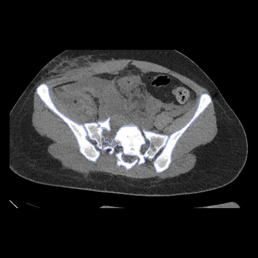
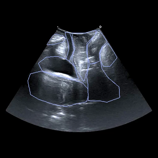
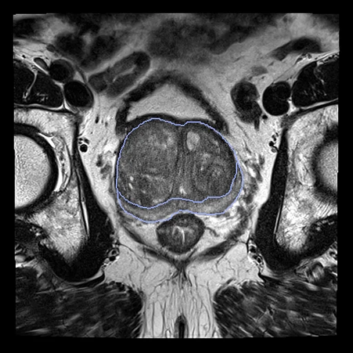
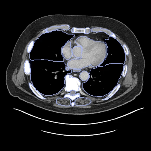
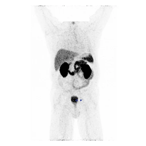
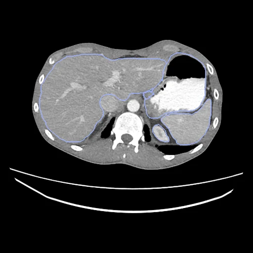
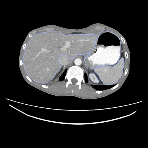
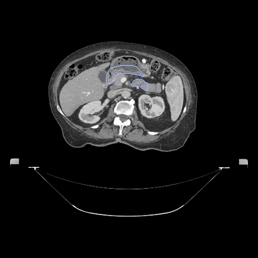
7725 test cases in 415 datasets. Each picture is the middle slice of the first test case, with the annotations results are checked against in colour; from a dataset's images, Next walks on through every dataset.
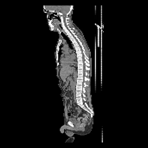
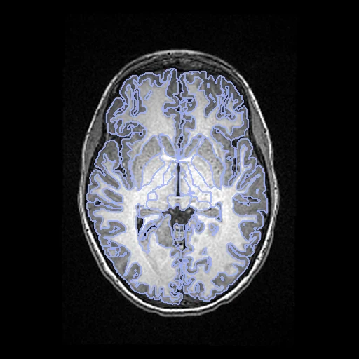
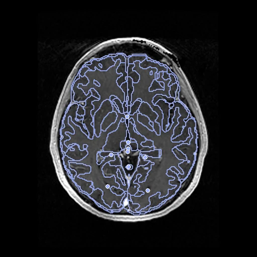
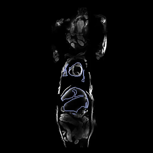
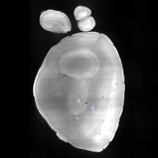
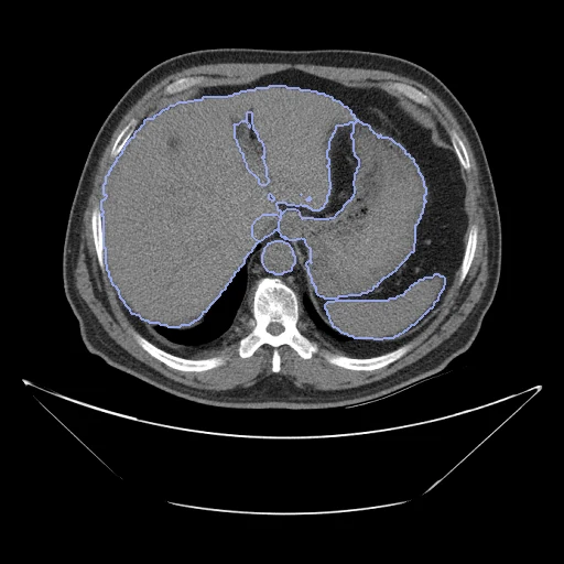
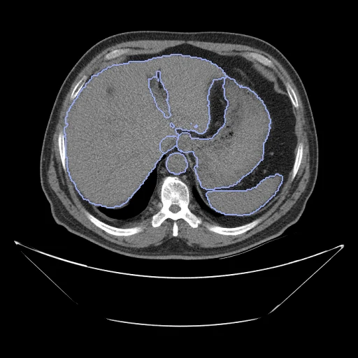
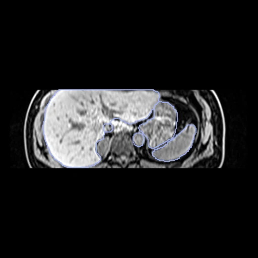
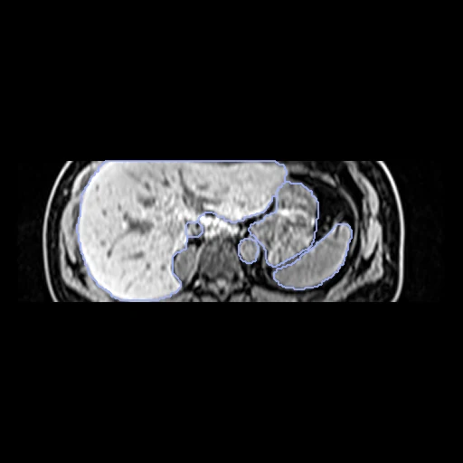
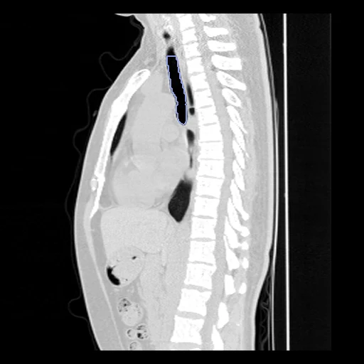
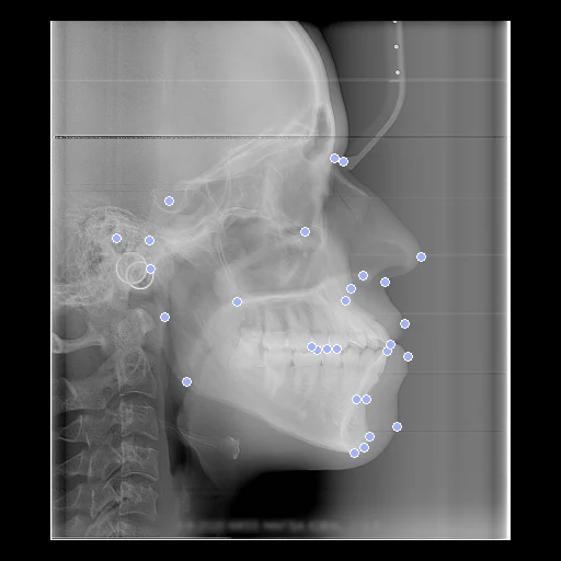
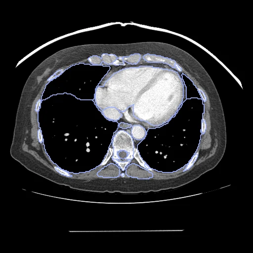
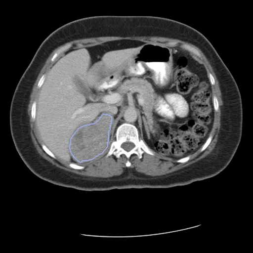
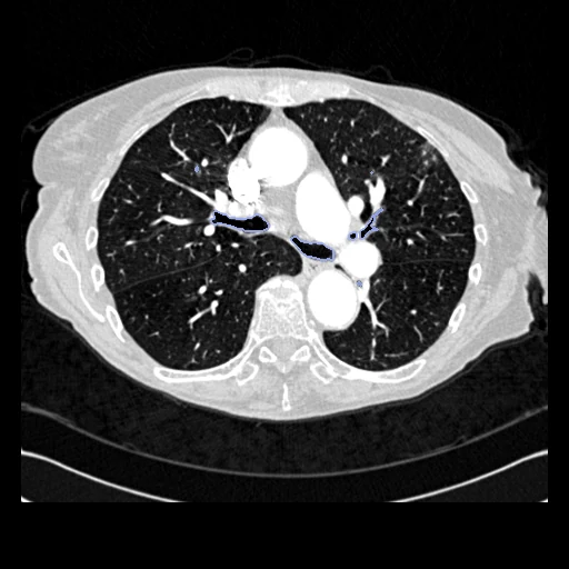
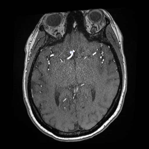
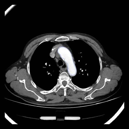
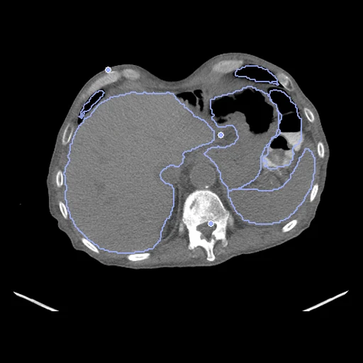
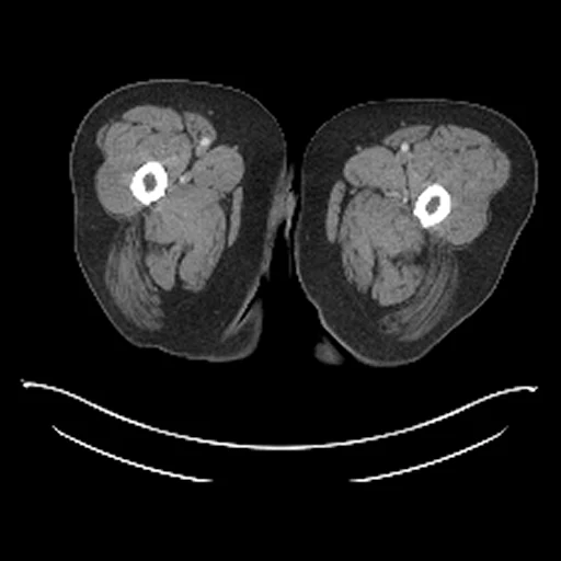
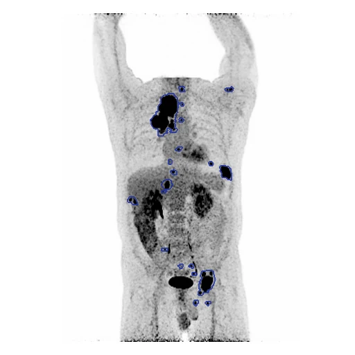
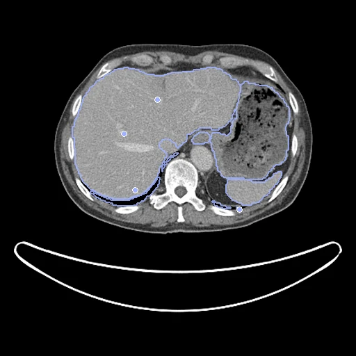
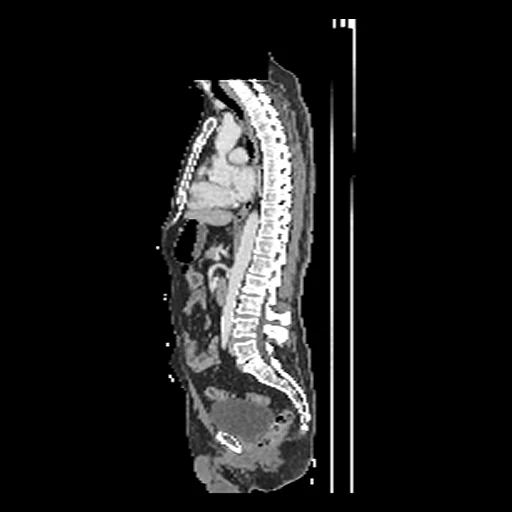
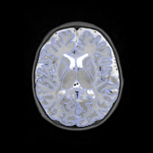
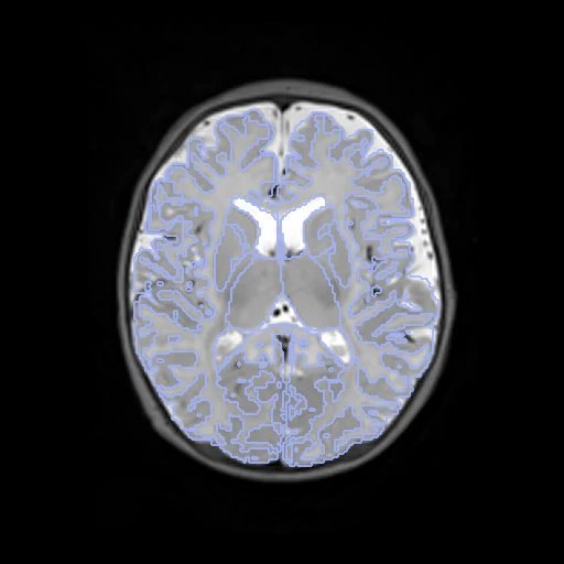
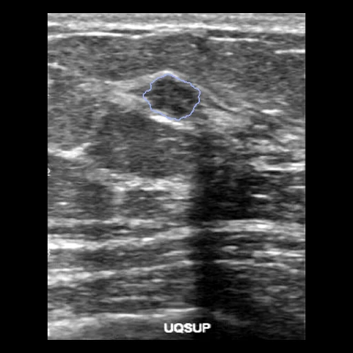
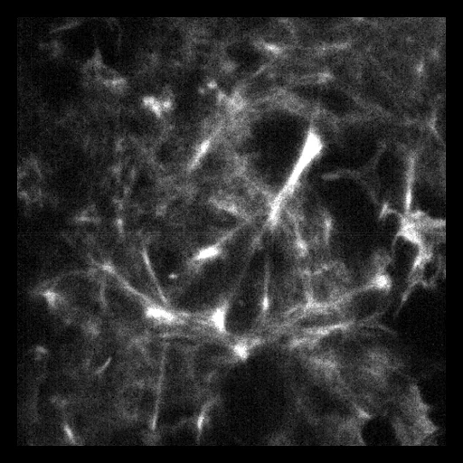
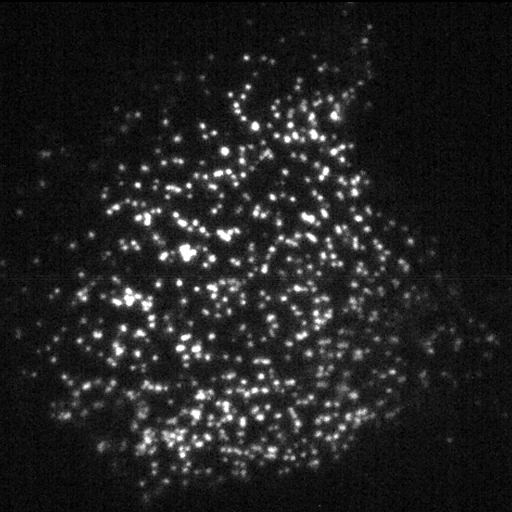
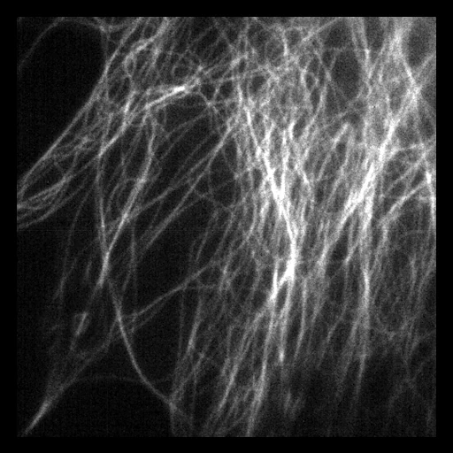
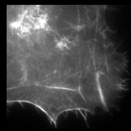
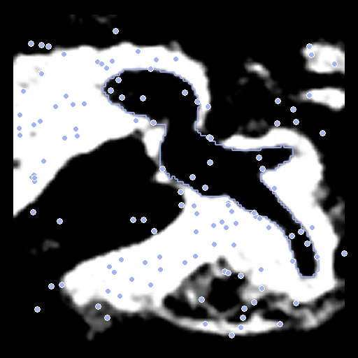
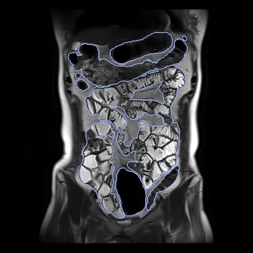
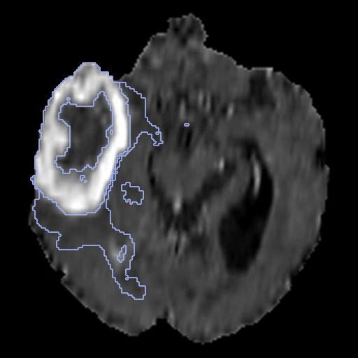
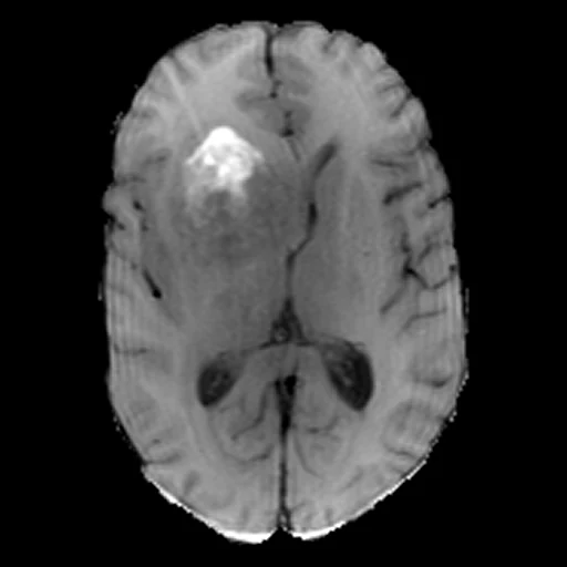
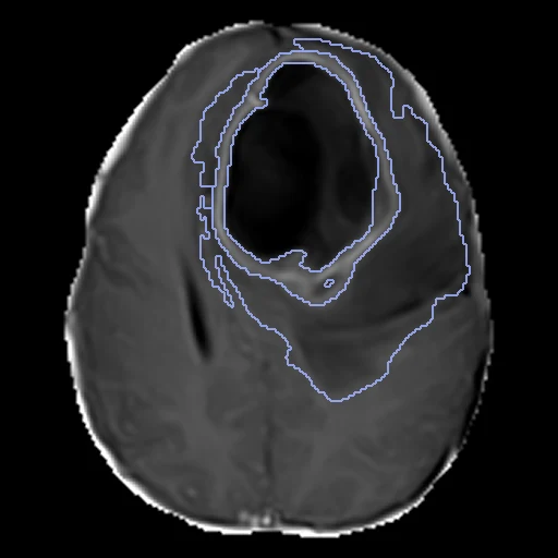
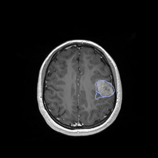
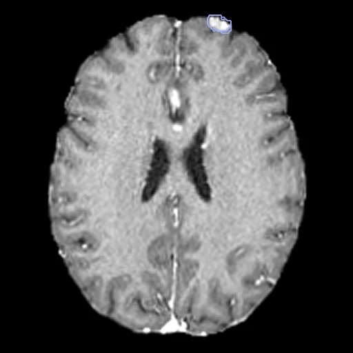
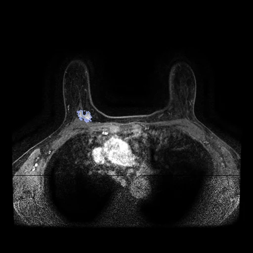
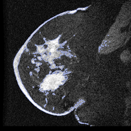
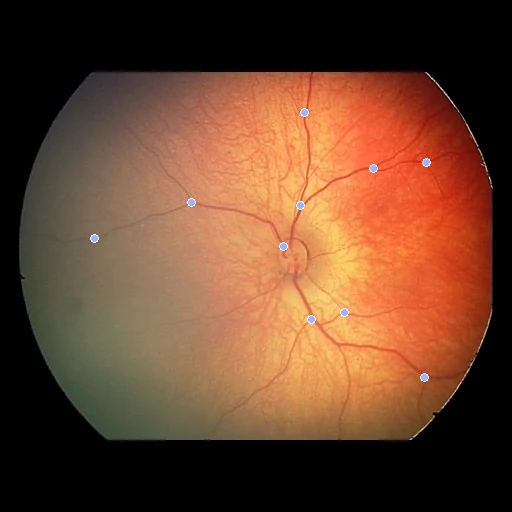
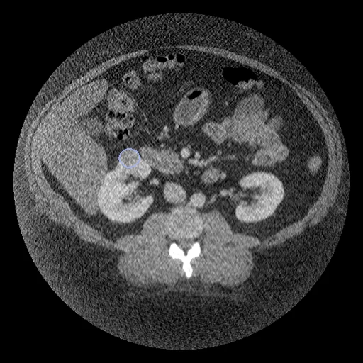
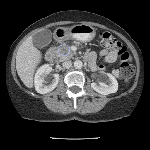
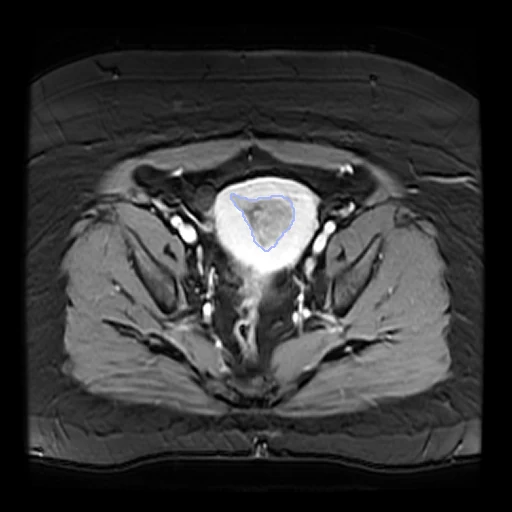
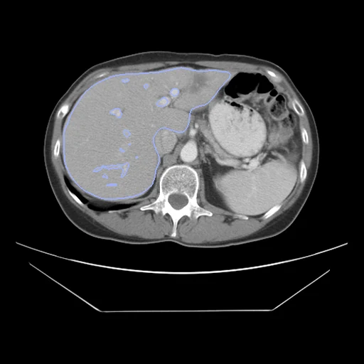
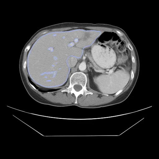
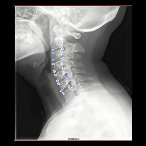
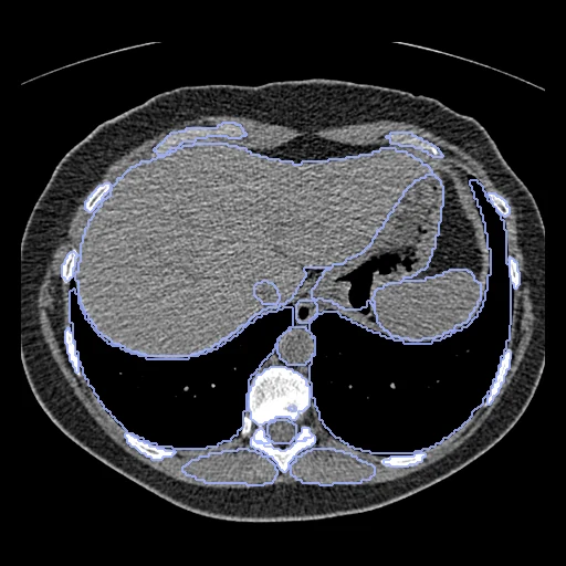
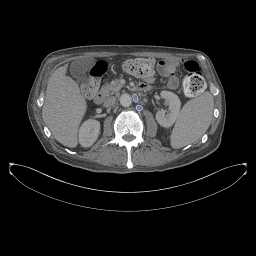
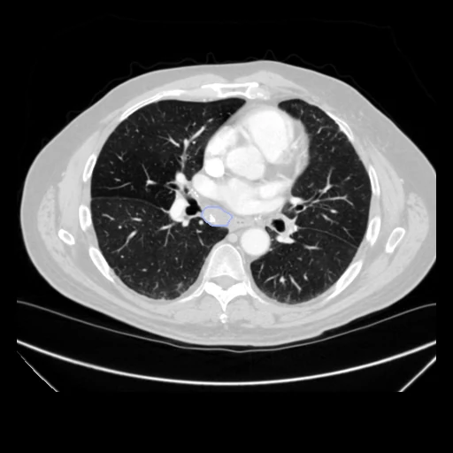
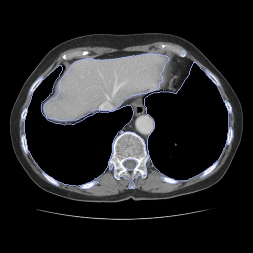
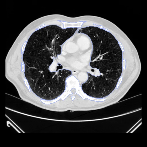
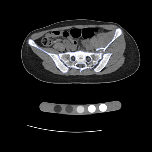
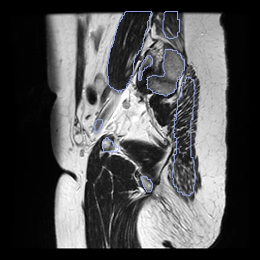
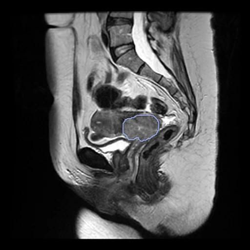
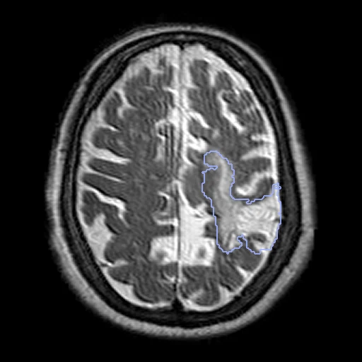
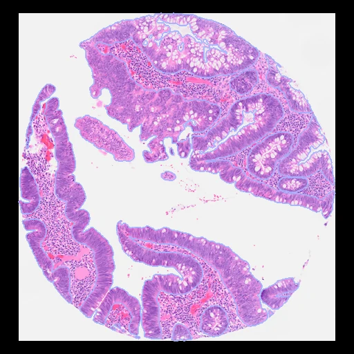
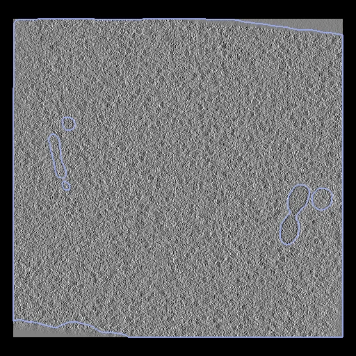
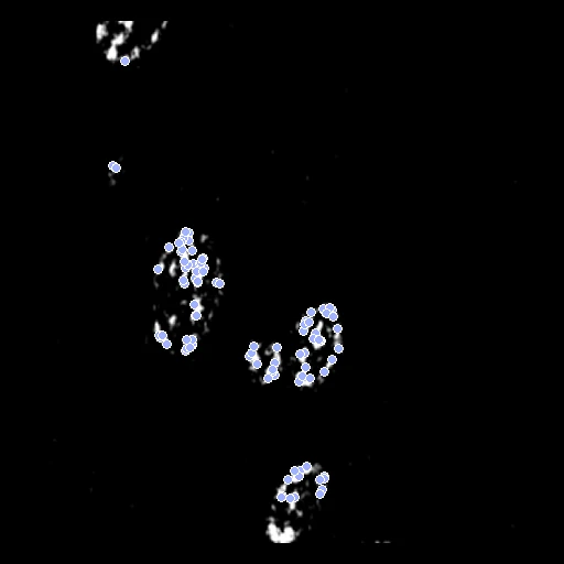
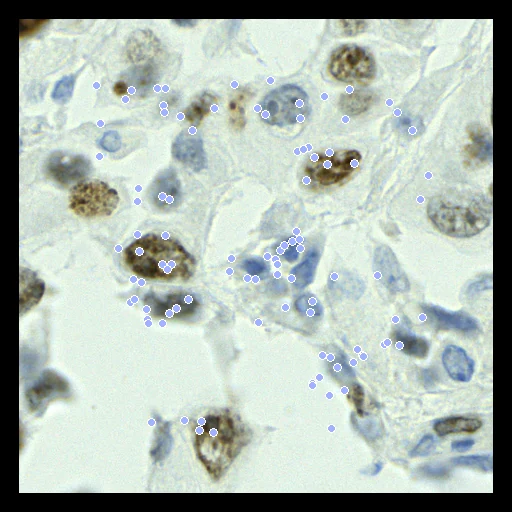
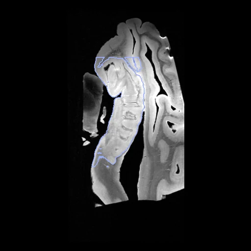
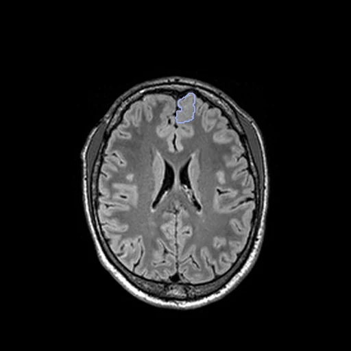
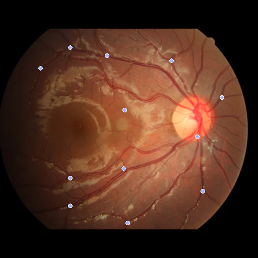
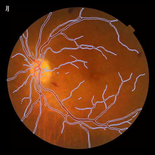
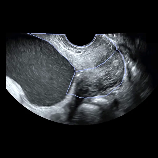
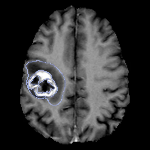
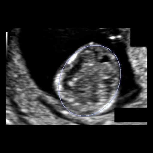
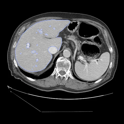
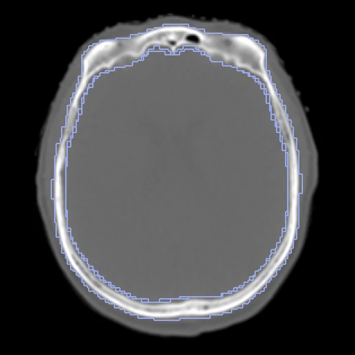
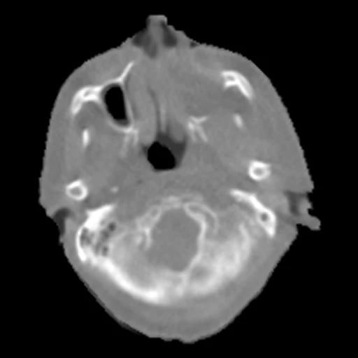
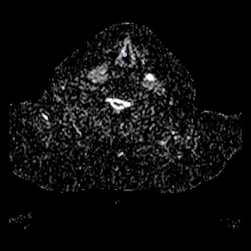
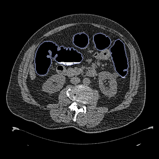
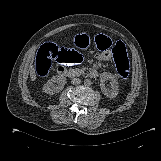
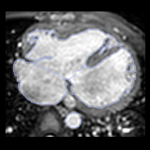
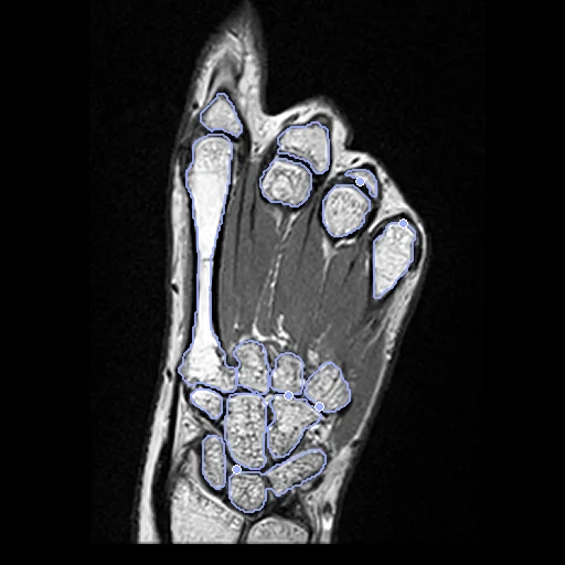
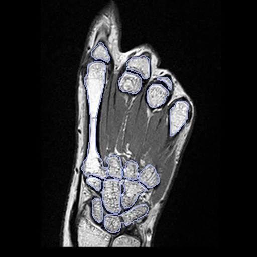
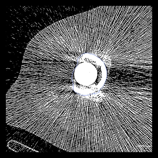
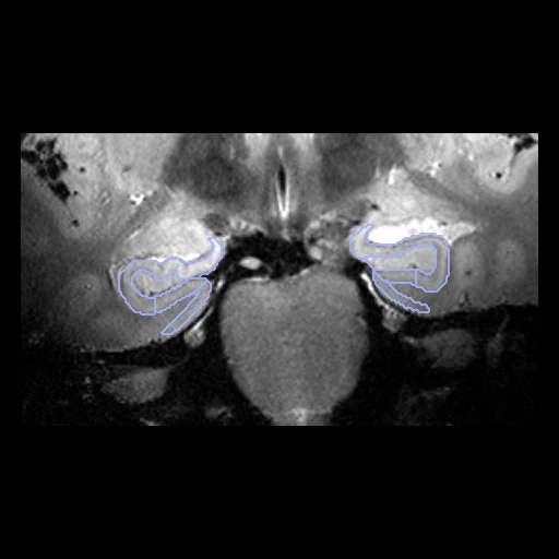
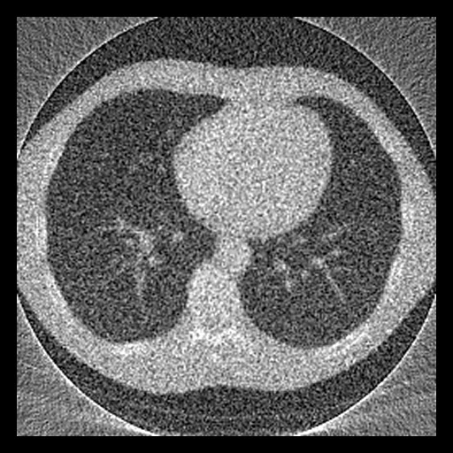
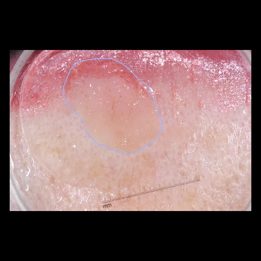
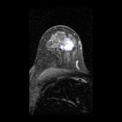
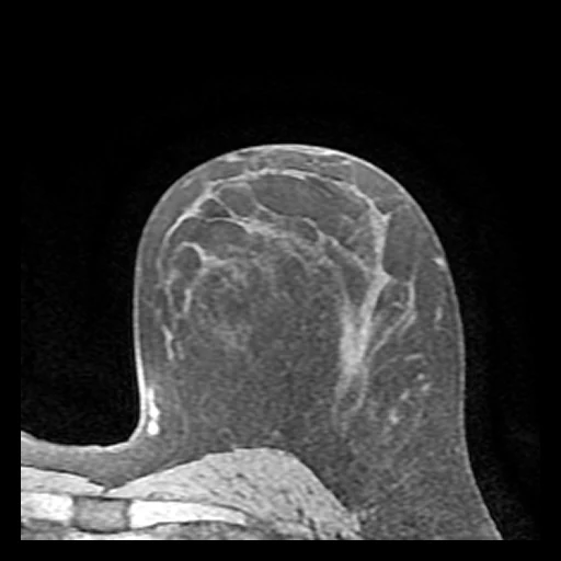
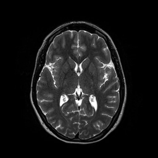
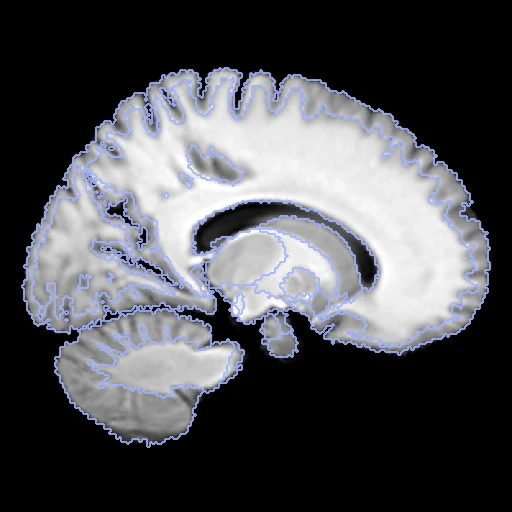
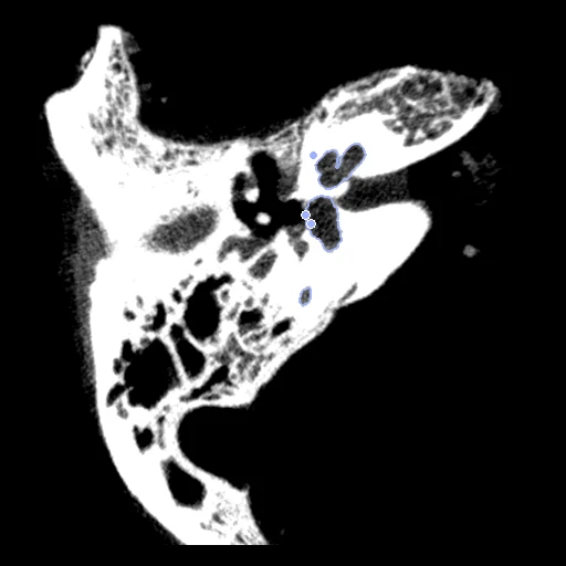
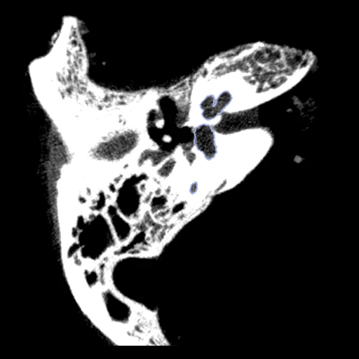
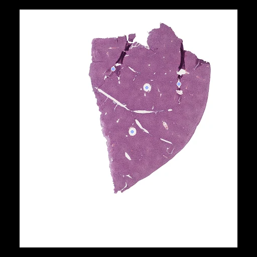
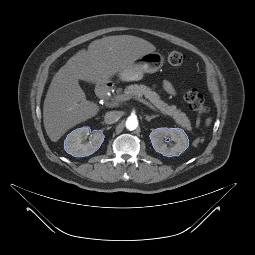
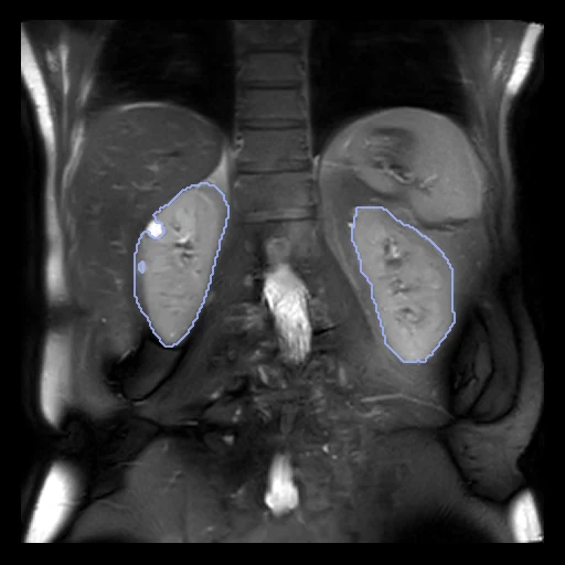
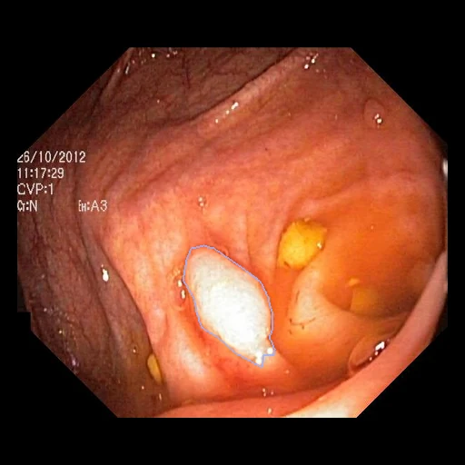
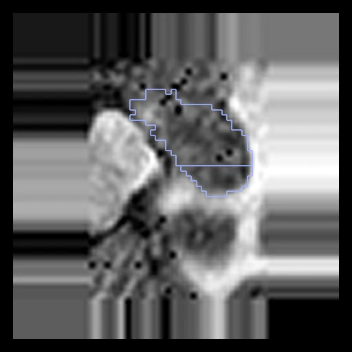
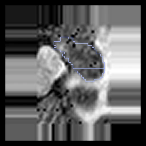
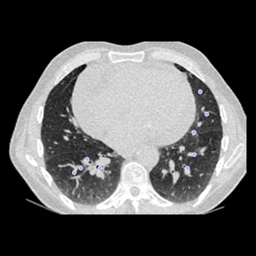
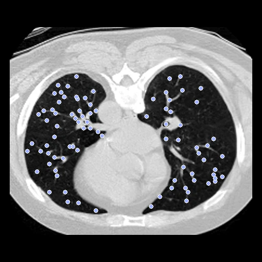
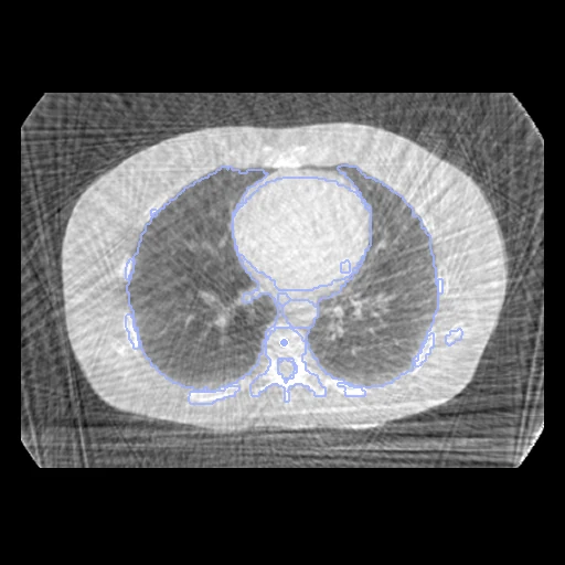
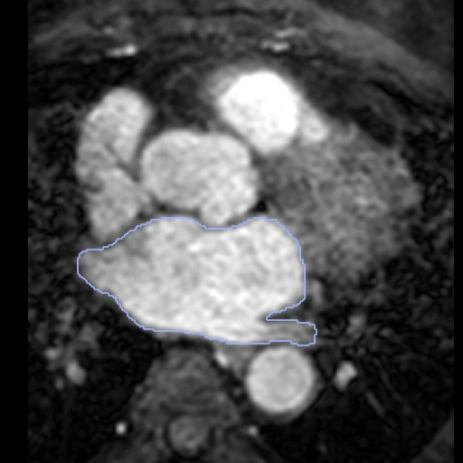
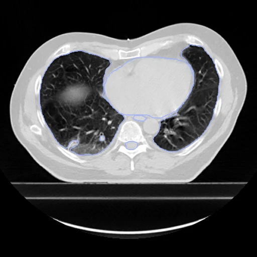
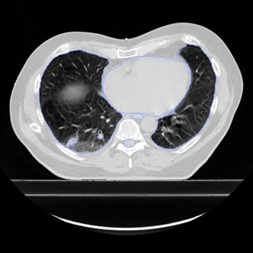
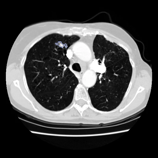
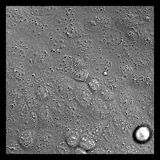
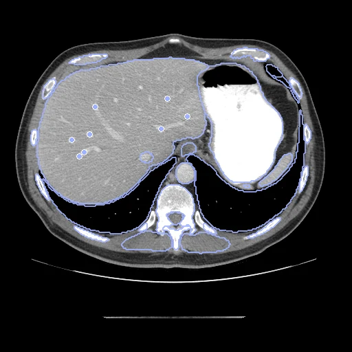
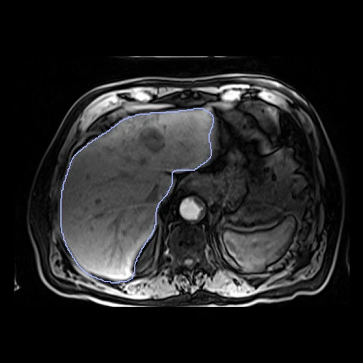
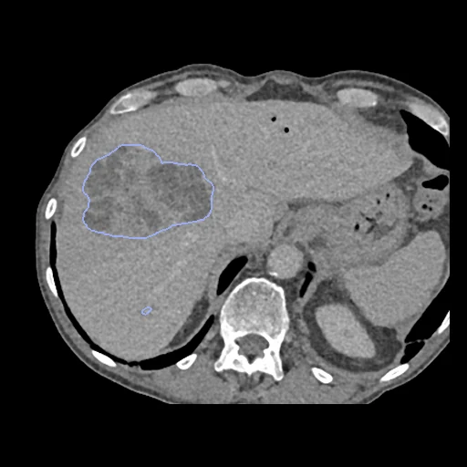
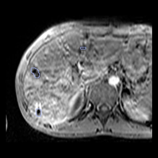
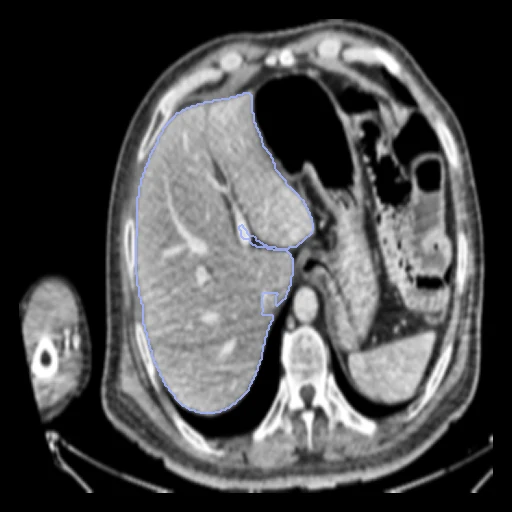
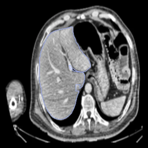
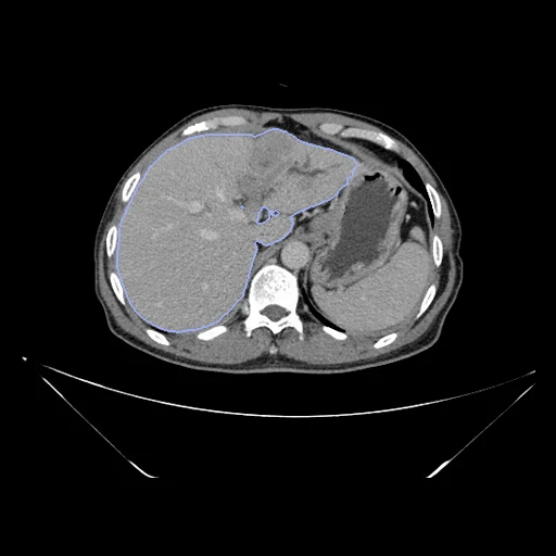
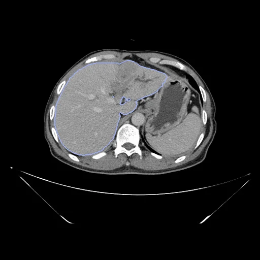
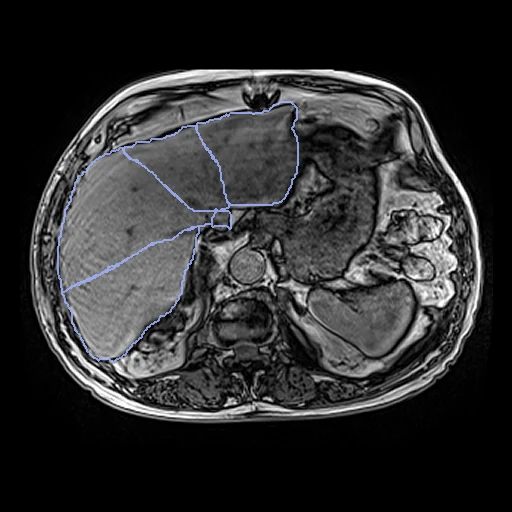
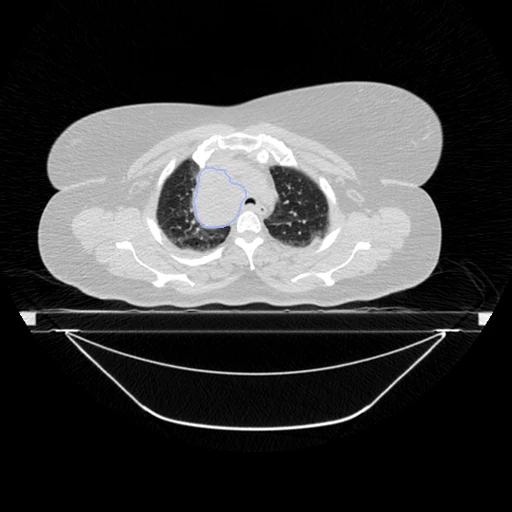
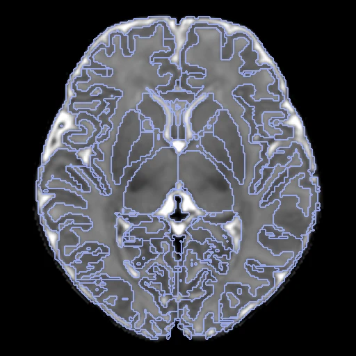
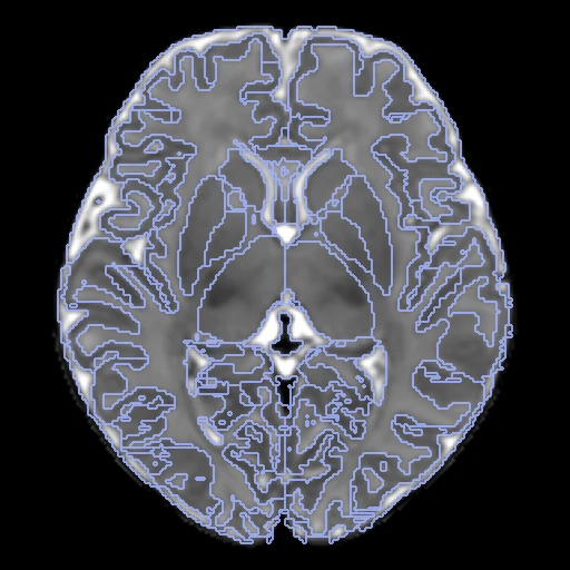
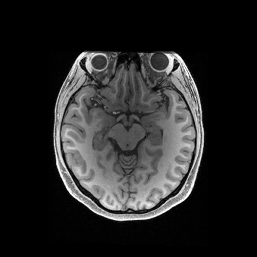
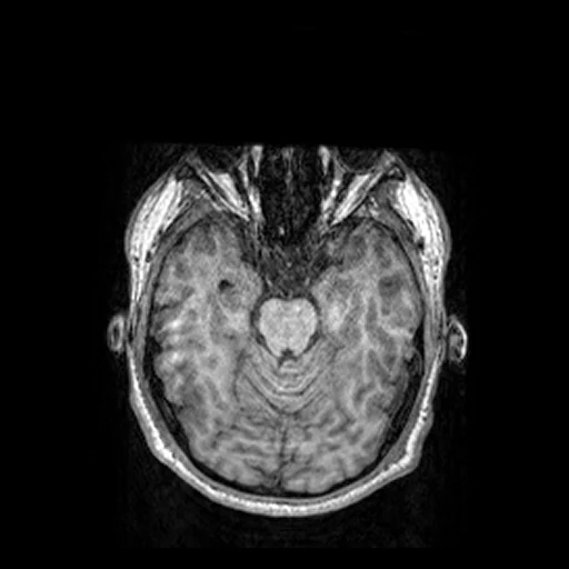
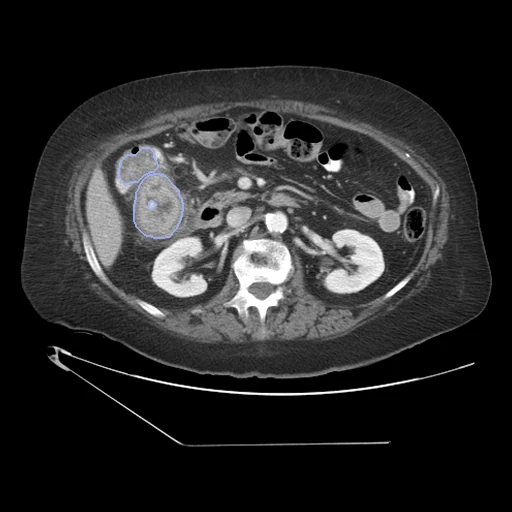
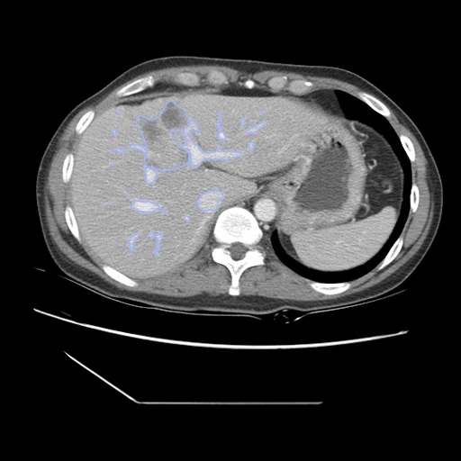
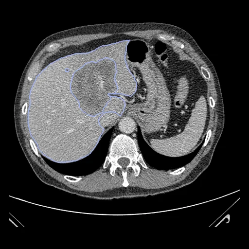
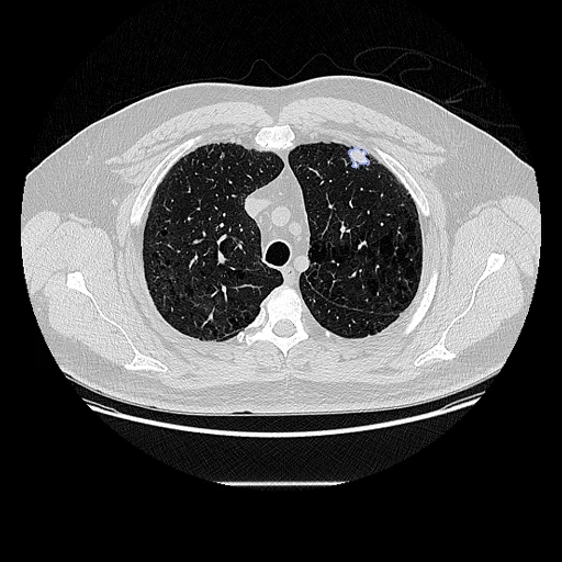
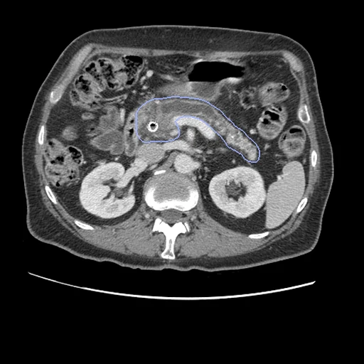
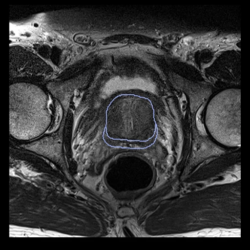
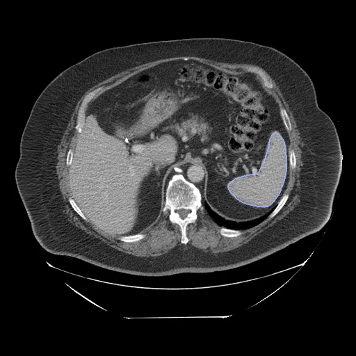
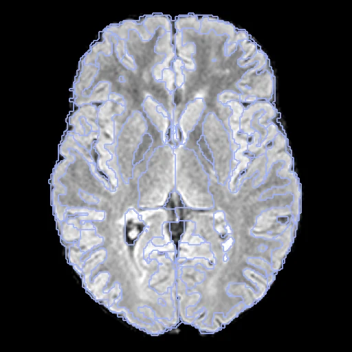
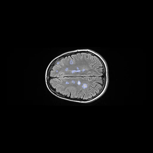
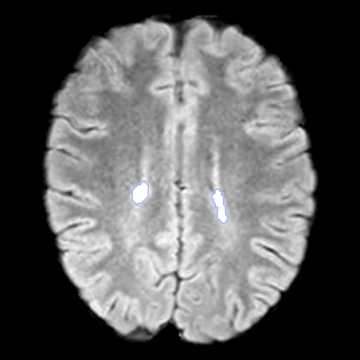
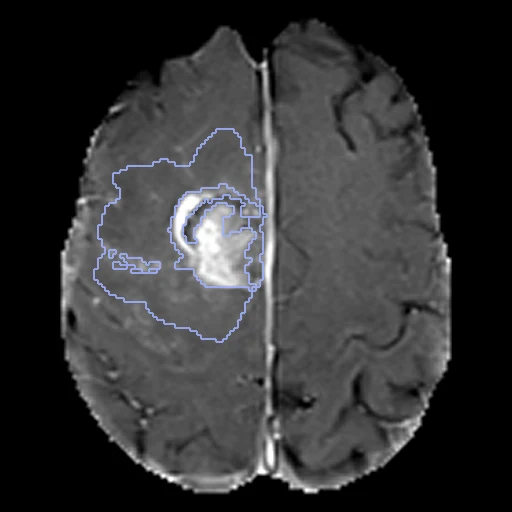
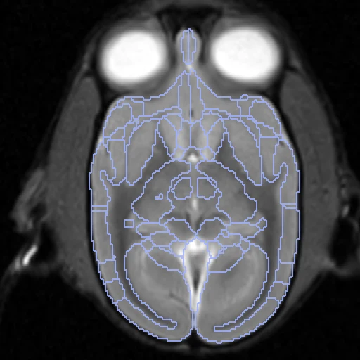
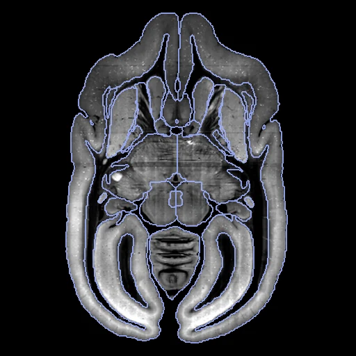
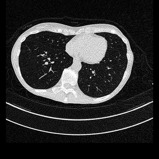
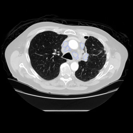
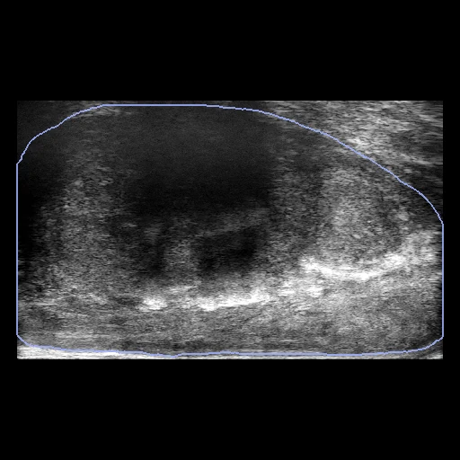
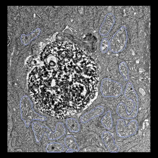
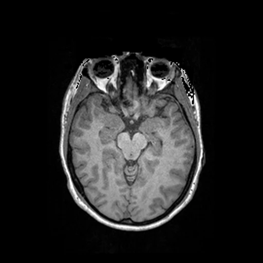
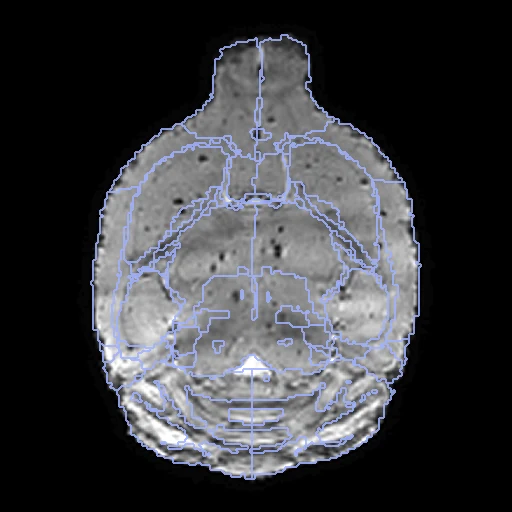
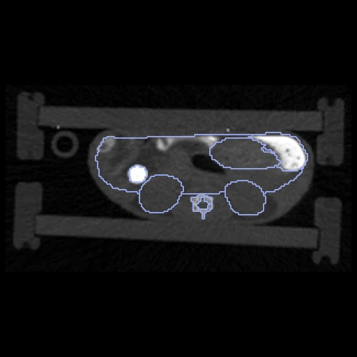
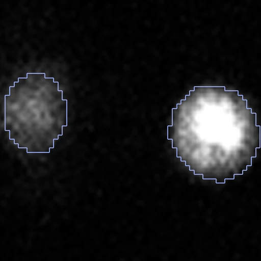
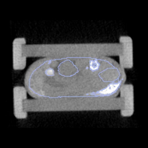
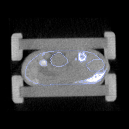
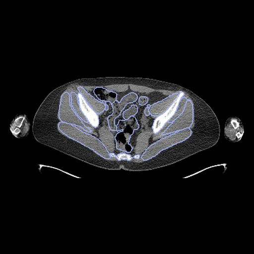
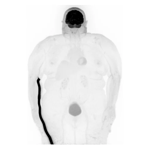
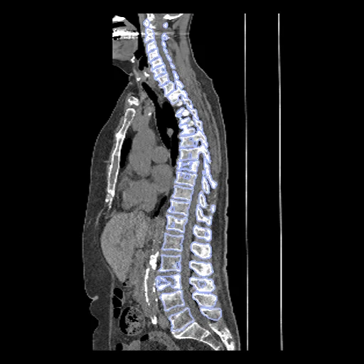
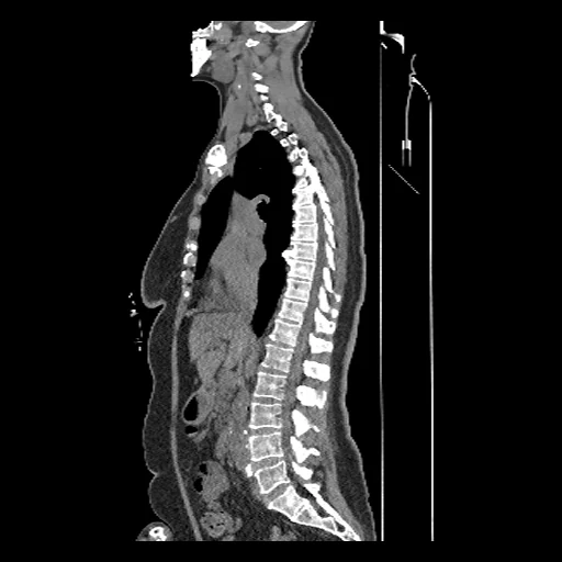
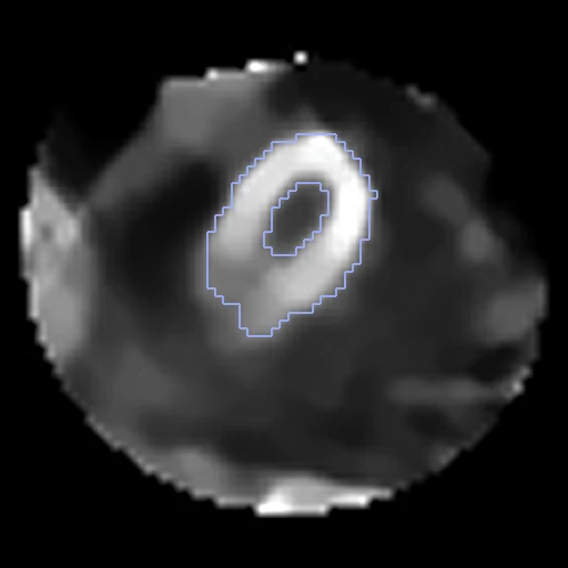
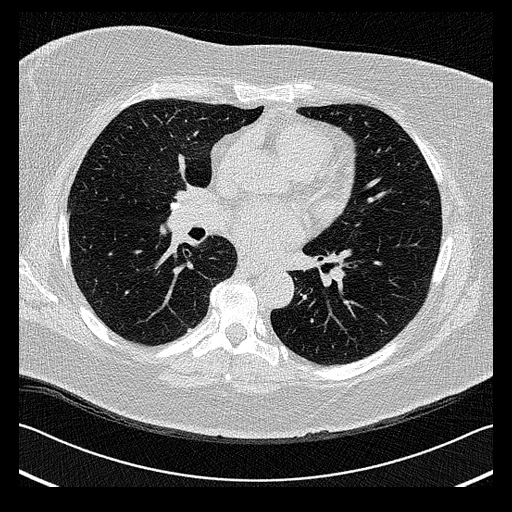
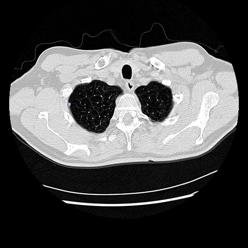
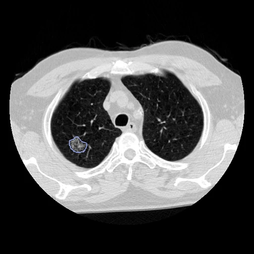
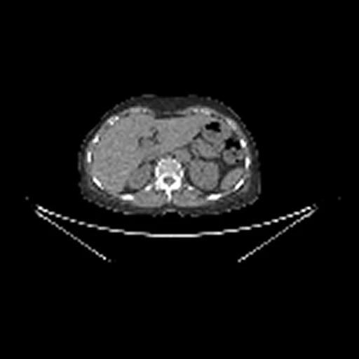
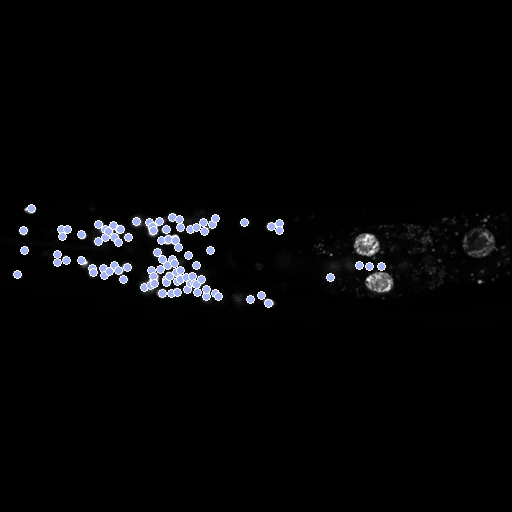
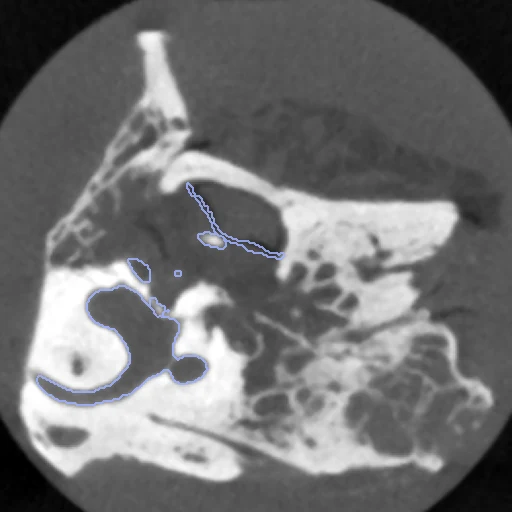
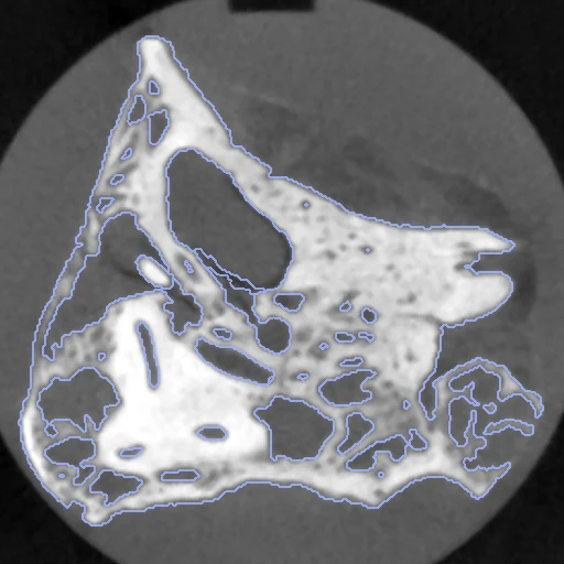
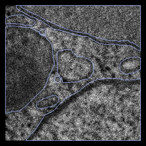
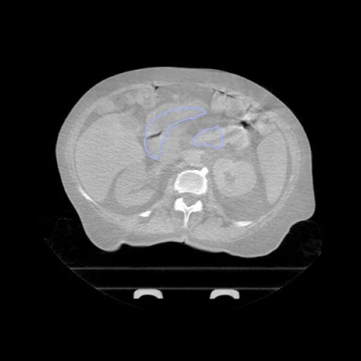
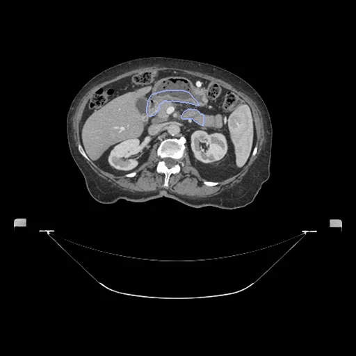
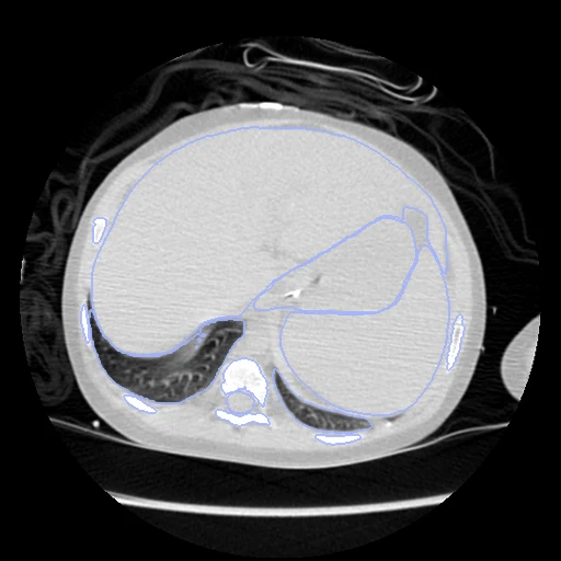
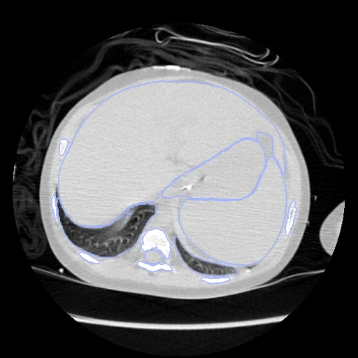
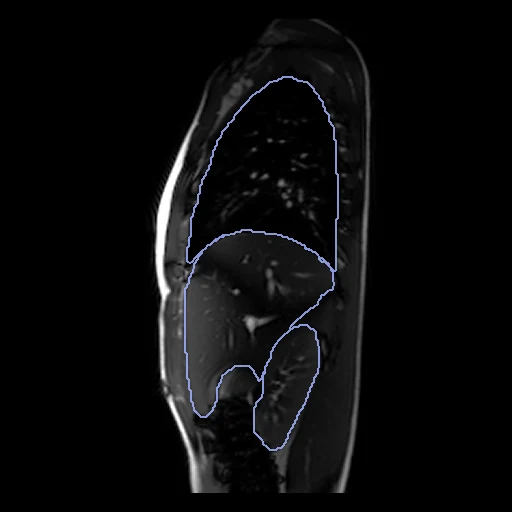
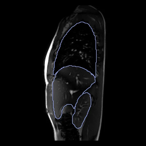
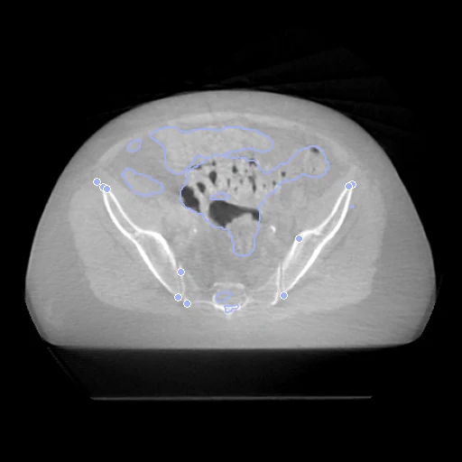
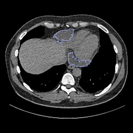
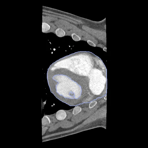
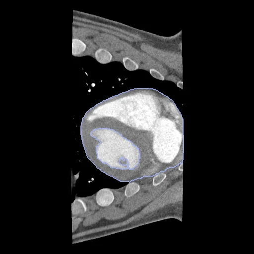
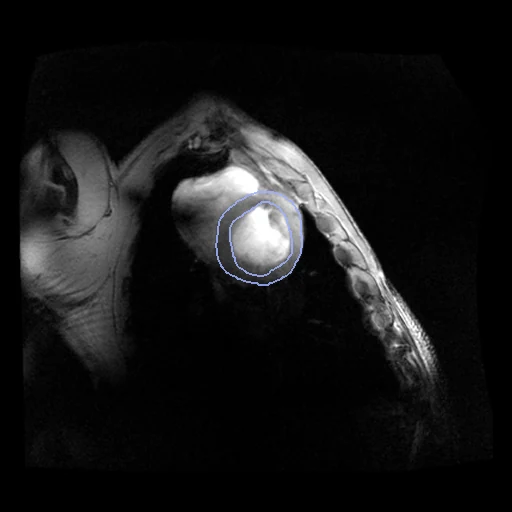
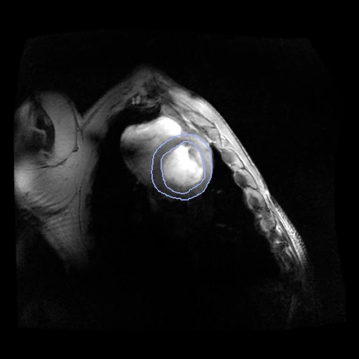
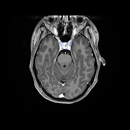
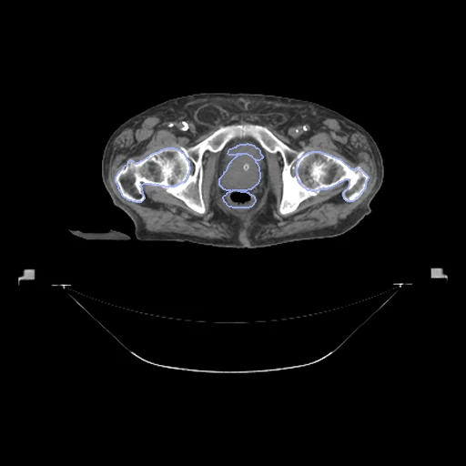
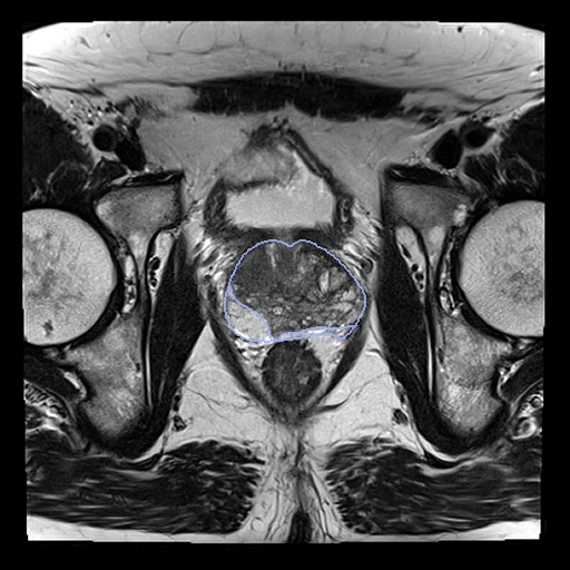
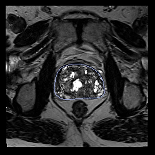
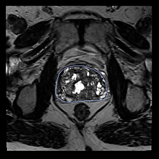
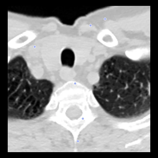
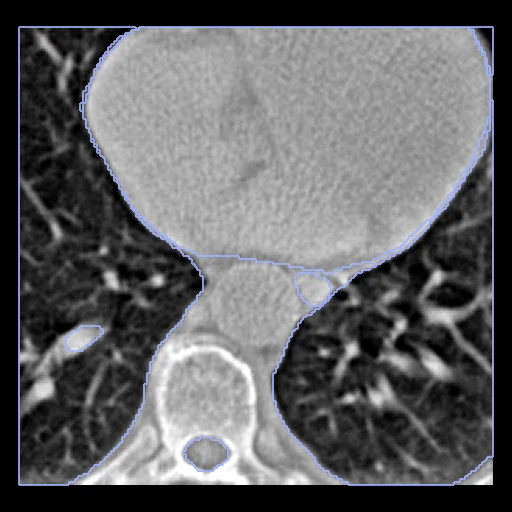
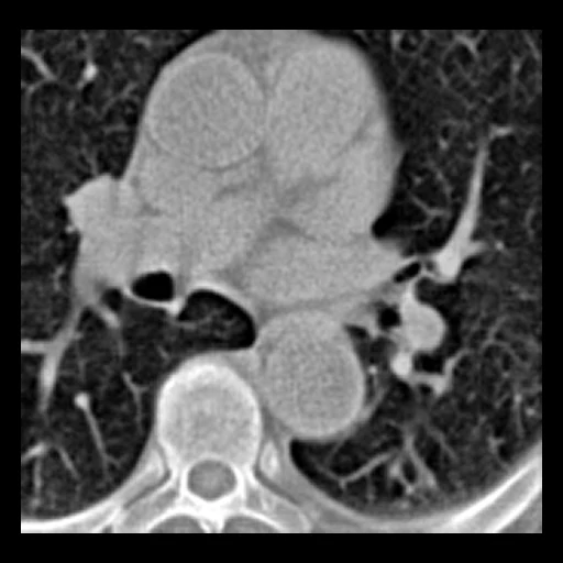
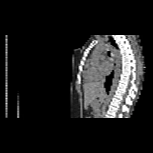
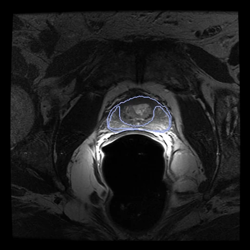
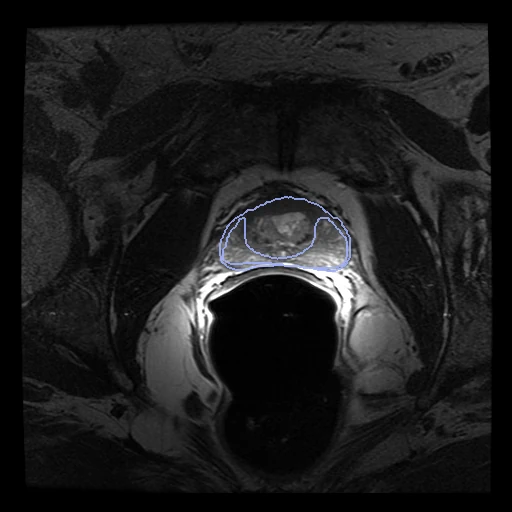
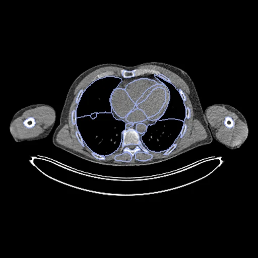
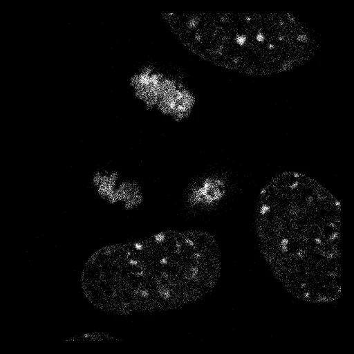
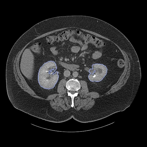
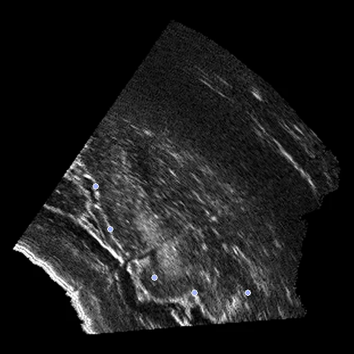
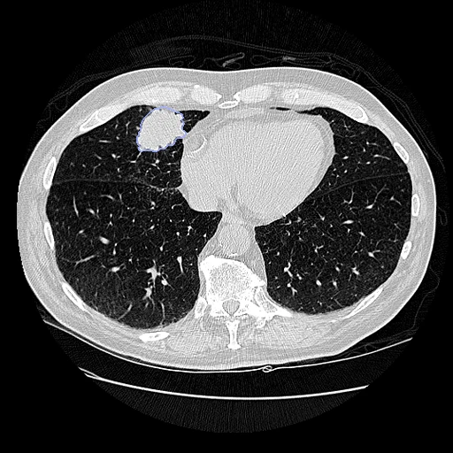
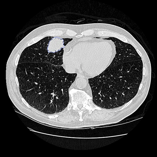
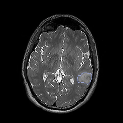
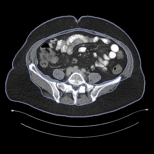
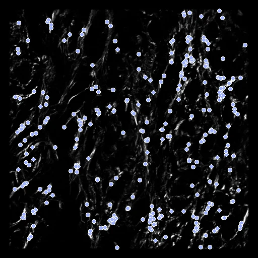
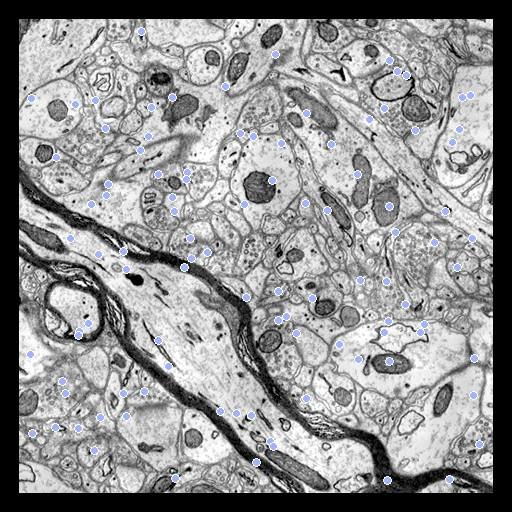
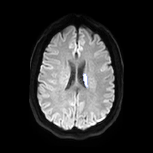
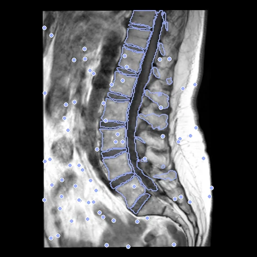
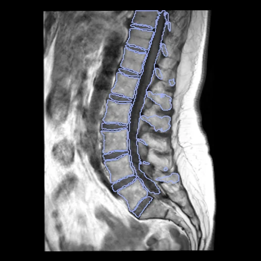
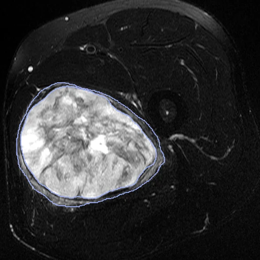
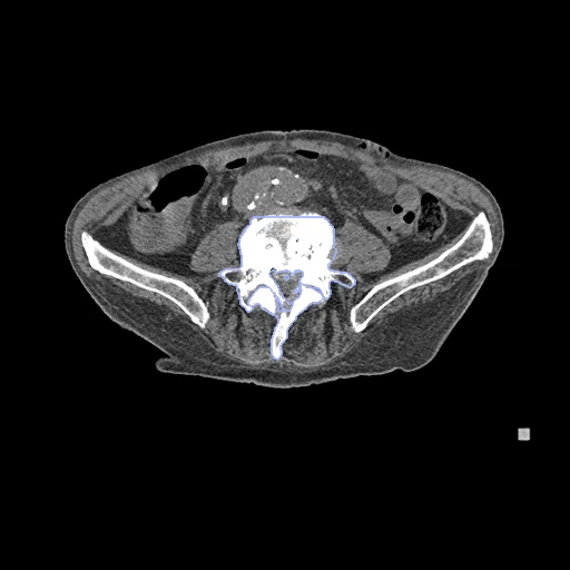
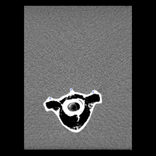
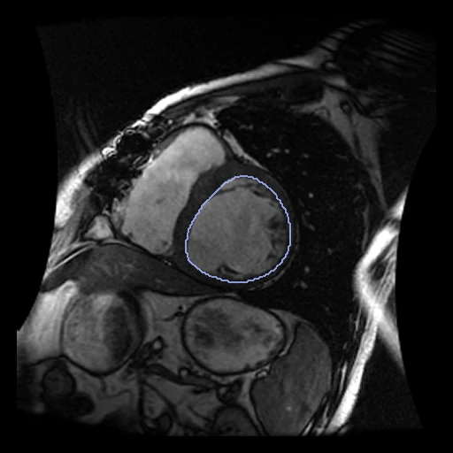
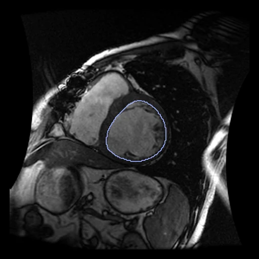
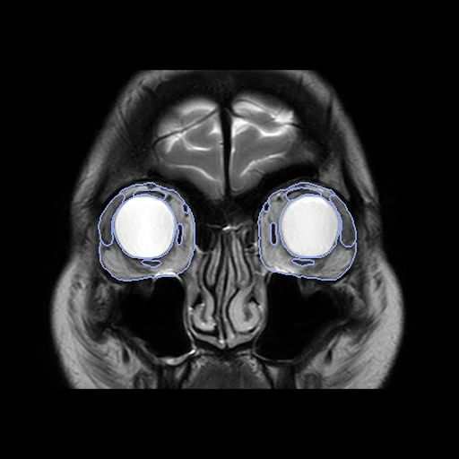
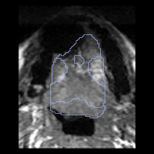
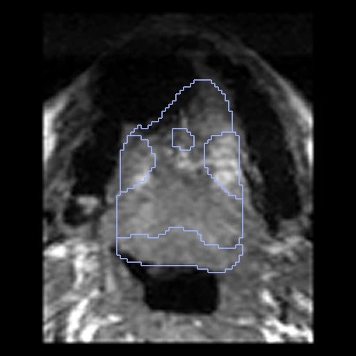
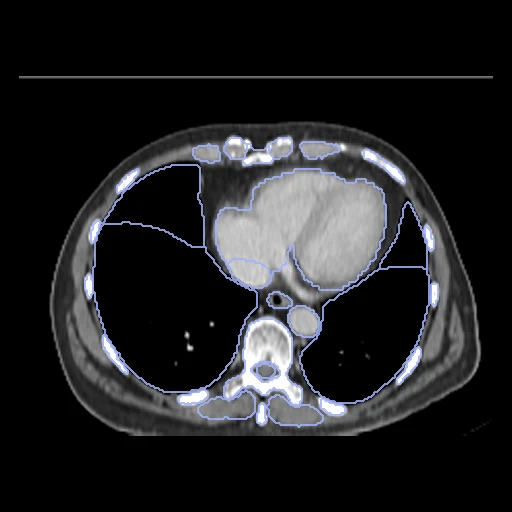
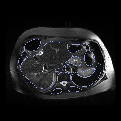
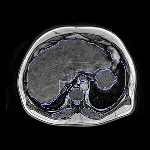
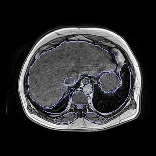
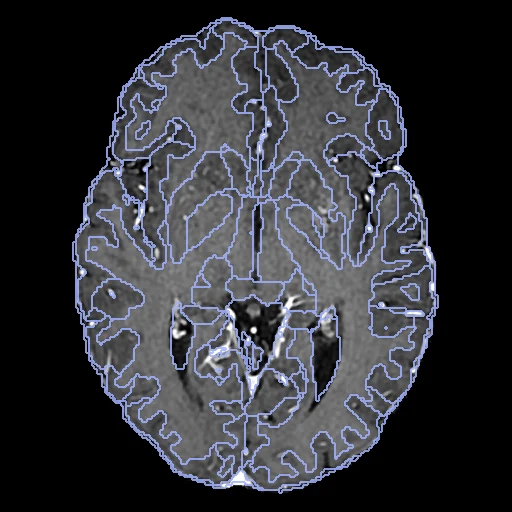
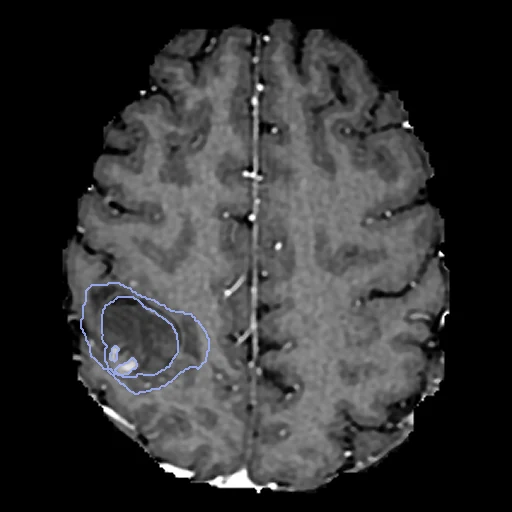
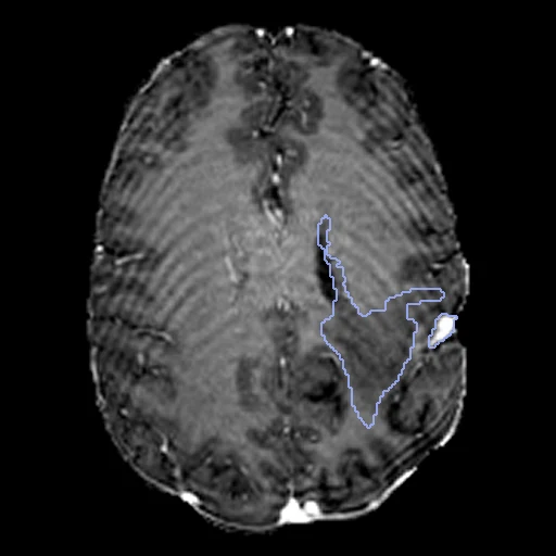
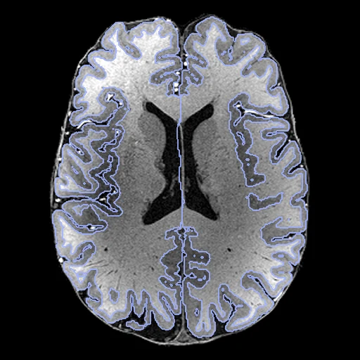
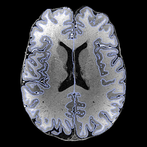
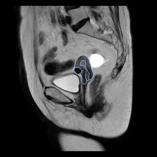
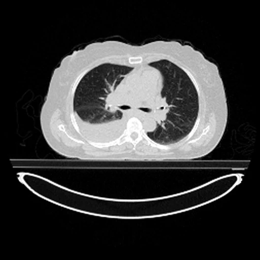
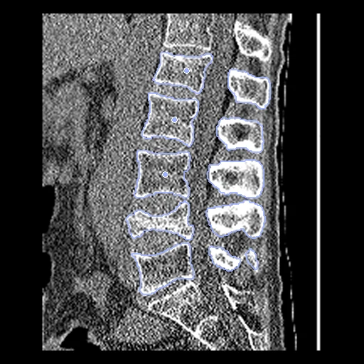
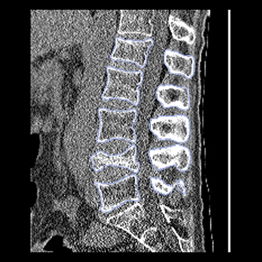
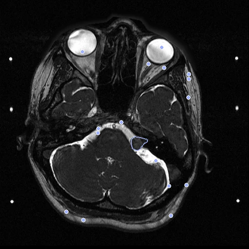
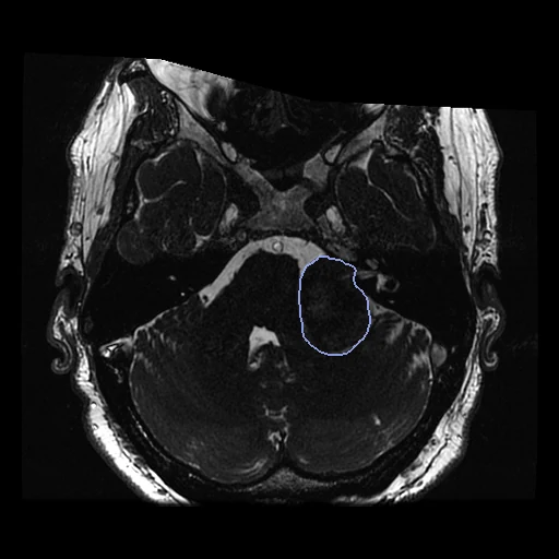
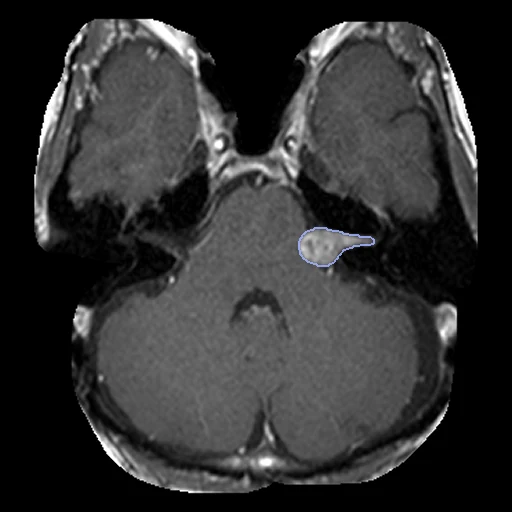
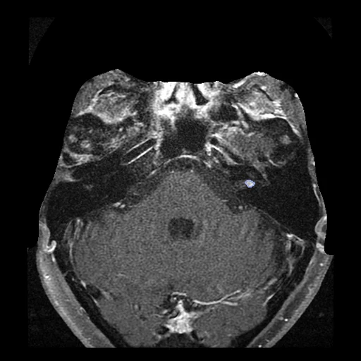
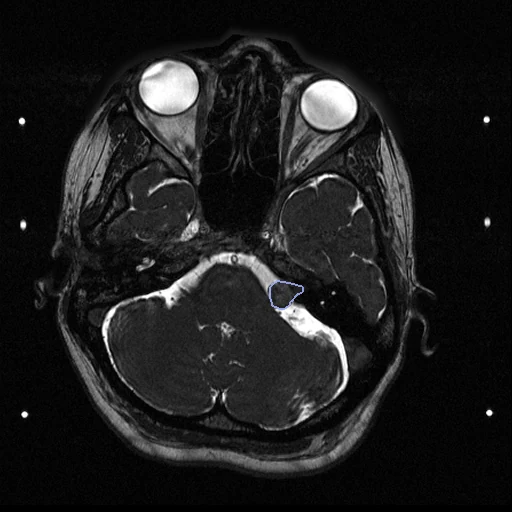
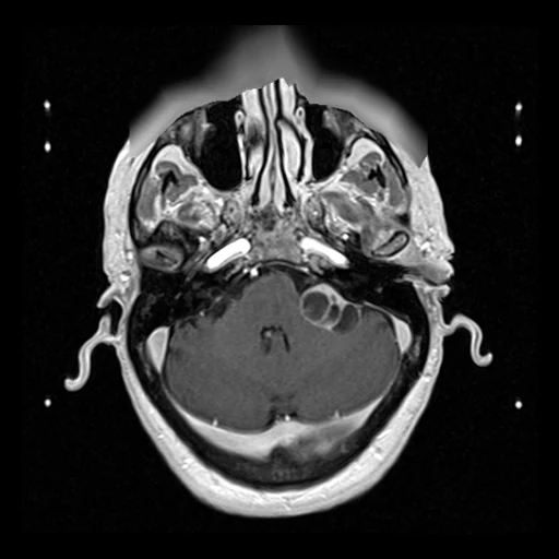
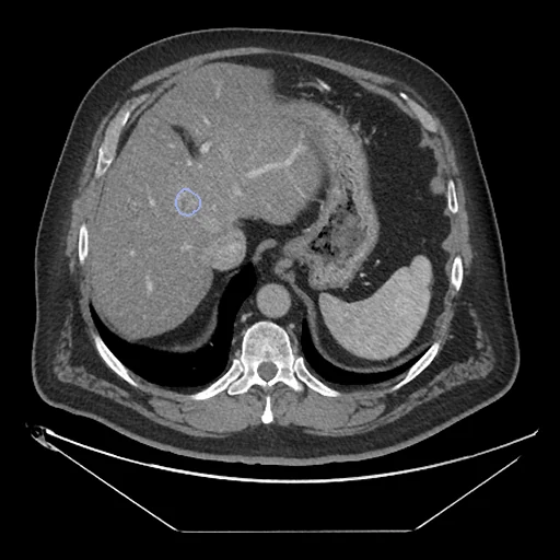
What results are checked against. by experts drawn or placed by people. by a program, verified by people found by a program, then checked. by a program the output of a program: a weaker check, and unfair to a method that relies on the same program. a known deformation two images aligned by construction, one of them deformed by a known smooth transform: exact, but on a deformation no body made. as released by a challenge, origin not documented.
Looked at for registration: 1616 public datasets, each with why it is in or out
Every public dataset found that could check a registration, 1616 so far. A dataset enters the main leaderboards when people drew, placed or verified its annotations, when it can be downloaded without a private agreement, and when no other dataset here already holds its images. Each is sorted, with its reason: to build (priority 1 for those that fill a gap of the corpus), waits for a tool (2D, 2D to 3D, video, meshes, images too large to read whole), truth to make, needs an account or a request, or left out.
Thorax · 43
| Dataset | Problem | Pairs | Checked against | Access | Licence | Status | Notes | To make the truth |
|---|---|---|---|---|---|---|---|---|
| 4D-Lung | lung 4D CT and CBCT, same patient | 20 patients | one oncologist contoured all 4D CT phases and a few CBCTsboth images | direct | CC BY 3.0open | left out | Learn2Reg ThoraxCBCT, in the corpus, already holds these images. status change: the collection page says 7 of 20 patients have gold coils implanted as fiducials in or near the tumour, visible on 4D CT and 4D CBCT; this gives point truth not noted before. Learn2Reg ThoraxCBCT holds these images. status change: Round 4 count: listed in 2 of the 38 round-4 surveys and benchmark papers (OncoReg 2025; Lung250M-4B 2023). Used in 1 of the 2D and microscopy registration papers read (PolyPose 2025 (as Learn2Reg ThoraxCBCT)). | threshold the coils on each phase of 4D CT and 4D CBCT, centroid per coil (or coil end points) = landmark, a person checks the match, 7 patients; limits: coils can migrate, motion blur on CBCT, few points per patient |
| ACRIN-NSCLC-FDG-PET (ACRIN 6668) | NSCLC: FDG PET/CT and CT before and after chemoradiation | 242 patients; 236 with >=2 studies | Central SUV reads only (no masks); BAMF AI labels in IDCone image | direct | CC BY 3.0open | truth to make no truth on both images; PET and CT of one PET/CT scan are aligned by the scanner, so a known warp on the CT gives exact PET-CT truth | Fails truth. | PET and CT of one PET/CT scan are aligned by the scanner, so a known warp on the CT gives exact PET-CT truth; for baseline vs post-treatment CT, two people place 20 landmarks (airway and vessel branches, vertebrae) on 10 pairs, TotalSegmentator labels on both as a weaker check. |
| Anti-PD-1_Lung / Anti-PD-1_MELANOMA | PET/CT before and during immunotherapy | Lung: 46 patients, 40 with 2 studies; Melanoma: 47 patients | Noneone image | direct | CC BY 3.0 (Lung); Melanoma NIH Controlled Data Access Policyopen | truth to make no truth on both images; PET and CT of one PET/CT scan are aligned by the scanner, so a known warp on the CT gives exact PET-CT truth | Fails truth. | PET and CT of one PET/CT scan are aligned by the scanner, so a known warp on the CT gives exact PET-CT truth; across visits, two people place 20 skeletal and vessel landmarks on 10 Lung pairs (Melanoma is controlled access), TotalSegmentator labels on both as a weaker check. |
| Coltea-Lung-CT-100W | Triphasic chest CT (native, arterial, venous), same session | 100 patients x 3 phases | Noneone image | direct | CC BY-SA 4.0 (code and data set)open | truth to make no truth on both images; Two people place 20 airway bifurcations and bone points on the native, arterial and venous phases of 10 patients | Fails truth criterion Licence checked on 2026-10-10 at https://github.com/ristea/cycle-transformer; derived test pairs shareable: yes; commercial use: yes. README 'License' section; share-alike applies to derived pairs if they are distributed | Two people place 20 airway bifurcations and bone points on the native, arterial and venous phases of 10 patients; phases are close in time, so this tests contrast change more than deformation. Check the licence first. |
| COPDGene full cohort | inspiratory and expiratory breath-hold CT, same subject, plus 5-year follow-up | about 10,000 subjects (unverified) | none beyond the 10 DIR-Lab pairs (300 landmarks each, covered) | controlled | application to COPDGene (unverified)ask | needs a request the data needs a request or a data use agreement | Unverified; source of DIR-Lab COPD. Large pool for self-made truth (e.g. Lung250M-style keypoints). status change: usage counted from papers: the training pool of the UNC foundation models; test always on the 10 DIR-Lab COPD pairs. status change: Round 4 count: listed in 6 of the 38 round-4 surveys and benchmark papers (Boveiri 2020 review; Bharati 2022 review; Zou 2022 review (Front Oncol); Chen X 2021 review (Prog Biomed Eng); uniGradICON 2024; multiGradICON 2024). | Lung250M-4B recipe on new inspiration/expiration pairs: corrField keypoints with a person verifying a sample on 10 pairs (favours corrField-like methods), plus 50 bifurcations by two people; lungmask lobes on both as a weaker check. |
| COVID-CTPRED (CREATIS) | chest CT of COVID-19 patients: non-contrast, contrast and inspiration/expiration at admission, plus follow-up CT | target 800 patients; same-day insp/exp and contrast pairs plus follow-ups (counts unverified) | none | hidden | not stated; algorithms run on CREATIS computing, images stay thereask | left out its images or its truth are not released | Rare same-day insp/exp pairs in a COVID cohort, but images never leave the host. | two people place 20 airway/vessel landmarks on 10 insp-exp pairs (DIR-Lab style) |
| CT Images in COVID-19 (serial subset) | Unenhanced chest CT, serial scans of COVID-19 patients | 29 patients, 121 CTs | Noneone image | direct | CC BY 4.0open | truth to make no truth on both images; Two people place 20 airway and vessel bifurcations away from the opacities on 10 serial pairs | Fails truth. | Two people place 20 airway and vessel bifurcations away from the opacities on 10 serial pairs; lungmask lobes on both as a weaker check. Opacities change between scans, so they cannot serve as landmarks. |
| CT-RATE | non-contrast chest CT with reports, some patients with more than one scan | 25,692 CTs from 21,304 patients (up to ~4,400 repeat scans; same-day vs follow-up unverified) | none | account | CC BY-NC-SA 4.0, gated; no redistributionnon-commercial | needs an account the download needs an account | Patient and scan IDs are in file names; repeat intervals must be checked. | TotalSegmentator lobes and airways on both (weaker); 20 airway bifurcations by two people on 10 pairs |
| CT-vs-PET-Ventilation-Imaging | inhale and exhale breath-hold CT, free-breathing 4DCT and Galligas PET/CT, same subject same session | 20 patients: one inhale/exhale BHCT pair, 4DCT phases and PET/CT each | none spatial (lung function tests only)one image | direct | CC BY 4.0open | truth to make no truth on both images; Two people place 50 airway and vessel bifurcations on the inhale/exhale breath-hold CTs of 10 patients (skip truncated scans) | Fails truth. Two BHCT pairs show little motion, some scans truncated. Candidate for self-made landmarks. status change: Round 4 count: listed in 1 of the 38 round-4 surveys and benchmark papers (Lung250M-4B 2023). | Two people place 50 airway and vessel bifurcations on the inhale/exhale breath-hold CTs of 10 patients (skip truncated scans); Galligas PET/CT is hybrid, so a known warp on its CT gives exact PET-CT truth. |
| DIR-Lab 4DCT and COPDgene | lung 4D CT and inspiration to expiration CT, same patient | 10 + 10 | 300 expert landmarks a pairboth images | request | non-commercial, password after a formnon-commercial | left out non-commercial terms with a password per requester and no redistribution, so the shared test images the corpus needs are impossible; two requests (2026-10) went unanswered, the lab having left Emory; Lung250M, LungDIRQA, POPI and L2R_NLST cover lung landmark registration | The historical reference. To add when the request is granted. Lung250M-4B already carries the COPDgene landmarks. status change: usage counted from papers: still the reference for lung landmark error, used by learning, implicit-neural and Gaussian methods alike. status change: Round 4 count: listed in 10 of the 38 round-4 surveys and benchmark papers (Fu 2020 review (PMB); Boveiri 2020 review; Bharati 2022 review; Zou 2022 review (Front Oncol); Chen X 2021 review (Prog Biomed Eng); Chen 2025 survey (MedIA); Learn2Reg 2020-21 (TMI 2022); uniGradICON 2024; multiGradICON 2024; Lung250M-4B 2023). | |
| DoseRAD2026 (0.35T MR-Linac MRI with registered CT) | 0.35 T MR-Linac bSSFP MRI and planning CT, thorax and abdomen, same patient | 115 patients: 75 released April 2026, 40 test held until 2030 | none for registration: the CT is already deformably registered to the MRI by the organisers, with manual quality controlone image | direct | CC BY-NC 4.0non-commercial | truth to make no truth on both images; the pairs are aligned by construction: apply a known smooth warp to the CT for exact MR-CT truth | data hosted on Hugging Face (link on the Zenodo record); a dose challenge, not a registration set; whether the unregistered CT is also shipped is unverified | the pairs are aligned by construction: apply a known smooth warp to the CT for exact MR-CT truth; two people place 15 landmarks on 10 pairs to check the given alignment |
| Duke Lung Cancer Screening Dataset 2024 (DLCS) | low-dose screening chest CT with nodule boxes | none: one (terminal) scan per patient; 1,613 public CTs | semi-automatic nodule 3D boxes | request | CC BY-NC-ND 4.0 (restricted access, request)non-commercial | needs a request the data needs a request or a data use agreement | Fails pairs (checked: paper says terminal exam only). Licence checked on 2026-10-10 at https://zenodo.org/api/records/13799069; derived test pairs shareable: unclear; commercial use: no. Zenodo record restricted; ND forbids sharing adapted material (resampled pairs) outside the team | not applicable (single time point); only usable for synthetic known-warp pairs |
| EMPIRE10 | lung CT, same patient | 30 | landmarks, fissures, kept by the organisersboth images | hidden | challenge use onlyask | left out | Scoring runs on their server only. status change: Round 4 count: listed in 8 of the 38 round-4 surveys and benchmark papers (Boveiri 2020 review; Bharati 2022 review; Zou 2022 review (Front Oncol); Chen X 2021 review (Prog Biomed Eng); Chen 2025 survey (MedIA); Li 2023 dataset collection (ACM CSUR); Learn2Reg 2020-21 (TMI 2022); Lung250M-4B 2023). | |
| Free-breathing lung MRI raw data (wave-CAIPI 4D; 3D UTE) | raw k-space free-breathing lung MRI with 8 breathing phases (wave-CAIPI) or UTE radial, single subjects | a few subjects (unverified) | noneone image | direct | CC BY 4.0open | truth to make no truth on both images; Reconstruct the 8 phases, then two people place 15 vessel bifurcations on end-inhale and end-exhale of each subject | Fails truth; raw data needs reconstruction. Found while checking 4D-MRI lung sets. Licence checked on 2026-10-10 at https://zenodo.org/api/records/3765167; derived test pairs shareable: yes; commercial use: yes. Zenodo record 3765167 (wave-CAIPI 4D); 3D UTE part not checked separately | Reconstruct the 8 phases, then two people place 15 vessel bifurcations on end-inhale and end-exhale of each subject; too few subjects for a test set, a demo case only. |
| FreeTune4D Digital4D phantoms | digital 4D-MRI phantoms with anatomy labels for motion reconstruction | Digital4D-900 benchmark, Digital4D-1M training | known motion and labels from the phantomsboth images | direct | none stated: abstract only; GitHub repos peilin-wang-git/FreeTune4D and Digital4D900 have no licence and no READMEunknown | left out no data download location found (the GitHub repos have no README or licence); digital phantoms with simulated motion. | Abstract says datasets are available; download location not found. Licence checked on 2026-10-10 at https://github.com/peilin-wang-git/Digital4D900; derived test pairs shareable: unclear; commercial use: unclear. data download location not found; JBHI paper not open | |
| INSPECT (Stanford pulmonary embolism CT) | CT pulmonary angiograms with EHR, same patient at several dates | 23,248 CTPA from 19,402 patients (project site); first AIMI release 20,078 CTs (~3,800 repeats, unverified) | none | account | research data use agreement, non-commercialnon-commercial | needs an account the download needs an account | Contrast-filled vessels help landmarking. | pulmonary vessel bifurcations found by a program then verified by a person on 10 pairs |
| Learn2Breath (Learn2Reg 2026 task 2) | Inspiration/expiration chest CT, same subject (exp moving, insp fixed) | 200 train + 10 val NLST pairs (1.5 mm, 224x192x224); 100 hidden COPDGene test pairs (>=2 L volume change) | Automatic lobe and fissure segmentations on both scans (organisers); test: lobes, fissures, costophrenic surfacesboth images | direct | no licence; Terms: participants must keep use consistent with the original dataset's data-use terms (named as COPDGene); released train/val files are NLST (TCIA NLST is CC BY 4.0)ask | in the corpuspriority 2 Its released images are L2R_NLST's; only the lobe/fissure labels are new (add them to L2R_NLST after checking checksums). CodaBench timed out from here: same files inferred from the 200+10 count and grid. | status change for NLST: re-released with lobe+fissure labels on both images. Dataset page says NLST, Terms page says COPDGene-derived (contradiction). Test data never released, Docker only Licence checked on 2026-10-10 at https://www.codabench.org/competitions/15955/; derived test pairs shareable: unclear; commercial use: unclear. Terms (COPDGene, controlled access) contradict the Dataset page (NLST); hidden COPDGene test set never released | |
| LOLA11 | single chest CT, lung and lobe segmentation | none | lung and lobe reference segmentations, hidden (submission only)one image | account | custom rules (Zenodo license.txt, 'other-open'): data only for preparing a LOLA11 entry; no other scientific use, no training of other algorithms; no sharing outside the registered teamask | needs an account the download needs an account | Fails: no pairs, hidden truth. Licence checked on 2026-10-10 at https://zenodo.org/api/records/4708800/files/license.txt/content; derived test pairs shareable: no; commercial use: no. scans now open on Zenodo 4708800 but the rules forbid using them for other purposes, incl. commercial products | No second image and hidden labels: no usable registration truth beyond a known smooth warp of a single CT (synthetic). |
| Longitudinal indeterminate lung nodules (NLST-derived) | low-dose CT, same patient, yearly T0 to T2, from NLST | 443 subjects, up to 3 scans | nodule locations per scan from NLST recordsboth images | controlled | CC BY 4.0 (CSV only)ask | needs a request the data needs a request or a data use agreement | Only a CSV of positions; images must come from NLST under agreement. | nodule centres as landmarks plus 20 vessel bifurcations per pair |
| Lung-Fused-CT-Pathology | pre-operative lung CT to whole-slide histology of excised ground-glass nodules, same subject | 6 patients | pathologist-marked invasive and in situ regions on histology, nodule segmentation on CT, creators' registration resultsboth images | direct | CC BY 3.0open | waits for a tool whole-slide histology (2D) to CT, and the CT-side correspondence is the creators' own registration; 6 patients. | Tiny; correspondence on CT is the creators' registration. WSI via Aspera. | |
| Lung-PET-CT-Dx | CT and FDG-PET/CT of lung cancer patients (same-session PET/CT, some separate diagnostic CT) | 355 subjects, 436 studies | tumour bounding boxes by one of five thoracic radiologists, reviewed by all fiveone image | direct | CC BY 4.0open | truth to make no truth on both images; warp CT by a known field and register PET to it: exact away from lung bases | Free breathing during PET and CT: lung tumours move between modalities. Boxes are on CT slices only. | warp CT by a known field and register PET to it: exact away from lung bases; for separate CT vs PET/CT-CT, two people place 20 airway/vessel landmarks on 10 pairs |
| MIDRAS common datasets (Brock 2010 multi-institution study) | lung 4D CT; liver 4D CT and exhale MR to CT; prostate repeat MR; same patient | one patient per task (4 tasks) | bifurcations and anatomic points localised by radiation oncologistsboth images | hidden | not releasedask | left out its images or its truth are not released | The reference multi-institution DIR comparison; its results fed the TG-132 tolerances. Data never public. | ask the Deformable Registration Accuracy Consortium (Brock); else vessel bifurcations by two people on public liver 4D CT/MR |
| MIDRC Data Commons | chest radiographs and CT (now also other modalities), multi-site US, many patients with longitudinal records | >135,000 public imaging studies; longitudinal subset size unverified | some annotations and labels (per study, unverified) | account | two MIDRC DUAs (University of Chicago): non-commercial research use, or a commercial-use DUA; both forbid distribution, publication or reproduction and derivative works (research results allowed); each user must sign separatelyask | needs an account the download needs an account | Part kept sequestered for validation. status change: Round 4 count: listed in 1 of the 38 round-4 surveys and benchmark papers (Foundation models review 2026). Licence checked on 2026-10-10 at https://www.midrc.org/midrc-data-use-agreement; derived test pairs shareable: no; commercial use: yes. registration on data.midrc.org; resampled pairs are derivative data, not shareable | CheXmask-style lung masks for CXR pairs (weak); 20 landmarks by two people on 10 CT pairs |
| Multi-breath 4DCT of lung cancer patients (PSI) | synthetic multi-breath 4DCT made by transferring motion across 4D-Lung longitudinal scans | 6 patients, 5 consecutive breaths each | motion fields used to generate the images (whether shared is unverified) | direct | CC BY 4.0open | truth to make six synthetic multi-breath 4DCTs, one archive per patient; the record neither lists nor mentions the generating motion fields | Derived from 4D-Lung (listed). Breath-to-breath variability is generated, not measured. | if the generating fields are included: dense known truth between breaths; else DIR-Lab style 300 landmarks on 2 patients by two people |
| NLST AI labels in IDC (TotalSegmentator-CT-Segmentations, BAMF-AIMI, nnU-Net-BPR) | Low-dose chest CT of NLST (T0/T1/T2 screens of the same participant) | TotalSegmentator labels for most NLST CTs (26,194 participants) | Automatic organ segmentations (TotalSegmentator v1.5.6) on every scan; BAMF adds AI lung/nodule labels (~10% radiologist-checked)both images | Zenodo manifests; files in IDC's public AWS and GCS buckets (idc-index or s5cmd, no login) | CC BY 4.0 (Zenodo); NLST CT images CC BY 4.0 (TCIA)open | left out TotalSegmentator v1.5.6 segmentations (DICOM SEG) and radiomics for most NLST CTs, in IDC's buckets through manifests (Zenodo record); BAMF's nnU-Net labels had about 10% of predictions graded by a radiologist, scan by scan (Zenodo 13244892). Labels from a network are what the corpus makes itself (automatic, ranked apart), and NLST pairs are already in the corpus (LUNA25, L2R_NLST, Lung250M). | status change: NLST (in corpus) now has AI organ labels on all timepoints via IDC; BAMF also covers QIN-BREAST, RIDER Lung PET-CT, NSCLC Radiogenomics, ACRIN 6668, Anti-PD-1 Lung, etc. AI labels, not expert truth. | |
| NLST-New-lesion-LongCT | Low-dose screening chest CT, same participant: baseline vs follow-up year | 126 follow-up scans from 122 participants, 152 new lesions | Radiologist point annotation of each new lesion on the follow-up scan; flag whether it was visible on baseline; elastix-registered baseline images providedone image | direct | CC BY 4.0open | truth to make no truth on both images; Use the original baseline, not the elastix-resampled one | Points on follow-up only (new lesions), so not a two-image landmark set; distinct from the Learn2Reg NLST subset. Published 2025-07. | Use the original baseline, not the elastix-resampled one; two people place 20 airway and vessel bifurcations on both scans of 10 pairs, and a reader marks each new lesion flagged visible on baseline at its baseline position. |
| NLST-Sybil (IDC) | Low-dose screening chest CT of NLST participants who developed cancer, several screening years per participant | 581 participants (pairs per participant not counted) | Bounding boxes of suspicious lesions by two thoracic radiologists, including precursor lesions at the cancer location on earlier scansboth images | Zenodo manifests; DICOM boxes and NLST CTs in IDC's public buckets, no login | CC BY 4.0 (Zenodo); NLST CT images CC BY 4.0 (TCIA)open | left out Left out on 2026-10-27 under Valentin's rule that the corpus takes only relevant, open sets (an open licence with commercial use, a truth made or checked by people, biomedical, a setting the corpus lacks): open, but not among the six minor sets kept. It was proposed as: Bounding boxes drawn jointly by two fellowship-trained thoracic radiologists on the LDCTs of NLST participants who developed cancer within a year, precursors at the cancer's location on earlier screens included (Zenodo record, Sybil README); the original JSON keys boxes by image, with no lesion ID across years. A pair would carry one or two box centres of a lesion that grows, under the median of ten landmark pairs; TotalSegmentator labels on both scans (automatic) would cover the lungs, as in LUNA25, which is non-commercial while this is CC BY 4.0. | Sparse lesion correspondences across years (box centres). Hosted by IDC, not TCIA. | |
| NLSTSeg (IDC) | Low-dose screening chest CT (NLST) | 601 participants; number with lesions on >1 year unverified | Expert volumetric segmentations of suspicious lesions + radiomics | direct | CC BY 4.0open | truth to make lesions segmented on one scan per participant (the positive screening year), none on the other NLST years; these CTs are also listed under NLST AI labels in IDC | Longitudinal coverage unverified. | First count participants with segmented lesions on two years; lesion centroids on both give sparse points, plus 20 bifurcations by two people on 10 pairs and lungmask lobes on both as a weaker check. |
| NSCLC Radiogenomics | chest CT and FDG-PET/CT of NSCLC patients, often both per patient | 211 subjects, 395 studies | tumour segmentations on CT (subset), semantic annotationsone image | direct | CC BY 3.0open | truth to make no truth on both images; CT to PET/CT-CT: two people place 20 landmarks on 10 pairs | Diagnostic CT and PET/CT are separate sessions: a real CT-CT/PET registration pair. AI organ labels exist via IDC (BAMF). | CT to PET/CT-CT: two people place 20 landmarks on 10 pairs; tumour mask warped from CT to PET/CT gives a lesion check; PET-CT within session via known warp |
| OncoReg | CT to CBCT, same patient | phase 2 private | landmarks and labelsboth images | hidden | public phase is Learn2Reg ThoraxCBCT; phase 2 data stay in hospitalsask | left out its images or its truth are not released | status change: Round 4 count: listed in 2 of the 38 round-4 surveys and benchmark papers (Learn2Reg 2024 overview; OncoReg 2025). | |
| Pseudo-CBCT from AAPM thoracic CT (Zenodo 5035494) | planning CT converted to simulated CBCT with geometric augmentation (synthetic CT-CBCT pairs) | 23 CT/psCBCT/organ-mask pairs per AAPM thoracic CT (patient count unverified) | organ masks (oesophagus, heart, lungs, cord) carried through the augmentation, on bothboth images | direct: Zenodo, two zips (4.7 and 17.3 GB) whose members read by HTTP ranges (listed and headers read that way) | CC BY 4.0 on the Zenodo record (the source LCTSC is CC BY 3.0 on TCIA)open | in the corpuspriority 3 The record holds the 60 LCTSC patients (24 test patients in test_psCBCT_AAPM.zip, 36 training patients in train_psCBCT_AAPM.zip), each as the planning CT resampled to 0.98 mm and cropped (159x163x239 for Test-S1-101), a pseudo-CBCT simulated from it on the same grid (OS-SART reconstructions at several settings) and the four LCTSC contours. The training set's geometric augmentations move CT, psCBCT and masks together and are not saved, so no CT-to-CBCT displacement exists: the truth would be a known warp drawn by the corpus (synthetic). The patients are the corpus's LCTSC (its draw pairs all 60, 40 of them in its test and development pairs) and the CBCT is simulated, so it ranks apart; real thorax CBCT/CT with people's points is L2R_ThoraxCBCT. | Synthetic CBCT; whether the geometric transforms are saved is unverified. | known warp: keep or re-run the published augmentation to get exact CT-CBCT displacement truth |
| Pulmonary tree labeling (airway, artery, vein point graphs) | pulmonary tree graphs from CT, between patients | between shapes | branch labelsboth images | direct | Apache-2.0 (repository)open | left out binary airway/artery/vein shapes and graphs, no CT images listed; branch labels would have to be mapped back to CTs. | Shape set; labelled branches could serve as point truth for lung CT registration. Round 4 count: listed in 1 of the 38 round-4 surveys and benchmark papers (Point-cloud shape survey 2025). | map branch labels back to the CTs: labelled bifurcations as landmarks between patients |
| Real-world multimodal longitudinal lung cancer dataset (MMIST-LUNG) | CT, PET and whole-slide images over follow-up, lung cancer | 1,365 patients, many missing modalities | noneone image | request | no licence on the MMIST repo; CT/PET come from TCIA TCGA-LUAD/LUSC (CC BY 3.0) and CPTAC-LUAD/LSCC (CC BY 3.0/4.0)open | needs a request the data needs a request or a data use agreement | Access conditions not stated. Licence checked on 2026-10-10 at https://multi-modal-ist.github.io/datasets/Lung/; derived test pairs shareable: yes; commercial use: yes. MMIST site says clinical variables harmonised from CPTAC and TCGA; take images straight from TCIA; one CPTAC-LSCC head CT is NIH controlled | lung and lobe labels from a public segmenter on both CTs (weaker) |
| RIDER Lung PET-CT | Repeat PET/CT | 244 subjects | Unverifiedone image | direct | CC BY 3.0open | truth to make no truth on both images; PET and CT of one PET/CT scan are aligned by the scanner, so a known warp on the CT gives exact PET-CT truth | Fails truth; BAMF AI labels exist in IDC. Licence checked on 2026-10-10 at https://www.cancerimagingarchive.net/collection/rider-lung-pet-ct/; derived test pairs shareable: yes; commercial use: yes. TCIA collection table; cite the DOI and follow the TCIA Data Usage Policy | PET and CT of one PET/CT scan are aligned by the scanner, so a known warp on the CT gives exact PET-CT truth; for repeat CTs, two people place 20 landmarks on 10 pairs, TotalSegmentator labels on both as a weaker check. |
| S0819-Tumor-Annotations (on S0819 (SWOG NSCLC)) | CT and PET/CT of advanced NSCLC, same patient across trial timepoints (baseline, on-treatment, follow-up) | 1,279 patients annotated; 1,126 with >=2 annotated studies; 3,744 lesions tracked (1,113 patients) | NCI annotation project: up to 5 RECIST/PERCIST lesions per scan segmented, same lesions tracked over time (DICOM Tracking ID/UID) plus seed points; radiologists, US board-certified reviewboth images | controlled | annotations CC BY 4.0; images NCTN/NCORP licence, now NIH Controlled Data Access Policyask | needs a request the data needs a request or a data use agreement | Sparse lesion correspondences (centroids/seed points with tracking IDs) and lesion masks on both scans; lesions change size so use as landmarks, not dense overlap. Largest tracked-lesion set found. NCTN trial images: TCIA lists them as "Unavailable" since July 2025 (moving to NCI CTDC, dbGaP request, trial-specific PHS); annotations themselves are public. | |
| SegTHOR (thoracic organs at risk CT) | chest CT, between patients | 40 training scans with labels (60 total) | manual heart, aorta, trachea, oesophagus labelsboth images | account | SegTHOR Data Use Agreement (accepted on CodaLab registration): research only, no commercial use, no passing to third parties; new purposes need CHB approval; no public postingnon-commercial | left out access by a signed agreement; the organisers' forum reads it as forbidding redistribution (checked 2026-10-16) | Segmentation set used for inter-patient overlap. Licence checked on 2026-10-10 at https://pagesperso.litislab.fr/cpetitjean/wp-content/uploads/sites/19/2021/11/DataUseAgreement_CHBDatabaseV3.pdf; derived test pairs shareable: unclear; commercial use: no. team members must each sign the DUA; CodaLab terms: 'You are not allowed to redistribute the data' | two people place 15 landmarks (carina, aortic arch) on 20 pairs |
| SPARE challenge (sparse-view 4D-CBCT) | 1-minute 4D-CBCT projections plus prior 4DCT, same subject; reconstruction with motion | Monte Carlo set 12 patients/32 scans; Varian 5 patients/30 scans; Elekta 6 patients/24 scans | ground-truth 4D-CBCT images and evaluation scripts; no landmarksone image | direct | no formal licence: 'Anyone is welcome to use the SPARE Challenge datasets for their research', cite Shieh et al.non-commercial | truth to make no truth on both images; In the Monte Carlo set the CBCT is simulated from a known 4DCT, so landmarks placed by two people on that CT and on the prior 4DCT carry exactly to the CBCT | Fails registration truth. Monte Carlo set built from TCIA 4D-Lung (covered). Repository hdl.handle.net/2123/34378 returned 403 to the fetcher. status change: Round 4 count: listed in 2 of the 38 round-4 surveys and benchmark papers (Hammoudeh 2023 survey; Chen X 2021 review (Prog Biomed Eng)). Licence checked on 2026-10-10 at https://image-x.sydney.edu.au/spare-challenge/; derived test pairs shareable: unclear; commercial use: unclear. data on Sydney eScholarship hdl.handle.net/2123/34378 (repository page blocked to scripts, rights field not read); commercial use not addressed | In the Monte Carlo set the CBCT is simulated from a known 4DCT, so landmarks placed by two people on that CT and on the prior 4DCT carry exactly to the CBCT; clinical sets get 15 bifurcations by two people per patient. |
| SPARK (TROG 15.01) | CT and kV projection images | – | not volume pairs | request | not readable: listed on Image X Institute 'Public Data' page, record on Sydney eScholarship (hdl 2123/31090) blocked to scriptsunknown | needs a request the data needs a request or a data use agreement | Licence checked on 2026-10-10 at https://image-x.sydney.edu.au/public-data/; derived test pairs shareable: unclear; commercial use: unclear. repository record returns 403 to curl and WebFetch; check the rights field in a browser | Implanted gold seeds show on the planning CT and on every kV projection: seed centroids on CT, projected through the recorded gantry geometry, give 2D/3D marker truth checked against the KIM traces. Licence unverified. |
| SPIROMICS | inspiratory and expiratory CT of COPD subjects, same subject, longitudinal | unverified | none known | controlled | unverifiedask | needs a request the data needs a request or a data use agreement | Unverified; listed as the other big inspiration/expiration COPD cohort. | Same recipe as Lung250M-4B: corrField keypoints on inspiration/expiration pairs with a person verifying a sample (favours corrField-like methods), plus 50 bifurcations by two people on 10 pairs; lungmask lobes as a weaker check. |
| SPREAD (Continuous Registration Challenge private set) | Longitudinal chest CT (lung densitometry study), same subject | Private set; size not on page (unverified) | Landmarks (per Continuous Registration paper, unverified)both images | hidden | Privateask | left out its images or its truth are not released | Named as the only private one of 8 evaluation sets; other 7 already covered status change: Round 4 count: listed in 2 of the 38 round-4 surveys and benchmark papers (Boveiri 2020 review; Zou 2022 review (Front Oncol)). | |
| Synthetic 4DCT Mid-Position benchmark | synthetic 4DCT made from 20 clinical 4DCT, with reference Mid-P images | 20 synthetic 4DCT | known synthetic motion and 4D tumour masksboth images | request | none found: paper (Phys Med 2023, CC BY article) abstract gives no data release or licenceunknown | needs a request the data needs a request or a data use agreement | Release not confirmed. Licence checked on 2026-10-10 at https://doi.org/10.1016/j.ejmp.2023.103144; derived test pairs shareable: unclear; commercial use: unclear. full text on ScienceDirect blocked to scripts; release not confirmed | |
| VAMPIRE challenge dataset (CT ventilation) | 4DCT inhale/exhale DIR to derive ventilation, compared with Galligas PET, DTPA-SPECT or Xenon-CT, same subject | 50 subjects (25 Galligas, 21 DTPA-SPECT humans, 4 Xenon-CT sheep) | reference ventilation images, lung masks, SIFT-detected landmark pairs (automatic)both images | request | none stated in the paper; no public download found (screening note: by request under IRB data-sharing arrangements)ask | needs a request the data needs a request or a data use agreement | Indirect truth (function correlation); landmarks are automatic, not expert. Licence checked on 2026-10-10 at https://pmc.ncbi.nlm.nih.gov/articles/PMC6605778; derived test pairs shareable: unclear; commercial use: unclear. paper (Kipritidis 2019, Med Phys) says an on-line dataset was intended but gives no URL or licence | |
| VESSEL12 | single chest CT, lung vessel segmentation | none (20 single scans) | vessel/non-vessel point labels, hidden except 3 example filesone image | direct | CC BY 4.0open | truth to make no truth on both images; No second image: no usable registration truth beyond a known smooth warp of a single CT (synthetic). | Fails: no pairs. Listed because it was asked. | No second image: no usable registration truth beyond a known smooth warp of a single CT (synthetic). |
Cardiac · 28
| Dataset | Problem | Pairs | Checked against | Access | Licence | Status | Notes | To make the truth |
|---|---|---|---|---|---|---|---|---|
| 5D cardiac MRI on a 1.5 T MR-linac (CMR-MOTUS k-space set) | ungated 3D cardiac MR k-space on an MR-linac, reconstructed cardiorespiratory motion states with deformation fields, 10 volunteers; compared with 2D cine | 10 subjects x many motion states | physical and digital phantom validation in the paper; in vivo only consistency with 2D cine motion; reconstructed DVFs are method outputone image | direct | CC BY 4.0open | truth to make no truth on both images; LV and RV labels on motion states by a public segmenter | Model DVFs are not independent truth; 2D cine acquired too | LV and RV labels on motion states by a public segmenter; two people mark mitral annulus and apex on end states |
| CAP LV Segmentation 2011 | cine MR | 200 | myocardium labelsboth images | request | CAP Data Use Agreement + challenge terms: data only for the cardiac segmentation challengeask | needs a request the data needs a request or a data use agreement | Licence checked on 2026-10-10 at https://www.cardiacatlas.org/lv-segmentation-challenge/; derived test pairs shareable: no; commercial use: unclear. FAQ: 'Can I use the data for other project than the cardiac segmentation? No.'; CAP user policy: no transfer of data or derivatives without CAP approval | |
| CETUS | 3D echo ED to ES, same patient | 15 to 45 | contours by 3 expertsboth images | account | CC BY-NC-SA 4.0 (zea docs)non-commercial | needs an account the download needs an account | ||
| CMRxMotion | cine MR ED to ES and breath-hold to free-breathing, same subject | 20 training volunteers | manual masks (only on good-quality scans)both images | account | CC BY non-commercialnon-commercial | needs an account the download needs an account | ||
| CMRxRecon (2023, 300 subjects) | cine MR (SAX stack and 2/3/4-chamber) and T1/T2 mapping, raw multi-coil k-space plus images, healthy volunteers, frames and weightings of the same slice | 300 subjects (120 train fully sampled) | manual chamber and myocardium labels by one radiologist on cine SAX/LAX and mapping (phases labelled unverified)both images | account | CC BY ('the CMRxRecon dataset follows the CC-BY license')open | needs an account the download needs an account | Also a motion-correction problem for T1/T2 mapping weightings with labels; later CMRxRecon2024/2025 add multi-centre data, labels unverified Licence checked on 2026-10-10 at https://repo-prod.prod.sagebase.org/repo/v1/entity/syn52965326/wiki; derived test pairs shareable: yes; commercial use: yes. Synapse wiki; free Synapse account, no application; CC-BY version not given | labels on mapping weightings give label truth; RV insertion points by two people on 30 subjects |
| CMRxRecon 2023, 2024, 2025 (cardiac multi-contrast k-space) | cardiac cine, T1/T2(/T2*) mapping, tagging, phase contrast, black blood, LGE, perfusion (2025); same subject same session | ~300 (2023) + 330 (2024) healthy volunteers, single centre; ~600 (2025) multi-centre with patients | none for registration | account | free for research and education; commercial use of the dataset prohibited (2024 paper)non-commercial | needs an account the download needs an account | Each sequence is a separate breath-hold, so slices of different sequences do not line up exactly. Within one mapping series the TI/TE-weighted images share slice and breath-hold. | within a T1 or T2 mapping series, warp one weighted image by a known 2D field and register another to it: exact multi-contrast truth; tagging vs cine of the same slice for a weaker check |
| Continuous-time 4D cardiac CT built by coronary-mask-guided registration | multiphase cardiac CT turned into artifact-reduced continuous-time 4D sequences | patient count and release unverified | pseudo ground truth made by registration (circular for registration tests)one image | hidden | unverifiedunknown | left out its images or its truth are not released | Release of the dataset not confirmed. | use as known-motion phantom only: the warps are known, so they give exact truth for re-registration |
| In-house 4D cardiac CT cohorts (AMC TAVI 1,509; MGH 61 4D CCTA; Ruijin 18) | ECG-gated 4D CCTA, 10-21 phases per patient, same patient over the cardiac cycle | AMC 1,509 patients (21,605 volumes); MGH 61 sequences x 20 phases; Ruijin 18 x 10 | AMC: 3D chamber labels at ES/ED for 12 patients, 2D slices in 4 phases for 81; MGH: LV/myocardium on 247 frames; Ruijin: LV cavity on all 10 phases by technician and clinicianboth images | hidden | in-houseask | left out its images or its truth are not released | public 4D cardiac CT remains almost absent (only the pig set and TIB listing found earlier) | if shared: chamber labels in every phase give label truth; valve annulus and coronary ostia points by two people |
| Kaggle Second Annual Data Science Bowl (cardiac MRI) | 2D cine MR short-axis stacks plus 2/4-chamber, 1,140 patients, frames of the same slice | 1,140 studies, ~30 frames per slice | only ED and ES LV volumes per patient; no contours releasedone image | account | Kaggle competition rules (accept to download)non-commercial | needs an account the download needs an account | Fails truth; huge and varied; volumes check only gross label overlap | public cardiac segmenter (nnU-Net on ACDC/M&Ms) on all frames, a weaker label check; two people click RV insertions on 30 subjects |
| LVQuan18 (DIG-Cardiac, processed) | 2D cine MR, single mid-ventricular short-axis slice, 20 frames over a cycle, patients from 3 Canadian hospitals | 145 training subjects x 20 frames (test 30 subjects without truth) | endo- and epicardial myocardium masks on every frame (one engineer, checked by two cardiac radiologists) plus wall thickness, dimensionsboth images | request | none stated (challenge site, data via Baidu/Dropbox links)unknown | needs a request the data needs a request or a data use agreement | Dense per-frame myocardium labels on a single 2D slice: good label truth for 2D cine registration; data via challenge contact (unverified now) Licence checked on 2026-10-10 at https://lvquan18.github.io/; derived test pairs shareable: unclear; commercial use: unclear. no licence or terms on the page | two people place 6 myocardial points (RV insertions, AHA segment borders) on ED and ES of 30 subjects |
| LVQuan19 | 2D cine MR, mid short-axis slice, 20 frames, original unprocessed images | 56 training subjects x 20 frames (1,120 images); 30 test without truth | myocardium masks on every frame plus LV indices (engineer, checked by two radiologists)both images | direct | none stated (training data by e-mail 'to count the number of participants', test data on Google Drive/Baidu)unknown | waits for a tool 2D single-slice cine MR; training data sent by e-mail. | Training data on Google Drive/Baidu per GitHub mirror (TiancongHua repo); same source cohort as LVQuan18 Licence checked on 2026-10-10 at https://lvquan19.github.io/; derived test pairs shareable: unclear; commercial use: unclear. no licence or terms on the page | RV insertion points clicked by two people on all 20 frames of 20 subjects |
| M&Ms and M&Ms-2 | cine MR ED to ES, same patient | 375 and 360 | manual ED and ES labelsboth images | account | M&Ms Dataset Research Use Agreement: research use only, sale prohibited, non-academic research needs case-by-case approval; no distribution or derivative works without written permissionask | left out M&Ms Research Use Agreement (read 2026-10-16), clause 5: 'You may not distribute, publish, or reproduce a copy of any portion or all of the M&Ms Dataset to others without specific prior written permission'; a written permission from the organisers (Lekadir, Campello) would reopen it | status change: Round 4 count: listed in 1 of the 38 round-4 surveys and benchmark papers (Chen 2025 survey (MedIA)). Licence checked on 2026-10-10 at https://www.ub.edu/mnms/documents/data_use_agreement.pdf; derived test pairs shareable: no; commercial use: no. same agreement for M&Ms-2; Canadian centre data (30 cases) only via collaboration | |
| MITEA | 3D echo ED to ES and scan-rescan, same subject | 134 subjects, 536 images | labels derived from cardiac MR (semi-automatic)both images | request | CC BY-NC-SA 4.0, no redistributionnon-commercial | needs a request the data needs a request or a data use agreement | ||
| MM-WHS | CT and MR, inter-subject | 20 + 20 training | 7 structures, manualboth images | request | registration form agreement: data disseminated for the MM-WHS 2017 analysis; recipients commit to not disseminate to any third partyask | left out MM-WHS terms (read 2026-10-16): 'The Recipient(s) commit to not disseminate the Data to any third party'; maintenance ended | status change: Round 4 count: listed in 6 of the 38 round-4 surveys and benchmark papers (Bharati 2022 review; Hammoudeh 2023 survey; Chen X 2021 review (Prog Biomed Eng); Chen 2025 survey (MedIA); Azad 2023 ViT review; Li 2023 dataset collection (ACM CSUR)). Licence checked on 2026-10-10 at https://zmiclab.github.io/zxh/0/mmwhs/; derived test pairs shareable: unclear; commercial use: unclear. signed form sent to organisers; purpose tied to the challenge | |
| MS-CMRSeg 2019 | bSSFP, LGE, T2 MR, heart, same patient | 45 | manual LV, RV, myocardiumboth images | request | signed Data Use Agreement Document; recipients commit to not disseminate the data to any third partyask | needs a request the data needs a request or a data use agreement | Licence checked on 2026-10-10 at https://zmiclab.github.io/zxh/0/mscmrseg19/; derived test pairs shareable: unclear; commercial use: unclear. data link sent after signed agreement | |
| Multi-view echocardiography sequences (Peressutti) | 3D echo of LV from different acoustic windows, same volunteer | 4 volunteers x up to 5 windows | Rigid transforms from optical tracking of the probeboth images | direct: Zenodo record 30999, one tar.gz a sequence (not readable by ranges, 30 to 80 MB each) | CC0 1.0 (Zenodo)open | left outpriority 2 The truth is optical probe tracking, not people, and the files contradict themselves: a pair's i_to_j and j_to_i transforms are not inverses under the authors' own convention (gaps up to 35 mm), and loops over three windows miss by 3 to 37 mm. | ||
| Multimodal STRAUS | simulated 3D US, cine MR, tagged MR of the same virtual patient | 18 patients, 90 sequences | reference meshes from an electromechanical model (synthetic), every frameboth images | direct | none stated (public CREATIS collection, no licence file)unknown | left out Left out on 2026-10-27 under Valentin's rule that the corpus takes only relevant, open sets (an open licence with commercial use, a truth made or checked by people, biomedical, a setting the corpus lacks): its licence is not shown to allow commercial use (none stated (public CREATIS collection, no licence file)). It was proposed as: Synthetic 3D echo, cine MR and tagged MR of 18 virtual patients with model meshes every frame: fills the cardiac US gap with simulated truth (mesh point correspondence not stated); no licence stated; 13 GB. | Licence checked on 2026-10-10 at https://humanheart-project.creatis.insa-lyon.fr/database/api/v1/collection/587de6f4e1af3f30a2980a58; derived test pairs shareable: unclear; commercial use: unclear. collection description and multimodalityStraus.html carry no terms | |
| Multiphase cardiac CT scans dataset (TIB listing) | ECG-gated multiphase cardiac CT, same subject across cardiac phases | 50 mitral valve prolapse + 50 coronary disease patients (unverified) | unverified | request | not readable: TIB LDM page and CKAN API behind a bot check (DOI 10.57702/rz053ven)unknown | needs a request the data needs a request or a data use agreement | Unverified: the page is behind a bot check. DOI 10.57702/rz053ven. Only 4D cardiac CT lead found. Licence checked on 2026-10-10 at https://service.tib.eu/ldmservice/dataset/multiphase-cardiac-ct-scans-dataset; derived test pairs shareable: unclear; commercial use: unclear. TIB LDM indexes datasets named in papers; the source host is not identified | Two people place 15 landmarks (coronary ostia and bifurcations, valve hinge points, apex) on end-diastole and end-systole of 10 patients; open TotalSegmentator heart labels on both phases as a weaker check. |
| MVAA 2026 (mitral valve CT, 3D TEE and surgical video) | cardiac CT, intraoperative 3D transesophageal echo and surgical video of the mitral valve | three separate segmentation tasks; whether the same patients appear in all modalities is unverified | expert mitral valve and leaflet segmentations per modality | direct | unverifiedunknown | waits for a tool the proposal pairs only 40 CTs with surgical video, with no shared truth (video to 3D: waits for a 2D/3D pose error); it names no CT-TEE pairs; data CC BY-NC | Training data on Google Drive (Springer community post). Likely fails pairs if cohorts differ. | if any patients have both CT and TEE, annulus landmarks (commissures, saddle points) placed by two people on both |
| MyoPS 2020 | bSSFP, LGE, T2 cardiac MRI, same patient | 45 patients, 2-6 slices each | LV/RV/myocardium/scar/edema labelsboth images | account | MyoPS Data Access Agreement: data for the sole purpose of the challenge analysis, cannot be used in another context; no third-party dissemination; delete within 36 months after Sept 2020ask | needs an account the download needs an account | Fails: sequences pre-aligned (MvMM) before release Licence checked on 2026-10-10 at https://zmiclab.github.io/zxh/0/myops20/files/DataAccessAgreementforParticipants_MyoPS.pdf; derived test pairs shareable: no; commercial use: no. signed by recipients and a permanent faculty member | |
| OCMR (open-access multi-coil k-space cardiac cine) | 2D cardiac cine MR raw k-space (fully sampled and prospectively undersampled), frames of the same slice | 165 fully sampled + 212 undersampled series (279 + 842 slices) | none (reconstruction dataset)one image | direct | OCMR terms and conditions: research and educational purposes only; derived datasets bound by the same terms; no selling or monetisingnon-commercial | truth to make no truth on both images; public cardiac segmenter labels on all frames | Fails truth; includes 0.55T data; useful to test registration on reconstructions with motion artefacts Licence checked on 2026-10-10 at https://www.ocmr.info/download/; derived test pairs shareable: unclear; commercial use: no. click-through terms before the download page | public cardiac segmenter labels on all frames; two people click RV insertions on ED/ES |
| PointCHD (congenital heart disease point clouds) | heart point clouds from CT, between patients | between shapes | part segmentationsboth images | request | dataset by e-mail from the author; README says source ImageCHD 'following the CC-BY license'ask | needs a request the data needs a request or a data use agreement | Shape set (JBHI 2024). Round 4 count: listed in 1 of the 38 round-4 surveys and benchmark papers (Point-cloud shape survey 2025). Licence checked on 2026-10-10 at https://github.com/Dinghow/PointCHD; derived test pairs shareable: unclear; commercial use: unclear. no LICENSE file, no data licence; contact dinghowyang@gmail for download | |
| PULSE (pulmonary artery deformation finite-element meshes) | right ventricular outflow tract and pulmonary artery surfaces from cardiac MR, reference and deformed shapes, same patient | 60 patients, 900 deformed configurations | finite-element displacement with point-to-point correspondenceboth images | direct | unverifiedunknown | waits for a tool finite-element meshes and point clouds only, no images. | Sci Data 2026. Meshes and point clouds only, no images; data host not opened. | render the meshes into synthetic volumes to make image pairs with exact displacement |
| STACOM 2014 perfusion MRI motion correction challenge | first-pass perfusion MR time series (one short-axis slice), rest and stress, same patient | 9-10 cases x 2 series x ~50 frames | manual myocardial contours by a single observer; perfusion indicesboth images | account | request form + CAP Policies for Data Distribution to Users (use solely for the approved Research Project; no transfer of data or derivatives; delete at project end)ask | needs an account the download needs an account | Listed in Li 2023. Small but a real 2D+time registration benchmark with evaluation code (Pontre 2017). Round 4 count: listed in 1 of the 38 round-4 surveys and benchmark papers (Li 2023 dataset collection (ACM CSUR)). Licence checked on 2026-10-10 at https://www.cardiacatlas.org/wp-content/uploads/2022/10/CAPPolicyStatementUsers.pdf; derived test pairs shareable: no; commercial use: unclear. challenge page links a request form; no licence named | a second observer redraws contours on 3 cases to measure observer error |
| STONE free-breathing myocardial T1 mapping (BIDMC) | 2D short-axis T1-mapping series (11 frames), same subject, groupwise motion correction | 210 subjects, 1,050 slices | expert LV and myocardium annotations; RV insertion points used for TREboth images | direct | no STONE download page found; the related MyoMapNet in-vivo T1 maps (MOLLI/LL4, BIDMC) on Harvard Dataverse are CC0 1.0unknown | waits for a tool 2D T1-mapping series, and no download page found. | Described as public in Any2Reg (El-Rewaidy, Nezafat et al.); download page not found. A related public MOLLI set is on Harvard Dataverse (MyoMapNet, doi:10.7910/DVN/5MZYAH). Licence checked on 2026-10-10 at https://dataverse.harvard.edu/api/datasets/:persistentId/?persistentId=doi:10.7910/DVN/5MZYAH; derived test pairs shareable: unclear; commercial use: unclear. CC0 applies to the Dataverse set doi:10.7910/DVN/5MZYAH, which is not the STONE free-breathing data; STONE itself has no terms found | |
| STRAUS 3D synthetic echocardiography (Alessandrini 2015) | simulated 3D echocardiography sequences (B-mode and RF) from an electromechanical heart model, healthy and ischemic, frame-to-frame | 8 sequences, one cycle each | ground-truth myocardial meshes over the cycle (exact simulated motion)both images | direct: Apache directory listing at http://www-sop.inria.fr/asclepios/data/STRAUS/ (Bmode/, meshes/, RF/), no account | none stated; the page says 'the use of such data is citation-ware' (cite Alessandrini et al., IEEE TMI 2015)ask | in the corpuspriority 2 The open directory www-sop.inria.fr/asclepios/data/STRAUS/ serves the 8 simulated 3D echocardiography sequences (normal, ladprox, laddist, lcx, rca, lbbb, lbbbsmall, sync: healthy, ischaemic and dyssynchronous) as B-mode volumes (94 to 125 MB zipped each), RF signals and the ground-truth myocardial meshes of each frame. The truth is the simulated motion (exact) but only 8 sequences exist, and the page states no licence, only that use 'is citation-ware' (cite Alessandrini et al., TMI 2015): ranked apart. | 3D, not 2D; distinct from Multimodal STRAUS (Zhou 2018) already listed; download at www-sop.inria.fr/asclepios/data/STRAUS/ | |
| Synthetic healthy and infarcted cine MR from STACOM 2011 (Duchateau 2018) | synthetic cine MR sequences generated from 15 real STACOM 2011 cases with model-driven motion (1 healthy + 30 infarct variants each), frame-to-frame | 15 x 31 synthetic sequences plus the real inputs | simulated meshes driving the image warps (exact known motion)both images | direct: Apache directory listing at http://www-sop.inria.fr/asclepios/data/STRAUS/SyntheticMRI/, no account | none stated; 'COPYRIGHT NOTICE: published reports ... should cite' Duchateau et al. 2018 and Tobon-Gomez et al. 2013ask | in the corpuspriority 3 The open directory www-sop.inria.fr/asclepios/data/STRAUS/SyntheticMRI/ holds 15 volunteers of STACOM 2011 (v1 to v16), each with the original cine short-axis sequence (InputImages) and 1 healthy plus 30 infarcted synthetic sequences made by warping it with model-driven motion (SyntheticData, VTK images and meshes). The truth is the simulated motion, 15 subjects cannot give ten test and ten development, and the page only asks to cite Duchateau et al. (TMI 2018) and Tobon-Gomez et al. (MedIA 2013). The volunteers are those of the corpus's STACOM2011 (tagged MR there, cine here), and cardiac cine is covered with people-made truth by ACDC, Sunnybrook and YorkCardiacMR. | Real image texture warped by known motion: exact cine registration truth; derived from STACOM 2011 (listed) but a distinct release | |
| Tagged MRI at 0.55T example dataset | 2D cardiac tagged MR at 0.55T, frames of the same slice | example subjects only (count unverified) | none stated; tag grid intersections are physical markers in the tissueone image | direct | CC BY 4.0open | truth to make no truth on both images; tag-line intersections tracked by two people (as Loecher 2021 did, 11-50 points per slice) on a few slices | One of very few public tagged-MR sets besides STACOM 2011; tags fade over the cycle | tag-line intersections tracked by two people (as Loecher 2021 did, 11-50 points per slice) on a few slices |
Breast · 23
| Dataset | Problem | Pairs | Checked against | Access | Licence | Status | Notes | To make the truth |
|---|---|---|---|---|---|---|---|---|
| ACRIN-FLT-Breast (ACRIN 6688) | FLT PET/CT before and during neoadjuvant chemotherapy | 83 patients; 74 with >=2 studies | Noneone image | direct | CC BY 3.0open | truth to make no truth on both images; PET and CT of one PET/CT scan are aligned by the scanner, so a known warp on the CT gives exact PET-CT truth | Fails truth. | PET and CT of one PET/CT scan are aligned by the scanner, so a known warp on the CT gives exact PET-CT truth; across visits, two people place 15 skeletal and vessel landmarks on 10 pairs, TotalSegmentator labels on both as a weaker check. |
| Auckland prone and supine breast MRI of volunteers (ABI breast biomechanics) | T1-weighted 1 mm MRI of the same women prone and supine | 110 volunteers (plus 86 prone-only patients) | soft-tissue landmarks tracked between poses by the group (theses); skin, rib cage, nipple segmentationsboth images | hidden | in-house, not releasedask | left out its images or its truth are not released | no public prone-supine breast MRI found anywhere; this is the largest known cohort | if shared: fibroglandular branch points and cysts clicked by two radiologists on 10 subjects |
| BREAST-CANCER-SCREENING-DBT (TCIA, re-checked) | DBT craniocaudal and mediolateral oblique views of the same breast | CC-MLO view pairs; lesion boxes for 201 patients | lesion bounding boxes by radiologists | direct | CC BY-NC 4.0non-commercial | left outpriority 3 One box centre a pair: 153 of the 180 breasts with boxes on both views have one box a view (boxes CSVs of the training, validation and test sets), and when there are several the boxes are not linked across views; too thin to rank methods. CC BY-NC. | No prior exams documented, so still no public temporal DBT pairs; only cross-view boxes. | CC and MLO are different projections of a compressed breast, so no dense truth; lesion box centres on both views give one matched point per lesion, a weak check only. |
| Dartmouth Prone to Supine Breast MRI trials (NCT03573804, NCT05131607) | diagnostic prone and supine breast MRI of the same patients for surgical planning | about 62 patients planned in the first trial | surgical and pathology correlation; skin markers (unverified) | hidden | trial data, not releasedask | left out its images or its truth are not released | also UPM Madrid prone-to-supine surface workflow on 18 cases (Alfano 2019), in-house | skin fiducials placed before both scans give exact surface points; lesion centres by a radiologist |
| DMID (Digital mammography Dataset for Breast Cancer Diagnosis Research) | mammograms with reports and lesion masks | no pairs found | lesion masksone image | direct | CC BY 4.0open | truth to make no truth on both images; Single exams: only between patients | MGRegBench: lacks paired images. Fails pairs: one image per subject, no repeat or second modality. Round 4 count: listed in 1 of the 38 round-4 surveys and benchmark papers (MGRegBench 2025). | Single exams: only between patients; a known smooth deformation on one mammogram, or pair CC and MLO views with lesion centres as weak points. |
| EA1141 | Dense-breast screening: abbreviated breast MRI and digital breast tomosynthesis, yearly rounds | 500 women; 498 with >=2 studies (360 with 4) | None seenone image | direct | CC BY 4.0open | truth to make no truth on both images; Biopsy clips, where present, are markers on later MRI rounds | Fails truth; MR/DBT multimodal and longitudinal pairs. Published 2026-05. | Biopsy clips, where present, are markers on later MRI rounds; otherwise two people place 10 landmarks (nipple, vessel branches) on 10 year-to-year MRI pairs. MRI vs DBT compression differs too much for dense truth. |
| HDMI breast photoacoustic + ultrasound CT (Dryad) | photoacoustic CT and ultrasound CT of the breast from one dual-modal system, same scan | 161 breast tumours (322 MAT files) | pathology diagnosis only | direct | CC0open | truth to make 322 MAT files, photoacoustic and ultrasound CT of 161 tumours from one ring array (co-registered by construction), pathology only; the page does not say whether a file is a slice stack or one slice | Both modalities come from one ring system at 10 Hz: aligned by construction (verify registration claim in the paper). | warp the ultrasound CT by a known field and register photoacoustic to it: exact multimodal truth (vessels vs tissue) |
| I-SPY1, ACRIN-6698, MAMA-MIA, Duke breast MRI | breast MR before and during chemotherapy | hundreds | tumour masks only; the tumour shrinks or a single time pointone image | direct | CC BY 3.0 / 4.0open | truth to make no truth on both images; Biopsy clips placed before chemotherapy show as signal voids on later MRIs: clip centroid per visit, checked by a person, gives marker truth | Biopsy clips placed before chemotherapy show as signal voids on later MRIs: clip centroid per visit, checked by a person, gives marker truth; two people add 10 vessel branches and the nipple on 10 pairs. | |
| ISPY2 (+ BreastDCEDL_ISPY2) | Longitudinal breast DCE-MRI during neoadjuvant therapy (unverified) | 719 patients | Unverifiedboth images | direct | CC BY 4.0 (images and supporting files)open | truth to make the row gives no truth on both images; 1.75 TB, beyond the laptop. | I-SPY1 covered; this is the larger follow-up trial Licence checked on 2026-10-10 at https://www.cancerimagingarchive.net/collection/ispy2/; derived test pairs shareable: yes; commercial use: yes. TCIA collection table; BreastDCEDL_ISPY2 derivative not checked separately | |
| KAU-BCMD (King Abdulaziz University mammograms) | mammograms, same breast and view across screening exams for some women | 1,416 cases, 2,206 JPG images; pairs in MGRegBench | none for registration (BI-RADS, density) | direct | CC0: Public Domain (Kaggle, uploaded by the author asmaasaad)open | truth to make no truth on both images; MGRegBench already adds expert landmarks to some pairs | Source of MGRegBench pairs. Repository found by search; official status unverified. Round 4 count: listed in 1 of the 38 round-4 surveys and benchmark papers (MGRegBench 2025). Licence checked on 2026-10-10 at https://www.kaggle.com/datasets/asmaasaad/king-abdulaziz-university-mammogram-dataset; derived test pairs shareable: yes; commercial use: yes. Kaggle API licenseName; GitHub repo Asmaaalsolami/KAU-BCMD is GPL-3.0 with an empty README; paper (Data 2021) article CC BY 4.0 | MGRegBench already adds expert landmarks to some pairs; more: two readers place 10 landmarks on 20 pairs |
| LAMIS-DMDB (digital mammography database) | mammograms | unverified | unverified | request | signed LAMIS-DMDB Dataset Transfer Agreement (Univ. Tébessa): research only, no commercial work; confidential, no disclosure or distribution to third parties without written consent; return or dispose after the researchnon-commercial | needs a request the data needs a request or a data use agreement | Named by MGRegBench as not publicly accessible. Round 4 count: listed in 1 of the 38 round-4 surveys and benchmark papers (MGRegBench 2025). Licence checked on 2026-10-10 at https://lab-lamis-univ-tebessa.dz/docs/agreemennt_LAMISDBM%20(1).docx; derived test pairs shareable: unclear; commercial use: no. agreement template found via a Law Insider listing; signed by the recipient organisation | Check for prior exams first; if any, two radiologists mark lesion centres and calcifications on 10 prior/current pairs. |
| Mammo-Bench | 19,731 mammograms from 6 public sets, ~6,500 patients | only where a source set has prior exams | lesion masks, BI-RADSone image | account | CC BY-NC-SA 4.0non-commercial | needs an account the download needs an account | Aggregate of public sets; MGRegBench had to inspect each source for pairs. Round 4 count: listed in 1 of the 38 round-4 surveys and benchmark papers (MGRegBench 2025). | Find patients with prior exams across the 6 sources; two radiologists mark masses and calcifications on 10 pairs. |
| MammoTracker | mammograms over time | unverified | lesion trackingboth images | controlled | needs EMBED accessask | needs a request the data needs a request or a data use agreement | ||
| MGRegBench | mammography prior to current, same view | 100 landmarked + 5,907 unlabelled | ~10 landmarks a pair by 2 radiologists, breast masksboth images | inherits INbreast, KAU-BCMD and RSNA termsunknown | left out | The repository given in the paper returns 404 (2026-10-10). status change: Round 4 count: listed in 1 of the 38 round-4 surveys and benchmark papers (MGRegBench 2025). | ||
| PAM3 hybrid photoacoustic + ultrasound breast tomography (Twente) | 3D multi-wavelength photoacoustic and speed-of-sound reconstructions from one scanner, phantom and in vivo | few phantoms and volunteers (unverified) | reference SOS model for a test object | direct | MIT (as on Zenodo)open | truth to make 3D photoacoustic and speed-of-sound volumes (MAT) of 8 volunteers and test objects from one scanner, aligned by construction; no registration truth, a known warp would make one | Reconstructions only (figure data); PA and SOS share geometry by construction. | warp the SOS volume by a known field and register a PA wavelength to it: exact truth; different wavelengths give extra contrasts |
| PriorNet DBT dataset (Duke) | DBT current exam to 1-year prior, same subject | 973 cancer and 7,123 non-cancer cases from 8 sites | lesion annotations on current exams and prior locationsboth images | hidden | unknown | left out its images or its truth are not released | No public access found; ask the authors. | |
| QIN Breast DCE-MRI | Breast DCE-MRI at visits during neoadjuvant chemotherapy | 10 patients, 2 visits each | None (pathological response only)one image | direct | CC BY 3.0open | truth to make no truth on both images; Two people place 10 landmarks (nipple, vessel branches, clips) on both visits of all 10 patients | Fails truth. | Two people place 10 landmarks (nipple, vessel branches, clips) on both visits of all 10 patients; pre- and post-contrast frames of one visit under a known warp give near-exact multi-phase truth. |
| QIN-BREAST | Breast cancer: prone FDG PET/CT (built for MRI registration) and quantitative prone MRI at 3 timepoints of neoadjuvant therapy | 68 patients, 216 studies | None from submitters (BAMF AI labels in IDC)one image | direct | CC BY 3.0open | truth to make no truth on both images; PET and CT of one PET/CT scan are aligned by the scanner, so a known warp on the CT gives exact PET-CT truth | Fails truth; good PET/CT-MRI prone multimodal pairs. | PET and CT of one PET/CT scan are aligned by the scanner, so a known warp on the CT gives exact PET-CT truth; for prone PET/CT to prone MRI, two people place 10 landmarks (nipple, vessel branches, sternum) on 10 pairs. |
| QIN-BREAST-02 | breast MR at several visits | – | no annotationsone image | controlled | restrictedask | needs a request the data needs a request or a data use agreement | Restricted access; if obtained, two people place 10 landmarks (nipple, vessel branches, biopsy clips) on 10 visit pairs. | |
| RIDER Breast MRI | Breast DWI MRI, coffee-break repeat and interval exams | 5 patients, 2 studies each | Noneone image | direct | CC BY 3.0open | truth to make no truth on both images; Coffee-break repeats have near zero motion: identity and inverse consistency checks only | Fails truth; tiny. | Coffee-break repeats have near zero motion: identity and inverse consistency checks only; 5 patients is too few for anything else. |
| RSNA Screening Mammography Breast Cancer Detection | screening mammograms, ~20,000 women | 54,705 images; same-breast pairs where prior exams exist | none for registration | account | Kaggle competition rules; not redistributable (MGRegBench)non-commercial | needs an account the download needs an account | Source of MGRegBench pairs (preprocessing script only). Round 4 count: listed in 1 of the 38 round-4 surveys and benchmark papers (MGRegBench 2025). | two readers place landmarks on 20 pairs |
| Sequential mammograms with breast masses (Loizidou, Cyprus) | prior and recent full-field mammograms, CC and MLO, same patient | 100 pairs | every mass delineated by two expert radiologists on both imagesboth images | direct | CC BY 4.0open | truth to make prior and recent mammograms (CC BY, Zenodo, 3.3 GB) whose truth is the masses drawn by two radiologists, one structure a breast; two readers place 10 landmarks on 20 pairs to reach a main board (mass masks kept as other labels) | MGRegBench names it as a candidate extension; sister set of the micro-calcification pairs already listed. Round 4 count: listed in 1 of the 38 round-4 surveys and benchmark papers (MGRegBench 2025). | mass masks match across time; add 10 landmarks per pair by two readers for a point check |
| Sequential micro-calcification mammograms | full-field mammograms prior to recent, same view, same patient | 200 | expert mask of every calcification on both images, no correspondence IDsboth images | direct | CC BY 4.0open | truth to make same Cypriot source and design; calcification masks carry no identities: match clusters by program, a person checks, or add reader landmarks; build after the masses set | weak truth status change: Round 4 count: listed in 1 of the 38 round-4 surveys and benchmark papers (MGRegBench 2025). |
Abdomen · 55
| Dataset | Problem | Pairs | Checked against | Access | Licence | Status | Notes | To make the truth |
|---|---|---|---|---|---|---|---|---|
| 20 MRI-CT pairs for colorectal liver metastasis ablation | liver MRI to CT, same patient | 20 pairs | unverified (no description) | request | CC BY 4.0 (restricted access)open | needs a request the data needs a request or a data use agreement | Restricted record with no description. | vessel and tumour-centre landmarks by two people |
| 3D-IRCADb-02 (respiratory cycle) | CT arterial phase in inhale to portal phase in exhale, same patient | 1 | organ, vessel and lesion segmentations and meshes on both scans (IRCAD)both images | direct | CC BY-NC-ND 4.0non-commercial | to buildpriority 3 One pair only (inhale arterial vs exhale portal CT); ND licence. | One pair only; ND clause may block derived labels. | |
| 4DCT-MRI liver digital phantoms (Ribeiro 2018, PSI/UMCG) | liver 4D CT made by warping a 3D CT with motion from 4D MRI, same patient | 9 data sets | ground-truth DVFs from 4D MRIboth images | hidden | not releasedask | left out its images or its truth are not released | Same 4DCT(MRI) idea as the listed Duetschler lung set; liver sets not found online. | request from PSI; the method itself makes exact truth from any 4D MRI plus CT |
| AbdomenAtlas 1.1 | CT, inter-patient | 9,262 CT | 25 structures labelled by a network then revised by radiologistsboth images | account | CC BY-NC-SA 4.0non-commercial | needs an account the download needs an account | ||
| AbdomenCT-1K | CT, inter-patient | 1,000 | 4 organsboth images | request | no data licence on repo (Apache-2.0 covers code); download via Google data usage tracking form; FLARE22 Zenodo calls AbdomenCT-1K labels 'research purpose only'ask | left out its images are mostly LiTS, KiTS19, MSD and NIH Pancreas-CT, already in the corpus; its tumour labels are a model's; the new 13-organ part is FLARE22 (Zenodo 7860267) | Licence checked on 2026-10-10 at https://github.com/JunMa11/AbdomenCT-1K; derived test pairs shareable: unclear; commercial use: unclear. images re-collected from MSD, KiTS, NIH Pancreas, LiTS, each with its own terms | |
| AHEP0731-Tumor-Annotations (on AHEP0731 (COG hepatoblastoma)) | CT, MR of pediatric liver tumours, same patient across trial timepoints (baseline, on-treatment, follow-up) | 79 patients annotated; 41 with >=2 annotated studies; 74 lesions tracked | NCI annotation project: up to 5 RECIST/PERCIST lesions per scan segmented, same lesions tracked over time (DICOM Tracking ID/UID) plus seed points; radiologists, US board-certified reviewboth images | controlled | annotations CC BY 4.0; images NIH Controlled Data Access Policyask | needs a request the data needs a request or a data use agreement | Sparse lesion correspondences (centroids/seed points with tracking IDs) and lesion masks on both scans; lesions change size so use as landmarks, not dense overlap. Images downloadable only with dbGaP approval (TCIA Data Retriever + CRDC key). | |
| AMPLIFAI (multi-phase liver CT, MICCAI 2026) | multi-phase contrast CT of the liver (non-contrast, arterial, portal venous, delayed), same patient same session | 590 public cases (531 train, 59 validation) with 2-4 phases; 174 private test cases | one radiologist lesion mask per case plus feature masks on specific phases (APHE on arterial, capsule on venous/delayed)one image | account | unverified (challenge terms of use)unknown | needs an account the download needs an account | Curated from WAW-TACE, HCC-TACE-Seg, TCGA-LIHC and PLC-CECT, so it overlaps listed sources but adds re-annotation. | two people place 10 vessel bifurcation pairs between arterial and venous phases on 20 cases; liver masks from a public segmenter on each phase as a weaker check |
| AREN0532-Tumor-Annotations (on AREN0532 (COG)) | CT, some MR of pediatric renal tumours (Wilms), same patient across trial timepoints (baseline, on-treatment, follow-up) | 540 patients annotated; 69 with >=2 annotated studies; 75 lesions tracked | NCI annotation project: up to 5 RECIST/PERCIST lesions per scan segmented, same lesions tracked over time (DICOM Tracking ID/UID) plus seed points; radiologists, US board-certified reviewboth images | controlled | annotations CC BY 4.0; images NCTN/NCORP licence, now NIH Controlled Data Access Policyask | needs a request the data needs a request or a data use agreement | Sparse lesion correspondences (centroids/seed points with tracking IDs) and lesion masks on both scans; lesions change size so use as landmarks, not dense overlap. NCTN trial images: TCIA lists them as "Unavailable" since July 2025 (moving to NCI CTDC, dbGaP request, trial-specific PHS); annotations themselves are public. | |
| AREN0533-Tumor-Annotations (on AREN0533 (COG)) | CT of pediatric Wilms tumour (incl. lung metastases), same patient across trial timepoints (baseline, on-treatment, follow-up) | 293 patients annotated; 238 with >=2 annotated studies; 455 lesions tracked | NCI annotation project: up to 5 RECIST/PERCIST lesions per scan segmented, same lesions tracked over time (DICOM Tracking ID/UID) plus seed points; radiologists, US board-certified reviewboth images | controlled | annotations CC BY 4.0; images NCTN/NCORP licence, now NIH Controlled Data Access Policyask | needs a request the data needs a request or a data use agreement | Sparse lesion correspondences (centroids/seed points with tracking IDs) and lesion masks on both scans; lesions change size so use as landmarks, not dense overlap. NCTN trial images: TCIA lists them as "Unavailable" since July 2025 (moving to NCI CTDC, dbGaP request, trial-specific PHS); annotations themselves are public. | |
| AREN0534-Tumor-Annotations (on AREN0534 (COG)) | CT, MR of pediatric bilateral/familial Wilms tumour, same patient across trial timepoints (baseline, on-treatment, follow-up) | 236 patients annotated; 227 with >=2 annotated studies; 513 lesions tracked | NCI annotation project: up to 5 RECIST/PERCIST lesions per scan segmented, same lesions tracked over time (DICOM Tracking ID/UID) plus seed points; radiologists, US board-certified reviewboth images | controlled | annotations CC BY 4.0; images NCTN/NCORP licence, now NIH Controlled Data Access Policyask | needs a request the data needs a request or a data use agreement | Sparse lesion correspondences (centroids/seed points with tracking IDs) and lesion masks on both scans; lesions change size so use as landmarks, not dense overlap. NCTN trial images: TCIA lists them as "Unavailable" since July 2025 (moving to NCI CTDC, dbGaP request, trial-specific PHS); annotations themselves are public. | |
| B-mode-and-CEUS-Liver (TCIA) | B-mode and contrast-enhanced ultrasound cine of liver lesions, same subject | 120 subjects | none spatial; pathology and cross-sectional imaging diagnosis onlyone image | direct | CC BY 4.0open | truth to make no truth on both images; If the cine is dual-display (B-mode and CEUS side by side in one plane), the two are aligned by construction and a known warp gives exact multimodal truth | Fails truth. Only useful for CEUS motion compensation if someone adds landmarks. | If the cine is dual-display (B-mode and CEUS side by side in one plane), the two are aligned by construction and a known warp gives exact multimodal truth; for frame-to-frame motion, two people track 5 vessel points on 10 clips. |
| BFH liver CT (Beijing Friendship Hospital, RCN) | abdominal CT, between patients | 92 scans | none | direct | none stated for the data (repo MIT covers code)unknown | truth to make no truth on both images; TotalSegmentator liver labels on both, weaker check | Fails truth; training pool only. Licence checked on 2026-10-10 at https://github.com/microsoft/Recursive-Cascaded-Networks; derived test pairs shareable: unclear; commercial use: unclear. README only asks to cite; preprocessed MSD+BFH on Google Drive | TotalSegmentator liver labels on both, weaker check |
| BTCV Multi-Atlas Labeling Beyond the Cranial Vault: Abdomen | portal-venous contrast CT of the abdomen, between subjects (label overlap) | 30 labelled training scans (870 directed pairs); 20 test scans with labels held by the organisers | 13 organs labelled by two trained students and verified by a radiologist (MIPAV)both images | account | no named licence: 'access under the conditions of the data sponsors'; register on Synapse and agree to the licence termsask | left out its images are in the corpus through Learn2Reg AbdomenCTCT | Source of the corpus's L2R AbdomenCTCT. Xu 2016 used 100 scans from the same two Vanderbilt trials; only 50 are released. Test labels stay with the organisers; scoring service status unverified. status change: source of Learn2Reg AbdomenCTCT (in the corpus) and pre-training data of its MR-CT task. Round 4 count: listed in 1 of the 38 round-4 surveys and benchmark papers (Learn2Reg 2020-21 (TMI 2022)). Licence checked on 2026-10-10 at https://repo-prod.prod.sagebase.org/repo/v1/entity/syn3193805/wiki/217753; derived test pairs shareable: unclear; commercial use: unclear. Synapse Data wiki and 2013 Data Description ('register and agree to the license terms... Do not send the direct link to anyone') | labels are expert: add vessel landmarks by two people on 20 pairs |
| CancerVerse | longitudinal CT, up to 26 studies, 13 abdominal cancers | thousands (unverified) | tumour masks reviewed by radiologists; matching across times not documentedboth images | direct | CC BY-NC-ND 4.0non-commercial | left out Left out on 2026-10-27 under Valentin's rule that the corpus takes only relevant, open sets (an open licence with commercial use, a truth made or checked by people, biomedical, a setting the corpus lacks): its licence is not shown to allow commercial use (CC BY-NC-ND 4.0). It was proposed as: 3 TB, beyond the laptop; the tumour masks change over time and their matching across visits is not documented; ND licence. | ND: derived labels cannot be shared | |
| CARE-Liver 2026 (LiQA multi-sequence liver MRI) | liver MRI with T2, DWI, T1 and Gd-EOB-DTPA arterial, venous, delayed and hepatobiliary phases, same patient, fibrosis | 800 patients (190 more than CARE 2025) | liver mask on the hepatobiliary phase only; other sequences unlabelled (organisers suggest registration-based segmentation)one image | account | unverifiedunknown | needs an account the download needs an account | status change: LiQA (CARE 2024, listed) grew to 800 patients and the 2026 segmentation task now scores non-contrast sequences, which rewards cross-sequence registration. Registration closed. | two people trace the liver on T2 and T1 for 20 patients and place 10 vessel landmarks per patient across sequences |
| CHAOS CT | abdomen CT and MR | – | manual liver and organ labels | direct | CC BY-NC-SA 4.0non-commercial | left out | CT and MR come from different patients; see CHAOS MR T1 and T2. status change: Round 4 count: listed in 1 of the 38 round-4 surveys and benchmark papers (Han 2022 GAN registration review). | CT and MR come from different patients: only inter-subject CT-to-MR, scored by the manual liver labels on both images (contour check). |
| CPTAC-CCRCC-Tumor-Annotations (on CPTAC-CCRCC) | CT, MR of clear-cell renal carcinoma, same patient across trial timepoints (baseline, on-treatment, follow-up) | 59 patients annotated; 7 with >=2 annotated studies; 14 lesions tracked | NCI annotation project: up to 5 RECIST/PERCIST lesions per scan segmented, same lesions tracked over time (DICOM Tracking ID/UID) plus seed points; radiologists, US board-certified reviewboth images | direct: TCIA NBIA API (open), annotations and source images series by series | CC BY 4.0 (annotations and source images, TCIA analysis-result page)open | in the corpuspriority 3 In the corpus as segmentation; only 7 patients have two annotated studies, too few for a registration set. | Sparse lesion correspondences (centroids/seed points with tracking IDs) and lesion masks on both scans; lesions change size so use as landmarks, not dense overlap. Few longitudinal cases. | |
| CPTAC-PDA-Tumor-Annotations (on CPTAC-PDA) | CT, MR of pancreatic cancer, same patient across trial timepoints (baseline, on-treatment, follow-up) | 99 patients annotated; 8 with >=2 annotated studies; 10 lesions tracked | NCI annotation project: up to 5 RECIST/PERCIST lesions per scan segmented, same lesions tracked over time (DICOM Tracking ID/UID) plus seed points; radiologists, US board-certified reviewboth images | direct: TCIA's NBIA API or Data Retriever manifest, no account | CC BY 4.0 (annotations and the CPTAC-PDA images, TCIA pages)open | in the corpuspriority 3 In the corpus as segmentation; 8 longitudinal patients only. | Sparse lesion correspondences (centroids/seed points with tracking IDs) and lesion masks on both scans; lesions change size so use as landmarks, not dense overlap. Few longitudinal cases. | |
| CT-ORG | CT, inter-patient | 140 | organ labelsboth images | direct: IBM Aspera public link on the collection page, no account (as CTORG's prepare.py fetches it) | CC BY 3.0 (TCIA page); the 20 3D-IRCADb-01 scans are CC BY-NC-ND 4.0 on IRCAD's page and stay outopen | in the corpuspriority 3 The corpus already holds CT-ORG as CTORG (segmentation, ten of the 21 test scans): a registration set reuses that download, but its moving images are resampled after the affine alignment, so it is built, not derived. The 21 test scans (140 patients, one scan each) have every organ drawn or corrected by hand and reviewed, enough for 10 test pairs of 20 patients, the training scans' lungs and bones (algorithm only) serving development; inter-patient abdominal CT is already covered by AMOS_CTCT, L2R_AbdomenCTCT, PancreasCT8 and PediatricCT. | ||
| CTpred-Sunitinib-panNET | Pancreatic NET: contrast CT before sunitinib and at first follow-up | 38 patients x 2 CTs | None (response class only)one image | direct | CC BY 4.0open | truth to make no truth on both images; Two people place 15 landmarks (vessel branches, vertebrae, kidney poles) on both CTs of 10 patients | Fails truth. | Two people place 15 landmarks (vessel branches, vertebrae, kidney poles) on both CTs of 10 patients; TotalSegmentator labels on both as a weaker check. |
| Digital-twin gastrointestinal motion | Synthetic 4D GI motion from static CT/MRI, same patient | 10 patients x 20 phases | Known DVFs, deformed masks (MSKCC pipeline)both images | direct: Zenodo record 17702490, one zip a patient, readable by ranges | CC BY 4.0 (Zenodo)open | left out Left out on 2026-10-27 under Valentin's rule that the corpus takes only relevant, open sets (an open licence with commercial use, a truth made or checked by people, biomedical, a setting the corpus lacks): open, but not among the six minor sets kept. It was proposed as: Zenodo (CC BY 4.0, 38.6 GB, one 2.2 to 5.5 GB zip a patient): 11 patients (six T2w MRI, two T1w 4D MRI, three contrast-enhanced CT, PMB 2025), each a static scan and 20 synthetic phases of analytical GI motion with their DVFs and deformed masks, float64 401x401x250 images at 1 mm. The truth is the provider's synthetic deformation, whose generator is published, and 11 patients cannot give ten test and ten development subjects; abdominal same-patient registration already has people-made landmarks (AbdomenDIRQA, LiverDIRQA). | ||
| Duke Liver MRI | multi-sequence liver MR, same patient | 310 series, 95 patients | manual liver masks | request | CC BY-NC-ND 4.0non-commercial | needs a request the data needs a request or a data use agreement | Sequences from one acquisition (Dixon in/opposed phase) are aligned by construction: a known warp gives exact multi-contrast truth; across breath-holds, liver masks where drawn on both plus MRSegmentator labels as a weaker check. | |
| ETH 4D liver MRI | 4D liver MR, same subject | 14 subjects | vessel landmarks for 7 subjectsboth images | request | no public release or terms found (ETH BMIC/CVL dataset pages return 404)unknown | needs a request the data needs a request or a data use agreement | Licence checked on 2026-10-10 at https://bmic.ee.ethz.ch/research/datasets.html; derived test pairs shareable: unclear; commercial use: unclear. von Siebenthal-type 4D liver MRI; likely by request from ETH authors | |
| FLARE22 | CT, inter-patient | 50 labelled CT | 13 organsboth images | direct | CC BY 4.0 (Zenodo); organ annotations from AbdomenCT-1K 'research purpose only'ask | in the corpuspriority 2 In the corpus as a segmentation dataset (FLARE22); its CTs are decathlon pancreas scans, and inter-patient abdominal CT registration is already AMOS_CTCT's, PancreasCT8's and L2R_AbdomenCTCT's. | Licence checked on 2026-10-10 at https://zenodo.org/api/records/7860267; derived test pairs shareable: yes; commercial use: unclear. Zenodo record says CC BY 4.0 but the description restricts AbdomenCT-1K labels to research; images from MSD (CC BY-SA 4.0) | |
| Free-breathing liver MRI with motion fields (PTB, MRpro) | 3D golden radial phase encoding liver MRI raw data, four respiratory states, one subject | 1 subject, 4 motion states | displacement fields between states (estimated by the authors) | direct | CC BY 4.0open | truth to make one subject's raw k-space (ISMRMRD, to reconstruct with MRpro) in four breathing states; the shipped displacement fields are the authors' estimates, not truth | Fields are estimates, not truth; k-space needs reconstruction. Earlier version Zenodo 15288250. | One subject: two people place 15 vessel bifurcations on each motion state to check the authors' fields; a demo case, not a test set. |
| H108M liver multiphase CT (Vietnam) | multiphase liver CT, same patient | 136 evaluation pairs | unverified | hidden | private (partner hospital)unknown | left out its images or its truth are not released | Used by TotalRegistrator 2025 (Pham et al. 2024 data). Round 4 count: listed in 1 of the 38 round-4 surveys and benchmark papers (TotalRegistrator 2025). | phases of one exam: liver and vessel masks by a program checked by a person |
| HCC-TACE-Seg | multi-phase CT, liver cancer, same patient | 105 | labels on the pre-procedure scan onlyone image | direct | CC BY 4.0open | truth to make no truth on both images; Two people place 15 vessel-branch and bone landmarks on pre- and post-procedure CT of 10 patients | Two people place 15 vessel-branch and bone landmarks on pre- and post-procedure CT of 10 patients; TotalSegmentator liver and vessel labels on both as a weaker check. | |
| ImFusion public ultrasound demo: liver sweep to MRI | tracked 2D B-mode liver sweep and MRI of one healthy volunteer, ultrasound-to-MRI | 1 volunteer | none (demo of ImFusion registration)one image | direct | CC BY 4.0 (per repo)open | truth to make no truth on both images; two people mark 10 vessel bifurcations in the sweep and the MRI | Single case demo; other folders of the repo may hold more US data | two people mark 10 vessel bifurcations in the sweep and the MRI |
| KiTS23 | CT, kidney, inter-patient | 489 | kidney, tumour, cystboth images | direct | CC BY-NC-SA 4.0non-commercial | in the corpuspriority 2 In the corpus, ranked apart: 11 test and 10 development pairs of the 226 patients neither KiTSCT nor KiTS23 holds, scored on TotalSegmentator's organs (automatic), the provider's kidneys shown. | status change: Round 4 count: listed in 2 of the 38 round-4 surveys and benchmark papers (Zou 2022 review (Front Oncol); Hammoudeh 2023 survey). | |
| LiQA (CARE 2024 liver fibrosis track) | multi-phase multi-sequence liver MRI (T2, DWI, arterial, portal, delayed, hepatobiliary), same subject, phases not registered, multi-centre | 440 patients, 6-7 sequences each | liver masks on hepatobiliary phase only (10 manual per centre, rest nnU-Net pseudo-labels); fibrosis stageone image | direct | CC BY 4.0 (per dataset paper)open | truth to make no truth on both images; Two people place 10 landmarks (portal and hepatic vein branches, liver dome, kidney poles) on the hepatobiliary and portal phases of 10 patients per centre | Real inter-phase misalignment by design; would need labels on a second phase. | Two people place 10 landmarks (portal and hepatic vein branches, liver dome, kidney poles) on the hepatobiliary and portal phases of 10 patients per centre; TotalSegmentator MRI liver labels on every phase as a weaker check. |
| LiTS (Liver Tumor Segmentation Challenge) | contrast CT of the liver, between patients | 131 training scans (131 x 130 pairs in RCN) | manual liver and tumour masksboth images | account | CC BY-NC-SA 4.0 (unverified)non-commercial | needs an account the download needs an account | Inter-patient liver overlap only; 131 cases overlap with MSD Task03 (liver). | two people place 6-10 liver landmarks (portal vein bifurcation, hepatic vein confluence) on 20 pairs, as RCN did on SLIVER07 |
| LLD-MMRI | multi-phase liver MR, same patient | 498 patients | lesion labels (per phase unconfirmed) | request | CC BY-NC-ND 4.0non-commercial | needs a request the data needs a request or a data use agreement | If lesion boxes exist on each phase, lesion centres give one point per phase; two people add 10 vessel-branch landmarks across phases on 10 patients, MRSegmentator liver labels as a weaker check. | |
| Longitudinal abdominal CT with expert-validated liver and metastasis masks (Zenodo 2026) | contrast CT, liver metastases under chemotherapy, same patient over time | 54 patients, several scans each (dates and intervals in metadata.csv) | liver and metastasis masks on every scan, expert-validatedboth images | direct | CC BY 4.0open | left out Already in the corpus as LiverMetsCT: the same 54-patient release (Dataverse TUW8FZ); this Zenodo copy is restricted. | found through the Zenodo API; lesions shrink or grow under therapy, so lesion masks are not a fixed reference; total size not listed | two people place 20 liver vessel bifurcations on 10 scan pairs, lesions excluded; liver mask overlap as a second check |
| Longitudinal pre- and post-treatment liver CT for image registration (Isfahan) | portal-venous CT before and after chemotherapy, same patient | 64 patients, 128 CT | expert-validated liver masks at both time pointsboth images | request | CC BY 4.0 (restricted access)open | needs a request the data needs a request or a data use agreement | Restricted record; overlaps the Harvard Dataverse set. | vessel bifurcation landmarks found by a program then verified |
| MCT-LTDiag (four-phase liver CT, 5 tumour types) | non-contrast, arterial, portal venous and delayed CT of the liver, same session | 517 patients | expert tumour masks (phase coverage unverified) | direct | unverified (Harvard Dataverse)unknown | truth to make tumour and liver masks on the portal venous phase only (case archive checked), the other phases registered to it with elastix B-spline per the paper; CC0 on Dataverse | Sci Data 2025. | two people place 10 vessel bifurcation pairs across phases on 20 cases |
| Merlin abdominal CT + Merlin Plus longitudinal metadata | abdomen-pelvis CT with reports (Stanford 2012-2018), same patient at several dates | 25,494 CTs from 18,317 patients; 3,830 patients with 2+ scans (Merlin Plus) | AI masks of 44 organs, vessels and sub-segments on all scans; 1,193 radiologist tumour masksboth images | account | CT: Stanford AIMI non-commercial research terms; Merlin Plus files: 'other' (unverified)non-commercial | needs an account the download needs an account | Largest longitudinal body CT pool found with labels on both sides; AI labels are weak truth. | two people place 20 landmarks (vessel branchings, vertebrae) on 10-20 pairs; AI organ labels give Dice on all 3,830 patients |
| Morfeus validation cohort (Velec 2017, Princess Margaret) | thoracic and abdominal 4D CT and MR, liver CT-MR, prostate MR; same patient | 74 patients | internal point landmarks (TRE) and structure surfaces (DTA)both images | hidden | not releasedask | left out its images or its truth are not released | One of the largest landmark-validated clinical DIR cohorts; not public. | request from UHN Toronto |
| OpenMultiModalLiverCirrhosisDataset | liver MRI, CT and ultrasound volumes stated as co-registered, per case | 1,080 cases | none for registration (fibrosis stage labels only) | direct | CC BY 4.0open | truth to make no truth on both images; none credible until provenance is confirmed | no institution, ethics or acquisition details on the record; 1,080 trimodal cases with 3D ultrasound in 1.3 GB looks synthetic; verify provenance before any use | none credible until provenance is confirmed |
| OVGU 4D liver MRI | 4D liver MR, same subject | several | none | direct | CC BY 4.0open | truth to make no truth on both images; Two people place 15 vessel bifurcations on end-inhale and end-exhale of each subject (inter-observer error reported) | Two people place 15 vessel bifurcations on end-inhale and end-exhale of each subject (inter-observer error reported); few subjects, extra test cases only. | |
| PANTHER | MR-linac T2 and diagnostic T1, pancreas | unknown whether fractions are paired | manual pancreas and tumour | request | CC BY-NC 4.0non-commercial | needs a request the data needs a request or a data use agreement | Check that MR-linac and diagnostic scans pair up; where they do, the manual pancreas and tumour on both are contours on both images, and two people add 10 landmarks (vessel origins, kidney hila) on 10 pairs. | |
| PanTrack | portal-venous CT series, 2 to 11 per patient, pancreas cancer and liver metastases | 116 matched lesion pairs, 45 patients | manual lesion masks with tracking IDs (one radiologist)both images | direct | CC BY-NC 4.0non-commercial | in the corpuspriority 2 PanTrack: the first linked pair of 20 patients (10 test, 10 development), ranked on the provider's TotalSegmentator organs, the lesion centres (median 1 a pair) shown; PanTrackCT takes the other 20 patients. | ||
| Princess Margaret liver SBRT breathing-motion dose accumulation cohort (Velec 2011) | free-breathing liver SBRT: static exhale planning CT and breathing-phase images (details unverified), same patient | 21 patients | none beyond clinical contours (dose accumulation study); MORFEUS registration | hidden | not releasedask | left out its images or its truth are not released | Not public. | request; vessel bifurcations on exhale and inhale by two people |
| Princess Margaret MR inhale/exhale volunteers (Brock 2005 MORFEUS) | thoracic and abdominal MR of healthy volunteers at inhale and exhale, same subject | a few volunteers (count unverified) | visible bifurcations tracked in lung and liverboth images | hidden | not releasedask | left out its images or its truth are not released | Not public. | request; vessel bifurcations by two people |
| Siemens/Mayo tracked ultrasound to CT, liver and kidney lesions (Wein 2008) | tracked freehand 3D ultrasound to contrast CT, liver and kidney, same patient | 25 patients | physician-defined ground truth registrations and target lesionsboth images | hidden | not releasedask | left out its images or its truth are not released | Classic CT-ultrasound study; not public. | request; vessel bifurcations on US and CT by two people |
| Simulated post-operative liver resection registration benchmark (Duke Liver MRI) | 3D liver MRI with a simulated resection cavity, same patient pre vs simulated post | 10 patients | known simulated resection and deformation (synthetic)both images | direct | CC BY 4.0open | left out only result tables and code deposited; the synthetic resections must be rebuilt on the Duke Liver Dataset. | only derived results and code are deposited; images must be rebuilt from the Duke Liver Dataset under its own terms | rebuild the simulation with the released code so the applied warp is known exactly |
| SLIVER07 | contrast CT of the liver, between patients | 20 training scans with masks (10 test, masks kept by the organisers) | manual liver masksboth images | account | site rules: data only for developing liver segmentation algorithms; other use needs the maintainers' explicit permission; no distribution outside the registered team; no commercial use of algorithms trained on itask | needs an account the download needs an account | Inter-patient liver only; mirrors on Zenodo and Hugging Face. status change: RCN (Zhao et al. ICCV 2019) reports 6 anatomical keypoints on every SLIVER07 training scan, placed by 3 expert doctors and averaged; preprocessed 128^3 crops with these landmarks are shared via the RCN repository's Google Drive link (licence of the derived files not stated). status change: Round 4 count: listed in 4 of the 38 round-4 surveys and benchmark papers (Bharati 2022 review; Hammoudeh 2023 survey; Chen X 2021 review (Prog Biomed Eng); Han 2022 GAN registration review). Licence checked on 2026-10-10 at https://sliver07.grand-challenge.org/Rules/; derived test pairs shareable: no; commercial use: no. registration benchmarking is 'other purposes' and needs permission | use the RCN 6-keypoint set after checking it; add 10 more vessel landmarks by two people on the 20 scans |
| SNMMI Lu-177 dosimetry challenge (multi-time-point SPECT/CT) | Lu-177 DOTATATE SPECT/CT at 4 time points over ~1 week, plus planar scans, baseline diagnostic scans, TIA maps | 2 patients x 4 SPECT/CT time points | organ (technologist) and lesion (radiologist) contours at each time pointboth images | direct | CC BY-NC 4.0 (Zenodo mirror, e.g. records/17271433)non-commercial | left out Left out on 2026-10-27 under Valentin's rule that the corpus takes only relevant, open sets (an open licence with commercial use, a truth made or checked by people, biomedical, a setting the corpus lacks): its licence is not shown to allow commercial use (CC BY-NC 4.0 (Zenodo mirror, e.g. records/17271433)). It was proposed as: SPECT/CT time points are new to the corpus, but only 2 patients (4 time points each). | Each SPECT has its own co-registered CT (hybrid, aligned up to breathing); no registration was done between time points in the released data. Very small. | CT-CT between time points: two people place 20 landmarks; organ contours give a Dice check; within one time point warp CT and register SPECT for exact truth |
| SynthRAD2025 | CBCT and planning CT, MR (incl. 0.35 T MR-Linac) and CT, same patient; head and neck, thorax, abdomen RT patients, 5 centres | 2362 cases: 1472 CBCT-CT, 890 MR-CT; training (about 65%) public | none; rigid alignment by organisers; deformed CT for validation and test held until 2030one image | direct | CC BY-NC 4.0 (centre D data: challenge-only licence, to be deleted after)non-commercial | truth to make no truth on both images; screen CT for metal markers (threshold, then a person confirms), centroid on CT and CBCT/MR = landmark | Paper says metal implants are present in a limited number of cases. Liver/pancreas fiducials, surgical clips and lung coils are likely in the abdomen and thorax subsets but unverified. 1x1x3 mm resampling. | screen CT for metal markers (threshold, then a person confirms), centroid on CT and CBCT/MR = landmark; count marker cases first; limits: 3 mm slices, CBCT streaks bias centroids, markers can migrate |
| TCGA radiology collections with repeat scans (TCGA-KIRC, -LIHC, -LUAD) | clinical CT and MR of cancer patients, same patient at several dates | KIRC: 46 of 237 CT patients with 2+ CTs; LIHC: 38 of 75 CT and 16 of 40 MR; LUAD: 34 of 67 CT (NBIA API) | none on pairs; BAMF AIMI automatic segmentations linked for TCGA-KIRC (others unverified) | direct | CC BY 3.0 (TCGA-KIRC page; other collections unverified)open | truth to make no truth on both images; TotalSegmentator or BAMF organ labels on both | Heterogeneous clinical protocols (phase, field of view). status change: Round 4 count: listed in 2 of the 38 round-4 surveys and benchmark papers (Foundation models review 2026; Learn2Reg 2020-21 (TMI 2022)). | TotalSegmentator or BAMF organ labels on both; two people place 20 landmarks on 10 pairs per organ |
| TriALS | multi-phase liver CT | unverified | unverified | account | CC BY-NC 4.0 (HF dataset marwankefah/TriALS, gated with manual approval); repo code Apache-2.0non-commercial | needs an account the download needs an account | Licence checked on 2026-10-10 at https://huggingface.co/api/datasets/marwankefah/TriALS; derived test pairs shareable: yes; commercial use: no. arXiv 2605.16572 points to github.com/xmed-lab/TriALS for data; uses MSD Liver images too | Check that the phases are not pre-registered; two people place 15 vessel-branch landmarks across phases on 10 patients, TotalSegmentator liver and vessel labels on all phases as a weaker check. |
| TRUSTED | 3D transabdominal ultrasound to CT of the kidney, same patient | 48 patients, 96 kidneys | 7 landmarks per kidney and kidney masks by 2 radiographers (STAPLE)both images | request | Data Usage Agreement (data_use_agreement.txt) to sign and e-mail before download; paper says researchers may use it freely to develop and validate methodsask | needs a request the data needs a request or a data use agreement | Licence checked on 2026-10-10 at https://pmc.ncbi.nlm.nih.gov/articles/PMC11993632; derived test pairs shareable: unclear; commercial use: unclear. Sci Data 2025; the agreement text is not reproduced in the paper | |
| UW-Madison GI tract MR-linac | 0.35 T MR-linac MR on different days, same patient | ~360 inter-day pairs (estimate) | stomach, small and large bowel on every scan, supervised by a radiation oncologistboth images | account | CC BY-NC 4.0non-commercial | needs an account the download needs an account | 2D PNG slices with RLE masks, must be rebuilt into 3D | |
| Vanderbilt laparoscopic-to-open liver deformation cohort (Heiselman 2018) | preoperative CT liver model to intraoperative sparse surface data at two insufflation pressures and open conversion, same patient | 25 patients plus a mock laparoscopic phantom | sparse tracked surface data; phantom subsurface targetsone image | hidden | not releasedask | left out its images or its truth are not released | Clinical part not public; the phantom line continues in the listed Image-to-Physical Liver Registration Sparse Data Challenge. | request; the sparse data challenge supplies the public analogue |
| WAW-TACE | multi-phase CT before and after chemoembolisation, same patient | 233 patients | tumour masks on one phase only; organs by TotalSegmentatorone image | direct | CC BY 4.0open | truth to make no truth on both images; Two people place 15 landmarks (hepatic and portal vein branches, vertebrae, kidney poles) on pre- and post-TACE CT of 10 patients | organ truth from a program | Two people place 15 landmarks (hepatic and portal vein branches, vertebrae, kidney poles) on pre- and post-TACE CT of 10 patients; the TotalSegmentator organs on both are a weaker check. |
| WORD | CT, inter-patient | 150 | 16 organs including rectum and bladder, manualboth images | request | GPL-3.0 and academic research onlynon-commercial | needs a request the data needs a request or a data use agreement | adds pelvic organs |
Pelvis · 29
| Dataset | Problem | Pairs | Checked against | Access | Licence | Status | Notes | To make the truth |
|---|---|---|---|---|---|---|---|---|
| AIDA CT pelvis landmarks | pelvic CT, between patients (from TCIA CT Lymph Nodes and CT Colonography) | 90 CT | 15 anatomical landmarks and 5 bone masks per scanboth images | controlled | AIDA license, controlled accessask | needs a request the data needs a request or a data use agreement | Inter-patient landmark truth; Swedish AIDA hub. | |
| BTCV Multi-Atlas Labeling Beyond the Cranial Vault: Cervix | radiotherapy planning CT of cervical cancer patients, between subjects (label overlap) | 30 labelled training subjects, 20 test subjects with hidden labels; 600 elastix registrations train to test supplied | bladder, uterus, rectum, small bowel delineated in clinical practice by a trained radiation oncologist; most patients carry four implanted markers around the cervixboth images | account | no named licence: 'access under the conditions of the data sponsors'; register on Synapse and agree to the licence termsask | needs an account the download needs an account | Erasmus MC. One planning CT per patient as far as the page says, so the markers give no intra-patient truth. Mix of prone (belly board) and supine; some organ contours missing. Licence checked on 2026-10-10 at https://repo-prod.prod.sagebase.org/repo/v1/entity/syn3193805/wiki/217753; derived test pairs shareable: unclear; commercial use: unclear. same challenge project as the Abdomen set | |
| COBRA2026 (pelvic CBCT projections) | Raw CBCT projections + planning CT deformably registered to daily CBCT, same patient | 867 cases, 6 centres (692/52/123) | Organiser deformable CT-to-CBCT registration (not independent truth); acquisition geometryone image | direct | CC BY-NC 4.0non-commercial | truth to make no truth on both images; threshold gold seeds on original CT and RTK CBCT, centroid = landmark, a person checks | Planning CT already warped to CBCT; unregistered CT availability unverified status change: the paper lists ct_original.mha, the planning CT without any registration, in every case, so CT-to-CBCT and CT-to-projection (2D/3D) registration can be scored. Metal implants and rectal balloons kept on purpose; prostate gold seeds likely but unverified. | threshold gold seeds on original CT and RTK CBCT, centroid = landmark, a person checks; seeds also visible on raw projections, so known geometry gives 2D/3D marker truth; limits: seeds migrate slightly, metal streaks, test set held by challenge |
| CPTAC-UCEC-Tumor-Annotations (on CPTAC-UCEC) | MR, CT of endometrial cancer, same patient across trial timepoints (baseline, on-treatment, follow-up) | 72 patients annotated; 17 with >=2 annotated studies; 9 lesions tracked | NCI annotation project: up to 5 RECIST/PERCIST lesions per scan segmented, same lesions tracked over time (DICOM Tracking ID/UID) plus seed points; radiologists, US board-certified reviewboth images | direct: TCIA's NBIA API or Data Retriever manifest, no account | CC BY 4.0 (annotations and the CPTAC-UCEC images, TCIA pages)open | in the corpuspriority 3 In the corpus as segmentation; 7 patients share a tracked lesion. | Sparse lesion correspondences (centroids/seed points with tracking IDs) and lesion masks on both scans; lesions change size so use as landmarks, not dense overlap. Few longitudinal cases. | |
| Fedorov MR-TRUS | MR to transrectal ultrasound, prostate, same patient | 3 | gland on both and fiducials (urethra base, verumontanum, apex, cysts)both images | direct | CC BY-NC-SA 4.0non-commercial | in the corpuspriority 3 Built: FedorovMRTRUS (track prostate-mrus-three-cases). | ||
| Gold Atlas | pelvis MR and CT, same patient | 19 | contours by five expertsboth images | request | restricted on Zenodoask | needs a request | ||
| Haukeland rectum | rectal contour meshes | – | contour meshes only, no imagesone image | direct | CC0open | truth to make no truth on both images; Contour meshes only, no images: no usable registration truth. | Contour meshes only, no images: no usable registration truth. | |
| Homologous Point Transformer prostate MR-histology | T2 MRI slice vs whole-mount H&E, same patient (slicing jig) | Patients x slices (unverified count) | Manually curated homologous control points on bothboth images | direct | CC BY 4.0open | waits for a tool 2D MR slice to 2D whole-mount histology. | 2D, 512x512 | |
| IMAG2 paediatric pelvis T2w and DWI (Necker, private) | T2w MRI to DWI b0, same session, children with pelvic tumours or malformations | 80 patients | hip bones, sacrum, L5 and bladder labelled on T2w and b0 by an expert and checked by two moreboth images | hidden | Proprietaryask | left out its images or its truth are not released | Bone labels on b0 transferred by a checked rigid registration. status change: Round 4 count: listed in 1 of the 38 round-4 surveys and benchmark papers (Multimodal DIR benchmark 2026). | |
| Low-resolution prostate MR on MR-Linac (AIDA LESSER) | repeated T2 MRI on an Elekta Unity MR-Linac, healthy volunteers, about 5 minutes apart, same subject (prostate, bladder filling, rectum motion) | 310 scans, 255 annotated | prostate (CTV), bladder and rectum on the initial and all high-resolution scans, single observerboth images | request | Controlled access: free for use in legal and ethical medical diagnostics research via a data sharing agreement; AIDA BY licence only within AIDAask | needs a request the data needs a request or a data use agreement | Partly fills the bladder-filling gap (minutes apart, volunteers). Licence checked on 2026-10-10 at https://datahub.aida.scilifelab.se/10.23698/aida/lesser; derived test pairs shareable: unclear; commercial use: unclear. request e-mail from an institutional account (PI with PhD), agreement template on the AIDA site | |
| LUND-PROBE | MR and synthetic CT, prostate | 432 | contours; the synthetic CT is made from the MRboth images | controlled | controlled accessask | needs a request the data needs a request or a data use agreement | circular pair | |
| MEGRE (prostate gold fiducial MRI, AIDA) | prostate multi-echo gradient-echo MRI (8 echoes) with 3 implanted gold seeds, one session per patient | none across sessions: 326 training + 40 test patients, one session each | centre of mass of each gold seed placed by hand (several observers), confirmed on CT that is not releasedone image | request | Controlled access: free for use in legal and ethical medical diagnostics research via a data sharing agreement; AIDA BY licence only within AIDAask | needs a request the data needs a request or a data use agreement | Fails pairs: one image session, CT not included. Same group's LUND-PROBE (listed) has MR + seeds + synthetic CT. Licence checked on 2026-10-10 at https://datahub.aida.scilifelab.se/10.23698/aida/megre; derived test pairs shareable: unclear; commercial use: unclear. request template says publications must include the dataset authors in the author list | only useful if the matching planning CT is obtained on request: seed centroids on CT vs MEGRE give same-week multimodal point truth (3 per patient); limits: seeds migrate slightly, calcifications mimic seeds on MR |
| MR-guided prostate biopsy pre/intra-procedural MRI registration evaluation dataset (Fedorov 2012) | pre-procedural endorectal-coil T2 MRI to intra-procedural T2 MRI during in-bore transperineal biopsy, same patient | 10 patients | gland, central and peripheral zone segmentations; 34 landmarks (3-4 per patient); 36 biopsy targets; 2 radiologistsboth images | hidden | unknownunknown | left out its images or its truth are not released | The cited URL na-mic.org/publications/item/view/2147 returns 404; ask Fedorov (SPL). NA-MIC 2016 mentions 250+ internal cases. | |
| MRgRT deformable dose accumulation comparison cases (Murr 2024, six centres) | online adaptive MR-guided RT fractions: prostate (2), cervix, liver, lymph node, plus a biomechanically deformed gold-standard case; same patient | 6 cases, many fractions | gold-standard case: known biomechanical deformation; clinical cases: contours onlyboth images | hidden | not releasedask | left out its images or its truth are not released | Shared among the six centres only. | request from Tübingen; the gold-standard idea (biomechanical warp of a real MR) gives exact truth |
| mu-RegPro (MR to TRUS registration for prostate, MICCAI 2023) | Pre-operative prostate MR to 3D transrectal ultrasound, same patient; volumes are sliced into 2D TRUS frames for slice-to-volume work (DreamReg, SCoPE-Reg) | 100+ paired MR-TRUS (train and validation released) | Expert-annotated corresponding landmarks and gland labels on both imagesboth images | direct | CC BY-NC-SA 4.0non-commercial | left out Already in the corpus as muRegPro (same train.zip); only its 12.7 MB val.zip is new and can join that entry. | Native task is 3D-3D; 2D TRUS-to-MR by slicing the TRUS volume (known slice pose) | Slice the TRUS volume at recorded poses for exact 2D-to-3D truth; MR-TRUS landmarks give the cross-modal check |
| NaF PROSTATE | 18F-NaF PET/CT, some double baselines within 14 days, follow-up at 6 and 12 months | 9 patients, 44 studies | Noneone image | direct | CC BY 3.0open | truth to make no truth on both images; PET and CT of one PET/CT scan are aligned by the scanner, so a known warp on the CT gives exact PET-CT truth | Fails truth; test-retest PET/CT. | PET and CT of one PET/CT scan are aligned by the scanner, so a known warp on the CT gives exact PET-CT truth; double baselines within 14 days are near identity for bone, so two people place 15 vertebra and pelvis landmarks on all 9 patients for a rigid check. |
| Odense prostate MR-linac structure propagation cohort (Christiansen 2020) | planning CT and MR to three daily T2 MR on a 1.5 T MR-linac, high-risk prostate, same patient | 12 patients x 3 fractions | target and organ contours delineated on planning MR, CT and fraction MRboth images | hidden | not releasedask | left out its images or its truth are not released | Not public. | request from Odense; gold seeds or calcifications as point truth if present |
| PENGWIN 2026 Task 3 (pelvic fracture reduction planning) | rigid reassembly of pelvic fracture fragments (meshes and point clouds from CT) to their anatomical position, same patient | 170 clinical cases with fragments; 480 simulated subjects x 20 fracture variants per bone | rigid reduction matrix per fragment: expert-made for clinical cases (method unverified), exact for simulated onesboth images | direct | CC BY 4.0 NonCommercial (Zenodo)non-commercial | waits for a tool fragment meshes and point clouds, not image pairs. | Shape/mesh registration, not image pairs; source CT is in Task 1/2 record 19732767 (500 cases, CC BY-NC-SA, 19.6 GB). Test phase hidden. | simulated fractures already give exact truth; for the 170 clinical cases two surgeons plan reductions independently to measure spread |
| PI-CAI public set (includes PROSTATEx) | prostate bpMRI: axial/sagittal/coronal T2w, high-b DWI, ADC of one exam | 1,500 exams (328 from PROSTATEx) | csPCa lesion delineations by trained readers under expert radiologists, all 1,500 cases human since 2025both images | direct | CC BY-NC 4.0non-commercial | truth to make one lesion map per exam, not one per image; a known warp between contrasts would make the truth (the known-warp track's kind); prostate MR already covered. | DWI and ADC come from one acquisition (aligned by construction); T2w vs DWI moves slightly and DWI is distorted. Some lesion masks were drawn on ADC/DWI, others on T2w. Do not combine with PROSTATEx (duplicate cases). | warp T2w by a known field and register ADC/DWI to it: truth exact up to EPI distortion; lesion masks warped along give a lesion-overlap check |
| Princess Margaret prostate MR-linac fractions (Malkov 2025) | 1.5 T MR-linac T2 MR: reference vs adapt, verify and beam-on MR of each fraction, same patient | 25 patients, 375 fraction images, 1,875 DIRs | CTV, bladder, rectum contoured on every image by a trained therapist, reviewed by an oncologist and a physicistboth images | hidden | data availability: 'will not be publicly available'ask | left out its images or its truth are not released | Intra- and inter-fraction prostate MR with large bladder changes; not public. | request from Princess Margaret; contours exist, landmarks (seeds, calcifications) by two people would add point truth |
| ProMotion LMU prostate motion | prostate position traces from 4D transperineal ultrasound during radiotherapy | 3,423 fractions, 191 patients (2024 edition) | positions from the ultrasound system's own registration | direct | CC BY 4.0open | left out no images: prostate position traces as CSV only (2019, 2022 and 2024 editions) | Fails: CSV positions only, no images (2019, 2022, 2024 editions). | not possible without images |
| Prostate Fused-MRI-Pathology | in vivo 3T prostate MRI (T1, T2, DWI, DCE) to whole-mount H&E histology of the prostatectomy specimen, same subject | 28 patients | MRI-histology slice correspondences, pathologist cancer annotations on histology, cancer extent mapped onto MRIboth images | direct | CC BY 3.0open | waits for a tool 2D histology to MRI slices, and the MRI cancer maps come from the creators' registration. | MRI cancer maps come from the creators' own deformable registration (circular for scoring); pseudo whole-mount sections stitched from quarters; 2D-to-3D slice problem. status change: Round 4 count: listed in 2 of the 38 round-4 surveys and benchmark papers (Hammoudeh 2023 survey; WSI registration review 2025). | |
| PROSTATE-MRI (NCI, TCIA) | Endorectal 3T prostate MRI and whole-mount prostatectomy histology cut in an MRI-based mould (MR/histology) | 26 patients | Slice correspondence by construction (specimen sectioned in the MRI plane using a patient-specific mould); no landmarks seenone image | direct | CC BY 3.0open | truth to make no truth on both images; The mould fixes slice correspondence | Truth limited to mould-based slice matching. | The mould fixes slice correspondence; two people place 8-10 landmarks (urethra, ejaculatory ducts, nodules, capsule points) on each MRI slice and its whole-mount section for 10 patients. |
| Prostate158 | T2, ADC, DWI prostate MR, same patient | 158 | gland zones and lesions | direct | none stated (Zenodo records 6481141/6592345 have no licence; repo MIT covers code)unknown | waits for a toolpriority 2 Waits for a tool: the pairs (a provider test study with a training study) would hold Prostate158's segmentation images, and fidopt's 'from' derives cases from a registration set's pairs, not pairs from a segmentation set. | Licence checked on 2026-10-10 at https://zenodo.org/api/records/6481141; derived test pairs shareable: unclear; commercial use: unclear. README: training data 'freely available'; paper data statement not reachable (Elsevier blocked) | Draw the gland and zones on ADC for 10 test cases (T2 is labelled): contours on both; DWI has EPI distortion, so a known warp on T2 gives only near-exact multi-contrast truth. |
| RAPSODI / Stanford radiology-pathology cohort | prostate MRI to whole-mount histopathology (3D-printed mold), same subject | 157 subjects in RAPSODI, 907 slice pairs; 152 in later Stanford work | 257 landmarks visible on both MRI and histology for 12 subjects (BPH nodules, ejaculatory ducts), prostate and urethra outlinesboth images | hidden | unknown | left out its images or its truth are not released | Only code is public (Slicer-RadPathFusion, ProsRegNet); data not released. Ask the authors. | |
| Robarts MR to TRUS prostate cases with intrinsic fiducials (Sun 2015) | T2 MR to 3D TRUS of the prostate, same patient | 20 patients | manually identified corresponding intrinsic fiducials in the whole gland and peripheral zoneboth images | hidden | not releasedask | left out its images or its truth are not released | Not public. | request from Western University (Robarts) |
| SynthRAD2023 | pelvic CBCT and planning CT (task 2), pelvic MR and CT (task 1), same patient; prostate, rectal and cervical RT patients (brain subsets too) | pelvis: 270 CBCT-CT and 270 MR-CT pairs (180 training each released first; validation and test later); brain the same | none; organisers aligned each pair rigidly with elastixone image | direct | CC BY-NC 4.0non-commercial | truth to make no truth on both images; in prostate cases, threshold the gold seeds on CT and CBCT (signal voids on MR), centroid per seed = landmark, 3 seeds per patient, a person checks the match | Resampled to 1x1x2.5 mm and cropped. Dutch prostate patients usually carry 3 gold seeds; whether seeds show in the released pelvis images is unverified (paper mentions hip implants and calcifications only). | in prostate cases, threshold the gold seeds on CT and CBCT (signal voids on MR), centroid per seed = landmark, 3 seeds per patient, a person checks the match; limits: 2.5 mm slices blur centroids, seeds can migrate a little, prostate deforms between days |
| UCL MR to TRUS prostate fusion cases (Hu 2012) | T2 MR to 3D transrectal ultrasound of the prostate, same patient | 8 patients | corresponding anatomical landmarks on MR and TRUSboth images | hidden | not releasedask | left out its images or its truth are not released | Not public; mu-RegPro (listed) is the public successor from the same group. | request from UCL |
| UCL SmartTarget MR-TRUS prostate pairs (Hu et al.) | T2 MRI to 3D transrectal ultrasound of the prostate, same patient | 108 pairs from 76 patients | gland labels and ad hoc landmarks (cysts, calcifications) by experts on bothboth images | hidden | private (UCL trials)unknown | left out its public successor, µ-RegPro, is in the corpus | Fails access. The public successor is mu-RegPro (already in the corpus). | use muRegPro (in the corpus) for public MR-TRUS |
Whole body · 29
| Dataset | Problem | Pairs | Checked against | Access | Licence | Status | Notes | To make the truth |
|---|---|---|---|---|---|---|---|---|
| AHOD0831-Tumor-Annotations (on AHOD0831 (COG Hodgkin lymphoma)) | CT and FDG PET/CT of pediatric Hodgkin lymphoma, same patient across trial timepoints (baseline, on-treatment, follow-up) | 165 patients; 164 with >=2 annotated studies; 517 lesions tracked | NCI annotation project: up to 5 RECIST/PERCIST lesions per scan segmented, same lesions tracked over time (DICOM Tracking ID/UID) plus seed points; radiologists, US board-certified reviewboth images | controlled | annotations CC BY 4.0; images NCTN/NCORP licence, now NIH Controlled Data Access Policyask | needs a request the data needs a request or a data use agreement | Sparse lesion correspondences (centroids/seed points with tracking IDs) and lesion masks on both scans; lesions change size so use as landmarks, not dense overlap. NCTN trial images: TCIA lists them as "Unavailable" since July 2025 (moving to NCI CTDC, dbGaP request, trial-specific PHS); annotations themselves are public. | |
| AIHub radiotherapy patients CBCT and EMR (Korea, dataset 71514) | CBCT of radiotherapy patients fused with pathology and treatment records; planning CT unverified | unverified | unverified | controlled | AIHub terms; online safe zone, Korean nationals onlyask | needs a request the data needs a request or a data use agreement | found on AIHub; built for prognosis, not registration | if several fractions exist: bony landmarks and fiducials across CBCTs, checked by a person |
| BodyParts3D / Anatomography | surface meshes of one reference body | none | part namesone image | direct | CC BY-SA 2.1 JP (unverified)open | truth to make no truth on both images; One reference body: no pair | Shape set; only point-cloud registration. Round 4 count: listed in 1 of the 38 round-4 surveys and benchmark papers (Point-cloud shape survey 2025). | One reference body: no pair; useful only as a shape template for a known deformation. |
| CALGB50303-Tumor-Annotations (on CALGB50303 (DLBCL lymphoma)) | CT and FDG PET/CT of lymphoma, same patient across trial timepoints (baseline, on-treatment, follow-up) | 152 patients annotated; 129 with >=2 annotated studies; 270 lesions tracked | NCI annotation project: up to 5 RECIST/PERCIST lesions per scan segmented, same lesions tracked over time (DICOM Tracking ID/UID) plus seed points; radiologists, US board-certified reviewboth images | controlled | annotations CC BY 4.0; images NCTN/NCORP licence, now NIH Controlled Data Access Policyask | needs a request the data needs a request or a data use agreement | Sparse lesion correspondences (centroids/seed points with tracking IDs) and lesion masks on both scans; lesions change size so use as landmarks, not dense overlap. NCTN trial images: TCIA lists them as "Unavailable" since July 2025 (moving to NCI CTDC, dbGaP request, trial-specific PHS); annotations themselves are public. | |
| Cancer Moonshot Biobank (CMB-AML, CRC, GEC, LCA, MEL, MML, OV, BRCA, PCA) | Clinical CT, MR, PET/CT, US over the course of treatment, same patient, plus WSIs | e.g. CMB-LCA: 129 patients, 89 with >=2 studies | Noneone image | direct | CC BY 4.0; head images NIH Controlled Data Access Policy (separate dbGaP phs002192)open | truth to make no truth on both images; Pick CT follow-up pairs (e.g. CMB-LCA) | Fails truth; longitudinal multi-modal clinical imaging. | Pick CT follow-up pairs (e.g. CMB-LCA); two people place 20 landmarks on 10 pairs, TotalSegmentator labels on both as a weaker check; PET/CT studies give exact PET-CT truth under a known warp. |
| DEEP-PSMA (MICCAI 2025) | PSMA PET/CT and FDG PET/CT of same patient, separate sessions | 100 train patients (both tracers), 10 val, 40 test | TotalSegmentator organs on both CTs + expert lesion (TTB) labels on both PETs; coarse rigid alignment providedboth images | account | CC BY-NC 4.0non-commercial | needs an account the download needs an account | Inter-session whole-body CT-CT and cross-tracer PET pairs with labels on both Licence checked on 2026-10-10 at https://zenodo.org/api/records/15281784; derived test pairs shareable: yes; commercial use: no. training data released on Zenodo 15281784 (24 Nov 2025 news on the challenge page) | |
| DeepLesion tracking (DLS) | CT follow-up sub-volumes, same patient | 3,891 lesion pairs | matched lesion centres, boxes and RECIST measurementsboth images | direct | annotations CC BY-SA 4.0; DeepLesion images unverifiedunknown | left out | Partial slabs, not whole volumes. status change: Round 4 count: listed in 1 of the 38 round-4 surveys and benchmark papers (Foundation models review 2026). | |
| DIR-DB radiotherapy re-planning database (Kanazawa University) | repeat radiotherapy planning CTs and setup CBCTs, same patient (head and neck, thorax, abdomen, pelvis, prostate brachytherapy) | 48 patients with 2 to 4 plans; CBCT for 17 | clinical contours on each planning CTboth images | controlled | access for users in Japan with IRB approvalask | needs a request the data needs a request or a data use agreement | URL is the describing paper (Takemura et al. 2020); the database site was not checked; contours change when targets are redefined | two people place 15 bony and soft-tissue landmarks on 10 plan pairs; contour overlap already available |
| Duke chest-abdomen-pelvis CT with organ labels (XCAT-to-CT, Segars 2013) | non-contrast CT to XCAT phantom, atlas to patient | 50 CT scans | manual organ labels (16 structures) by students, corrected by a radiologistboth images | hidden | private (Duke)unknown | left out its images or its truth are not released | Fails access. | |
| EAY131-Tumor-Annotations (on EAY131 (NCI-MATCH)) | CT, some PET/CT and MR, many tumour types, same patient across trial timepoints (baseline, on-treatment, follow-up) | 2,407 patients annotated; 73 with >=2 annotated studies; 61 lesions tracked (27 patients) | NCI annotation project: up to 5 RECIST/PERCIST lesions per scan segmented, same lesions tracked over time (DICOM Tracking ID/UID) plus seed points; radiologists, US board-certified reviewboth images | direct: TCIA's NBIA API or Data Retriever manifest, no account | CC BY 4.0 (annotations and EAY131 images, TCIA pages)open | left outpriority 3 TCIA warns the EAY131 volumes have missing slices (found Summer 2026, fix pending); build as tumours-same once corrected. | Sparse lesion correspondences (centroids/seed points with tracking IDs) and lesion masks on both scans; lesions change size so use as landmarks, not dense overlap. Images public (CC BY 4.0) but few longitudinal annotations. | |
| ENHANCE.PET whole/total-body FDG PET/CT with 130 CT regions | whole- and total-body FDG PET/CT, healthy and diseased, between subjects (PET and CT same session) | 1,683 PET/CT studies | 130 CT regions segmented by software then corrected by physicians in trainingboth images | direct | CC BY 4.0 (Careggi, Leipzig); CC BY-NC 4.0 (autoPET part)non-commercial | to build The Sci Data 2026 article of ENHANCE.PET 1.6k, open on AWS (s3://enhance-pet-1-6k) without a request: no request or agreement is needed. Its Leipzig and Careggi CTs and their 127 corrected structures are built for segmentation as EnhancePETBodyCT, and Leipzig CT to PET as ENHANCEPET_CTPET_Syn (other patients); an inter-subject registration set would use other patients of the same release. | Sci Data 2026. Access route not found on the article page (unverified); TotalSegmentator, SAROS and WORD sources are open. | two people place 20 skeletal landmarks on 20 subjects for inter-subject TRE |
| Fetal pose keypoints in EPI time series (MIT, Xu 2019) | Fetal BOLD EPI MR volumes over time (25-35 weeks); same subject, motion between frames | 1705 labelled volumes from 70 pregnancies (pairs within a series) | 15 manual keypoints (joints, bladder, eyes) per labelled volumeboth images | hidden | not released (no public link found)unknown | left out its images or its truth are not released | Would be a strong intra-subject motion set if released; ask MIT (Golland/Adalsteinsson). | |
| Healthy-Total-Body-CTs | low-dose total-body CT from uEXPLORER PET/CT at several time points the same day, same subject | 30 subjects (15 x 3 time points over 3 h, 15 x 6 over 12 h) | MOOSE automatic segmentations of 37 structures at the 90-minute time point onlyone image | controlled | CT images NIH controlled access; segmentations and clinical CC BY 4.0ask | needs a request the data needs a request or a data use agreement | Truth on one time point; silver labels could be made by running the same tool on the others. | Run MOOSE on every time point for labels on both (a weaker check); two people place 20 skeletal and vessel landmarks on 10 same-day pairs. |
| J-MID (Japan Medical Image Database) | CT and MRI with reports from 7 Japanese university hospitals | unverified | annotations being made for AI (unverified) | controlled | Japan Radiological Society agreement (unverified)ask | needs a request the data needs a request or a data use agreement | Japan-only access; no registration truth. | two radiologists mark landmarks on 20 follow-up pairs |
| JHU chest-abdomen-pelvis CT (TransMorph) | CT, between patients | unverified | organ labels | hidden | not publicunknown | left out its images or its truth are not released | Listed by Azad 2023 (TransMorph); private. Round 4 count: listed in 1 of the 38 round-4 surveys and benchmark papers (Azad 2023 ViT review). | use TotalSegmentator-labelled public CT (e.g. AbdomenCT-1K) |
| Lesion Tracking Dataset for 3D TBP (Huang et al. 2024, on 3DBodyTex) | Textured 3D total-body scans (photographic texture on surface meshes) of the same person in two poses; match skin lesions across scans | 198 subjects, about 25,000 annotated lesion pairs (JHU); Skin3D adds 25,000+ lesion boxes with multi-annotator test set | Manually annotated corresponding lesions on both meshes (triangle id and xyz), ordered row-to-rowboth images | request | annotations: no licence file; meshes: 3DBodyTex.v1 signed agreement (Univ. Luxembourg)ask | needs a request the data needs a request or a data use agreement | Surface-to-surface with texture, not photo-to-volume; both scans same session in different poses, not longitudinal Licence checked on 2026-10-10 at https://github.com/weilunhuang-jhu/LesionTrackingDatasetTBP3D; derived test pairs shareable: unclear; commercial use: unclear. skin3d README: 'You need to sign an agreement in order to access the data' | |
| Mayo proton therapy longitudinal CT (private) | planning CT to repeat CT in proton therapy, many sites | 1,222 pairs, 553 patients | clinical CTV and OAR contours and dose (no landmarks) | hidden | Institutionalask | left out its images or its truth are not released | Largest DIR cohort reported; not shared. | two people place landmarks on 20 pairs |
| MedPointS | medical point clouds from segmentations | between shapes | part labels | direct | MIT tag on the HF datasets (wlsdzyzl/MedPointS-cls/-cpl/-seg), but the point clouds are built from MedShapeNet shapes whose sources carry mixed licences (some NC/ND)ask | waits for a tool point clouds only (built from MedShapeNet), 1,020 per-patient multi-structure samples in its segmentation set: waits for mesh and point-cloud scoring | Named in the point-cloud survey; page not found. Round 4 count: listed in 1 of the 38 round-4 surveys and benchmark papers (Point-cloud shape survey 2025). Licence checked on 2026-10-10 at https://flemme-docs.readthedocs.io/en/latest/medpoints.html; derived test pairs shareable: unclear; commercial use: unclear. MIT is the creators' tag; upstream source licences may still apply per shape | Point clouds derived from segmentations, no image pairs: no registration truth possible from it alone. |
| MedShapeNet | 100,000+ anatomical shapes from public segmentations | between shapes | shape labels | direct | per-source licences listed by the maintainers: CC BY 4.0 (e.g. TotalSegmentator, VerSe, SkullBreak, AMOS), CC0 (CT-ORG), but also CC BY-NC-SA/NC-ND (EMIDEC, LNDb, 3DTeethSeg), TCIA Restricted (GLISRT), HCP terms, and blanksask | waits for a tool meshes only, from public segmentations; structure names are the only correspondence: waits for mesh scoring; mixed source licences | Shape set for point-cloud registration; no images. Round 4 count: listed in 1 of the 38 round-4 surveys and benchmark papers (Point-cloud shape survey 2025). Licence checked on 2026-10-10 at https://github.com/GLARKI/MedShapeNet2.0; derived test pairs shareable: unclear; commercial use: unclear. mixed: only the CC BY/CC0 sources are open; download site medshapenet.ikim.nrw shows no licence | Shapes, not images: only a known deformation of a shape; no image registration truth. |
| QUADRA_HC total-body FDG PET/CT test-retest (Zenodo 16686025) | total-body FDG PET/CT (Biograph Vision Quadra), 48 healthy adults, test and retest scans, same subject | 48 test-retest pairs (CT and PET) | MOOSE automatic organ segmentations, sanity-checked (presence on both sessions unverified) | direct: Zenodo, one 20.2 GB zip whose members read by HTTP ranges (listed and NIfTI headers read that way) | CC BY 4.0 on the Zenodo record and in the Scientific Data descriptoropen | in the corpuspriority 2 48 healthy adults scanned twice on a Biograph Vision Quadra 38 +/- 10 days apart (22 to 98), arms down, low-dose CT of 512x512 at 1.52 x 1.52 x 2 mm (531 slices for HC_003) with PET; all 48 have both sessions. The truth is MOOSE 3.0.13's 135 automatic structures on each CT in eight multi-label files, which the authors did not verify one by one (sanity checks of laterality, presence and gross shape only), so it ranks apart as automatic. No corpus dataset holds these subjects; whole-body same-patient CT in the corpus is LongitudinalCT (non-commercial). | Open whole-body CT rescan with organ labels; test-retest interval unverified. Different from Healthy-Total-Body-CTs (listed). | MOOSE or TotalSegmentator labels on both (weaker); two people place 20 skeletal and vessel landmarks on 10 pairs; PET/CT hybrid gives exact multimodal pairs under a known warp |
| RadImageNet | CT, MR, US slices with diagnostic labels | none | image labelsone image | request | unverifiednon-commercial | needs a request the data needs a request or a data use agreement | Fails pairs: one image per subject, no repeat or second modality. Round 4 count: listed in 1 of the 38 round-4 surveys and benchmark papers (Foundation models review 2026). | Unpaired slices with diagnostic labels: no usable truth. |
| RUMC whole-body longitudinal CT (TotalRegistrator) | thorax-abdomen-pelvis CT, same patient over time | 591 train, 104 evaluation pairs | TotalSegmentator labels (automatic)both images | hidden | in-houseunknown | left out its images or its truth are not released | Training and test data of TotalRegistrator 2025; private. Round 4 count: listed in 1 of the 38 round-4 surveys and benchmark papers (TotalRegistrator 2025). | |
| Skin3D lesion annotations on 3DBodyTex | 3D textured total-body meshes, same subject two poses, lesion boxes in 2D texture images and lesion IDs across scans | 10 subjects with longitudinal lesion IDs (~20 pairs each); 25,000+ lesion boxes overall | manual lesion boxes (multiple annotators on test) and IDs linking the same lesion across two scansboth images | request | repo licence NOASSERTION; meshes need the signed 3DBodyTex.v1 agreementask | needs a request the data needs a request or a data use agreement | Superseded in size by the Huang 2024 lesion-pair set on the same meshes Licence checked on 2026-10-10 at https://github.com/sfu-mial/skin3d; derived test pairs shareable: unclear; commercial use: unclear. README: sign an agreement at cvi2.uni.lu/datasets | use the Huang 2024 pairs |
| Slicer Registration Case Library (NA-MIC RegLib) | about 30 real registration problems: brain tumour and MS follow-up, DTI to T2, breast and prostate MR pre/post, abdominal MR-CT, PET/CT follow-up, knee, vertebra, mouse brain, wood micro-CT | one pair or a few per case (case IDs 1 to 50, about 30 published) | mostly none: example fiducials in a few cases (C15, C17, C48, C49, C50) and the authors' solution transforms | direct | data licence not stated; repository text Apache-2.0, Slicer wiki case pages CC BY-SA (page text)unknown | left out its download links point to the retired slicer.kitware.com MIDAS server, which answers 404, and no mirror was found | Data were served from NA-MIC MIDAS; whether the .mrb links still work is unverified. Fails the truth criterion except for a few fiducial sets. Licence checked on 2026-10-10 at https://github.com/Slicer/SlicerRegistrationLibrary; derived test pairs shareable: unclear; commercial use: unclear. MRB files linked to the retired slicer.kitware.com/midas3 server | two people place 10 to 20 landmarks per case; for C17/C48 abdominal MR-CT the example fiducials can be completed |
| TotalSegmentator CT | CT, inter-patient | 1,228 scans | 117 classes from a network, corrected in a loopboth images | direct | CC BY 4.0open | left out | Truth largely made by a program. | |
| UDPET (Ultra-low dose PET challenge 2022-2024) | total-body FDG-PET (Siemens Vision Quadra, United Imaging uEXPLORER), full dose and simulated low dose from list mode | 1,447 subjects, dose reduction 4-100x | none; low-dose images subsampled from the same list mode, perfectly aligned | request | signed Data Transfer Agreement (Univ. Bern / Ruijin): academic purposes in an approved research plan only; no commercial use; no distribution or disclosure to third parties; team members bound toonon-commercial | needs a request the data needs a request or a data use agreement | Monomodal, no CT provided. Exact alignment across noise levels: a clean known-warp test with controllable noise. Licence checked on 2026-10-10 at https://drive.google.com/file/d/1-dGYEB6lWSJeY8eI5XFtT3MtJGOqXAX1/view; derived test pairs shareable: unclear; commercial use: no. DTA linked from udpet-challenge.github.io/dataset.html; manuscripts must be sent to UNIBERN before submission | warp the full-dose PET by a known field and register the low-dose PET to it: exact truth with noise level as a difficulty knob |
| UK Biobank, NAKO, raw OAI | large cohorts | very many | mostly automatic | request | access only through each resource's application and access/data use agreement (UK Biobank site blocks scripts; terms not re-read)ask | needs a request the data needs a request or a data use agreement | status change: usage counted from papers: UKBB Dixon fat/water pairs are aligned by acquisition, a free multimodal truth under a known warp. status change: Round 4 count: listed in 8 of the 38 round-4 surveys and benchmark papers (Bharati 2022 review; Zou 2022 review (Front Oncol); Hammoudeh 2023 survey; Chen X 2021 review (Prog Biomed Eng); Foundation models review 2026; Point-cloud shape survey 2025; uniGradICON 2024; multiGradICON 2024). Licence checked on 2026-10-10 at https://www.ukbiobank.ac.uk/; derived test pairs shareable: no; commercial use: unclear. none of the three allows passing data to people outside the approved project; NAKO and OAI (NDA) have separate DUAs | Needs an application; on a pulled subset of repeat-visit pairs, two people place 20 landmarks on 10 pairs, TotalSegmentator labels on both as a weaker check; data cannot be redistributed. |
| VISCERAL Anatomy3 (and Gold Corpus) | whole-body CT, contrast thorax-abdomen CT, whole-body T1 MR, contrast fat-saturated abdominal T1 MR; between subjects | 120 volumes, 30 per modality (CADS re-hosts 40 CT; MR availability unverified) | up to 20 organs and up to 53 named landmarks per volume by radiologists (VISCERAL chapter: 1,920 structures, 4,860 landmarks; the CADS card says 1,295 and 1,760)both images | request | 'Academic research use only; Non-commercial use only; Internal use only (not transferable, no sublicensing, no redistribution of original images)'; original images via an application form, labels opennon-commercial | needs a request the data needs a request or a data use agreement | visceral.eu is down; CADS says it obtained renewed permission. Whether any patient has both CT and MR is unverified. Landmarks are the same named points across subjects, so they give inter-subject point truth. | |
| Whole-body MRI of neurofibromatosis patients (Synapse, MGH) | whole-body STIR and T1 MRI of the same patient, NF1, NF2 and schwannomatosis | 48 patients, 96 stacks | tumour annotations unverified | account | unverifiedunknown | needs an account the download needs an account | same-session contrasts, so mostly a multicontrast test with small motion | vertebral and joint landmarks on both contrasts of 10 patients by two people |
Brain · 195
| Dataset | Problem | Pairs | Checked against | Access | Licence | Status | Notes | To make the truth |
|---|---|---|---|---|---|---|---|---|
| 250 µm in vivo 7T human phantom (Lüsebrink 2017, 2021) | One healthy adult, 7T: eight 250 µm T1 MPRAGE runs over five sessions, 150 µm TOF, 330 µm QSM, 450 µm T2, back-to-back 700 µm T1, DTI, >120 T1/PD volumes over ten years; same subject | Many same-subject pairs (between sessions, between contrasts, over 10 years) | None; authors registered all contrasts to the 250 µm T1 (registrations archive)one image | direct | CC0 1.0 (OVGU repository; Dryad 10.5061/dryad.38s74 CC0)open | truth to make no truth on both images; Rigid truth by construction between same-day runs (head is rigid): check non-rigid methods return near-identity | Prospective motion correction; brain shape stable over sessions, so inter-session truth is close to rigid. | Rigid truth by construction between same-day runs (head is rigid): check non-rigid methods return near-identity; landmarks (AFIDs) placed twice on 250 µm and 700 µm T1 to test multi-resolution and long-term pairs |
| 3T and 7T paired T1w/T2w MRI (UNC, 2023) | 3T and 7T T1w and T2w, same healthy subject | 10 subjects (unverified) | tissue annotation maps and cortical surfaces; images pre-aligned by a programboth images | direct | CC BY 4.0open | truth to make 10 subjects already aligned by a program for synthesis, so only a known warp would make a truth; tissue maps not stated as manual. | Different from the Chu 2025 hippocampal-subfield set already listed. | two people place 20 landmarks on 10 subjects |
| 7T multimodal ex vivo human brains (6 Chinese donors) | ex vivo 7T structural, diffusion, QSM, T1 and T2 maps of the same brains; templates across brains | 6 brains | segmentation labels; within-brain maps share a session | direct | unverified (figshare)unknown | to buildpriority 2 Inter-subject brain with manual labels is covered (Mindboggle-101, UltraCortex); adds ex vivo 7T, 6 hemispheres (15 pairs); who drew the label maps is not stated | Sci Data 2025. Small; between-brain registration with labels. | two people place 20 landmarks on all 6 brains |
| ABCD Study (release 6.0) | T1, T2, DWI, fMRI of children 9-16 at 21 sites, same subject every 2 years | 11,868 participants; imaging at baseline, 2 and 4 years and about half of the 6-year visit | FreeSurfer outputs (automatic)both images | controlled | NBDC Data Use Certificationask | needs a request the data needs a request or a data use agreement | Largest longitudinal brain pool; growth plus scanner changes; no redistribution. status change: Round 4 count: listed in 1 of the 38 round-4 surveys and benchmark papers (multiGradICON 2024). | two raters place the 32 AFIDs fiducials (published protocol) on both scans of 20 pairs; SynthSeg labels on both |
| ABIDE I/II | T1 MR and rsfMRI, ~2,200 subjects (autism and controls) | Inter-subject only | None manual | direct | CC BY-NC-SA (INDI)non-commercial | truth to make no truth on both images; Inter-subject only: AutoAFIDs places the 32 AFIDs on 20 subjects and a person checks them | Fails truth. status change: usage counted from papers: training pool only. status change: Round 4 count: listed in 9 of the 38 round-4 surveys and benchmark papers (Fu 2020 review (PMB); Boveiri 2020 review; Bharati 2022 review; Zou 2022 review (Front Oncol); Hammoudeh 2023 survey; Chen X 2021 review (Prog Biomed Eng); Chen 2025 survey (MedIA); Han 2022 GAN registration review; LUMIR 2025). | Inter-subject only: AutoAFIDs places the 32 AFIDs on 20 subjects and a person checks them; SynthSeg labels on both as a weaker check. |
| ACNS0332-Tumor-Annotations (on ACNS0332 (COG medulloblastoma/PNET)) | MR (few CT) of pediatric brain tumours, pre-op to relapse, same patient across trial timepoints (baseline, on-treatment, follow-up) | 85 patients; 76 with >=2 annotated studies; 116 lesions tracked | NCI annotation project: up to 5 RECIST/PERCIST lesions per scan segmented, same lesions tracked over time (DICOM Tracking ID/UID) plus seed points; radiologists, US board-certified reviewboth images | controlled | annotations CC BY 4.0; images NCTN/NCORP licence, now NIH Controlled Data Access Policyask | needs a request the data needs a request or a data use agreement | Sparse lesion correspondences (centroids/seed points with tracking IDs) and lesion masks on both scans; lesions change size so use as landmarks, not dense overlap. NCTN trial images: TCIA lists them as "Unavailable" since July 2025 (moving to NCI CTDC, dbGaP request, trial-specific PHS); annotations themselves are public. | |
| ACRIN-DSC-MR-Brain (ACRIN 6677 / RTOG 0625) | Recurrent glioblastoma MRI at baseline and post-treatment | 107 evaluable patients with baseline + >=1 follow-up | Central neuroradiologist 2D/3D enhancement and FLAIR volume measurements only (no masks seen)one image | controlled | NIH Controlled Data Access Policyask | needs a request the data needs a request or a data use agreement | Fails truth (no masks or landmarks). Images moved to dbGaP controlled access (phs004225, head images) in July 2025. | Two raters place the 32 AFIDs, skipping those in the tumour, on 10 baseline/follow-up pairs; SynthSeg labels on both as a weaker check. |
| ACRIN-FMISO-Brain (ACRIN 6684) | Glioblastoma: MRI, 18F-FMISO PET and low-dose CT, before and after treatment (PET/MR) | 45 patients | MR enhancing-tumour masks and PET hypoxia masks; hypoxia masks built after registering FLAIR to PETone image | controlled | NIH Controlled Data Access Policy (images); CC BY 3.0 (clinical)ask | needs a request the data needs a request or a data use agreement | Masks are different structures and derived through registration: weak check. Images moved to dbGaP controlled access (phs004225, head images) in July 2025. | PET and low-dose CT of the PET/CT are aligned by the scanner, so a known warp gives exact PET-CT truth; for MR to CT, two people place 15 skull and ventricle landmarks on 10 patients. Skip the hypoxia masks (made by registration). |
| ADHD-200 | T1 brain MRI of children with and without ADHD, between subjects | 949 scans used by RCN; about 970 subjects | none manual; papers use FreeSurfer labels | direct | INDI sharing terms, non-commercial (as for ABIDE; not re-read for ADHD-200)non-commercial | truth to make no truth on both images; SynthSeg or FreeSurfer labels on both, a weaker check | Fails truth. Training pool only in every paper seen. | SynthSeg or FreeSurfer labels on both, a weaker check; two raters place AFIDs on 20 subjects for point truth |
| ADNI | Longitudinal T1 MR (with back-to-back repeat scans), AD/MCI/controls | Thousands of longitudinal and repeat pairs | None manual except the HarP subsetone image | controlled | ADNI DUA, no redistributionask | needs a request the data needs a request or a data use agreement | Fails access. status change: usage counted from papers: mostly a training pool or a zero-shot target; ICNet added 5 ventricle landmarks per scan by a radiologist (not released). status change: Round 4 count: listed in 13 of the 38 round-4 surveys and benchmark papers (Haskins 2020 survey; Fu 2020 review (PMB); Boveiri 2020 review; Bharati 2022 review; Zou 2022 review (Front Oncol); Hammoudeh 2023 survey; Chen X 2021 review (Prog Biomed Eng); Abbasi 2022 scoping review; Chen 2025 survey (MedIA); Han 2022 GAN registration review; LUMIR 2025; rethink-reg 2024; ATOM 2024). | Back-to-back repeats as near-identity, inverse and transitivity checks; two raters place AFIDs on 10 longitudinal pairs. No redistribution, so each user pulls the test images. |
| Aerobic Glycolysis Imaging PET-MRI (Ruijin, OpenNeuro ds004054) | simultaneous FDG-PET and MRI (T1w, T2 map, T2* map, ASL CBF) on Biograph mMR; temporal lobe epilepsy and controls | 42 subjects (24 patients, 18 controls) | none | direct | CC0open | truth to make no truth on both images; warp the T2 map by a known field and register PET, T1w or T2* map to it: exact truth if the volumes are still in scanner space (check first) | Several quantitative MR maps plus PET from one simultaneous session: rich multimodal set. Small size suggests processed volumes; native vs standard space not checked. | warp the T2 map by a known field and register PET, T1w or T2* map to it: exact truth if the volumes are still in scanner space (check first) |
| AFIDs on OASIS-1 and HCP | T1 MR, inter-subject | many | 32 expert fiducials a brainboth images | account | only the SNSX and LHSCPD placements are CC BY 4.0 on OpenNeuro (already in the corpus)ask | needs an account | status change: the AFIDs protocol is now the point truth of the main brain registration challenge (LUMIR), not only a fiducial-placement study. status change: Round 4 count: listed in 2 of the 38 round-4 surveys and benchmark papers (Learn2Reg 2024 overview; LUMIR 2025). | |
| AHEAD (in vivo 7T) | 7T MP2RAGEME quantitative maps (R1, R2*, QSM...), 105 adults 19-80 y | 105 subjects | Manual STN/SN/RN/GPi/GPe masks were drawn but only MNI probability maps are releasedone image | direct | CC BY 4.0 (figshare)open | truth to make no truth on both images; R1, R2* and QSM maps come from one MP2RAGEME acquisition and are aligned by construction: a known warp gives exact multi-contrast truth | Fails truth: individual manual masks not shared (readme). | R1, R2* and QSM maps come from one MP2RAGEME acquisition and are aligned by construction: a known warp gives exact multi-contrast truth; inter-subject pairs get AFIDs by two raters on 10 pairs. |
| AHEAD post-mortem brains (Alkemade 2022) | Whole post-mortem brains: 7T qMRI (200-400 µm) vs blockface vs 200 µm sections, 5 stains | Several brains (7 planned), many sections each | Authors' co-registration in blockface spaceone image | direct | CC BY 4.0 (UvA figshare records)open | truth to make no truth on both images; Two people place 10 landmarks (vessels, sulcal points, tissue edges) on 10 section/blockface pairs and on blockface vs MRI | Host and licence not confirmed (likely UvA figshare/DataverseNL). Licence checked on 2026-10-10 at https://api.figshare.com/v2/articles/16844500; derived test pairs shareable: yes; commercial use: yes. paper: datasets 'shared without any restrictions'; stains, 3D reconstructions, parcellations on figshare DOIs 10.21942/uva.* | Two people place 10 landmarks (vessels, sulcal points, tissue edges) on 10 section/blockface pairs and on blockface vs MRI; the authors' co-registration is a reference only. |
| AIBL (Australian Imaging, Biomarkers and Lifestyle) | T1 MR and PiB PET, ageing and Alzheimer's, same subject at baseline, 18 and 36 months | imaging subset released at 3 time points; subject count unverified | noneone image | controlled | AIBL Data Use Agreement via LONI IDA applicationask | needs a request the data needs a request or a data use agreement | Same access route as ADNI; no redistribution. | two raters place the 32 AFIDs fiducials (published protocol) on both scans of 20 pairs; hippocampus traced by a person (HarP protocol) on both visits |
| AIHub brain metastasis diagnosis and treatment imaging (Korea, dataset 517) | contrast brain MRI with brain CT and MRI, metastasis location and size | unverified | metastasis annotations (on which images unverified) | controlled | AIHub terms; online safe zone, Korean nationals onlyask | needs a request the data needs a request or a data use agreement | found on AIHub | AFIDs on CT and MRI of 10 patients by two raters |
| AIHub Parkinson's multimodal MR and PET (Korea, dataset 516) | MR and PET DICOM of Parkinson's and atypical parkinsonism patients, same patient | unverified | polygon or circle annotations of 8 anatomical regions (on MR, PET or both unverified) | controlled | AIHub terms; online safe zone, Korean nationals onlyask | needs a request the data needs a request or a data use agreement | found on AIHub | if regions are drawn on both MR and PET: region overlap; else striatum and midbrain landmarks by two people |
| AIHub stroke follow-up imaging (Korea, dataset 71747) | stroke follow-up imaging for thrombectomy decisions, same patient over time (modalities unverified) | unverified | unverified | controlled | AIHub terms; online safe zone, Korean nationals onlyask | needs a request the data needs a request or a data use agreement | found on AIHub; analysis only inside the online safe zone, no download | lesion masks at each time point if present; two people place 15 landmarks on 10 pairs |
| AIHub stroke MR-CT synthetic pairs (Korea) | synthetic brain MRI (DWI, SWI) and non-contrast CT pairs made by a model trained on real stroke pairs | 28,000 processed pairs of slices | lesion segmentations on CT and MRI (JSON polygons)both images | account | AI-Hub terms: 'only Korean nationals can apply for the data'; health data opened through the AI-Hub safe zone (analysis without download)ask | needs an account the download needs an account | Synthetic PNG pairs; real raw pairs stay in Korea's secure zone. Licence checked on 2026-10-10 at https://aihub.or.kr/aihubdata/data/view.do?aihubDataSe=data&dataSetSn=71964; derived test pairs shareable: no; commercial use: unclear. foreign company access not possible as stated; v1.1 opened 2026-06-30 | synthetic pairs are aligned by construction: apply a known warp |
| ALBERTs neonatal atlases (Gousias) | Neonatal T1/T2 MR at term-equivalent age (15 preterm, 5 term); inter-subject | 20 neonates = 380 directed pairs | Manual 50-region segmentation by I. Gousiasboth images | request | Registration form; academic non-commercial research only; no redistributionnon-commercial | needs a request the data needs a request or a data use agreement | Link-out only, like Hammers n30r95 (same site). status change: Round 4 count: listed in 3 of the 38 round-4 surveys and benchmark papers (Fu 2020 review (PMB); Zou 2022 review (Front Oncol); Han 2022 GAN registration review). | |
| Allen Human Brain Atlas (6 donors): MRI and sample coordinates | T1/T2/DTI MRI of six post-mortem donors with tissue-sample positions in native MRI and MNI space; between subjects to MNI | 6 donor-to-MNI pairs | Sample positions in native MRI (Allen) and three competing MNI coordinate sets (Allen, alleninf-style correction, 2025 refined set)both images | direct | Allen Terms of Use (non-commercial by default)non-commercial | truth to make the samples' MNI coordinates come from registrations (three published sets disagree), no independent truth; 6 donors. | Ding 2016 hemisphere and ds003590 already listed; this is the six-donor microarray set. | Hundreds of sample points per donor in native MRI act as landmarks: map them with each method to MNI and compare against the refined 2025 set; two people check 30 points per donor |
| Allen Human Brain Reference Atlas (Ding 2016) | One left hemisphere: 7T 200 µm MRI and DWI vs 641 Nissl + 287 parvalbumin sections, blockface | 1 specimen, ~900 sections to one MRI | Expert annotations of ~860 structures on Nissl sectionsone image | direct | Allen Terms of Use: non-commercial by default, written permission for commercialnon-commercial | truth to make no truth on both images; Two people place 10 landmarks (vessels, sulcal fundi, nucleus edges) on 10 Nissl section/MRI slice pairs, using blockface to find the slice | A search result claimed CC BY 4.0 since 2022; the Allen terms page (2022) says non-commercial. Treat as NC. | Two people place 10 landmarks (vessels, sulcal fundi, nucleus edges) on 10 Nissl section/MRI slice pairs, using blockface to find the slice; non-commercial licence. |
| Amsterdam IMAGO glioma dataset | adult glioma MRI (time points unverified) | 1,700 adults | unverified | controlled | CC BY-NC-ND 4.0 (article); access via EUCAIM catalogueask | needs a request the data needs a request or a data use agreement | Longitudinal content not checked. | Check for follow-up scans first; if present, two raters place AFIDs away from the tumour on 10 pairs, SynthSeg labels on both as a weaker check. |
| AOMIC (ID1000, PIOP1, PIOP2) | 3T T1w, DWI and fMRI of one healthy subject in one session | 928 + 216 + 226 subjects | none (FreeSurfer/fMRIPrep derivatives, automatic) | direct | CC0open | truth to make no truth on both images; warp T1w by a known field and register b0 (distortion-corrected) or mean EPI to it | Large, open, same-session. T1w vs b0/DWI is multimodal but EPI distortion and motion break exact alignment. | warp T1w by a known field and register b0 (distortion-corrected) or mean EPI to it; exact truth only after field-map correction, else a known-warp test on top of a residual misalignment |
| APIS | NCCT and ADC MR of stroke, same patient | 60 | masks on ADC onlyone image | account | CC BY-NC-SA 4.0non-commercial | needs an account the download needs an account | Two people place 15 landmarks (ventricle horns, AFIDs points) on NCCT and ADC of 10 patients; SynthSeg labels on both (it reads CT and MR) as a weaker check. | |
| B(RAIN)2 (DBS and iEEG resource, DABI) | DBS patients: pre-op MRI (T1, T2, FGATIR, DWI), intra-op O-arm CT or fluoroscopy, pre- and post-op CT, ECoG/LFP; same patient | unverified number of subjects | DBS lead and ECoG contact coordinates (Lead-DBS, manually adjusted) in native and MNI spaceone image | account | not readable: DABI pages are rendered client-side, no terms in the served HTMLunknown | needs an account the download needs an account | DABI page says the repository is under review for modification. Access terms unverified. Licence checked on 2026-10-10 at https://dabi.loni.usc.edu/brain2/about; derived test pairs shareable: unclear; commercial use: unclear. DABI account needed to download; check its data use terms in a browser | leads and ECoG strip contacts are bright on intra-op O-arm CT and post-op CT: contact centroids = landmarks between the two CTs; a stereotactic frame may also show on O-arm (unverified); limit: lead bending, brain shift |
| B-Q MINDED test-retest cross-scanner brain MRI | Healthy volunteers, two 3T scanners, repeat sessions | Unverified count | Noneone image | direct | none stated (open access record, citation requested)unknown | truth to make no truth on both images; Scan-rescan and cross-scanner pairs: near-identity, inverse and cycle consistency | Scan-rescan; truth fails Licence checked on 2026-10-10 at https://zenodo.org/api/records/6473268; derived test pairs shareable: unclear; commercial use: unclear. Zenodo open record with no licence field; description asks for two citations only | Scan-rescan and cross-scanner pairs: near-identity, inverse and cycle consistency; two raters place AFIDs on 10 cross-scanner pairs for point truth. |
| Baby Connectome Project (BCP) | Longitudinal infant/child T1/T2/dMRI MR, birth to 5 years | Many longitudinal pairs | None manual (the labelled part is released as BOBs) | controlled | NIMH Data Archive DUAask | needs a request the data needs a request or a data use agreement | Fails access; use BOBs instead. | Use BOBs for labels; on BCP pairs, two raters place 20 landmarks (AFIDs adapted to infants) on 10 longitudinal pairs. NDA terms block redistribution. |
| BCBM-RadioGenomics | T1-post MRI of breast-cancer brain metastases at repeat Gamma Knife sessions, same patient months apart | 165 patients, 268 studies (about 100 repeat studies) | Expert-reviewed masks of tumour, resection cavity and nearby structures on each studyboth images | direct: TCIA public IBM Aspera faspex package (no account), files one at a time; unstripped heads controlled | CC BY 4.0 (skull-stripped images, segmentations, radiomics, clinical data); head images NIH Controlled Data Access Policyopen | left out Left out on 2026-10-27 under Valentin's rule that the corpus takes only relevant, open sets (an open licence with commercial use, a truth made or checked by people, biomedical, a setting the corpus lacks): open, but not among the six minor sets kept. It was proposed as: BCBM-RadioGenomics: 268 skull-stripped T1-post NIfTI of 165 patients, 60 of them with two or more Gamma Knife sessions, CC BY 4.0, 5.4 GB public Aspera package; the masks were created and reviewed by a neurosurgeon and a radiation oncologist. The collection's radiomics table shows they are almost all tumours and treatment targets, which change between sessions, while brainstem, chiasm or optic nerves are drawn on two sessions of only 8 patients, so the corpus would label the anatomy itself as for UCSDBrainMets, the same setting already ranked apart. | Single sequence; repeat sessions often treat new lesions, so label overlap across time is partial. | |
| Booth lab ADHD MRI datasets (Lytle et al. 2020, 2021) | T1 MP-RAGE, children with ADHD, between subjects | 47 children (LUMIR zero-shot) | LUMIR-made labels (automatic)both images | direct: OpenNeuro, public S3 bucket s3://openneuro.org, file by file, no account | CC0 (dataset_description of ds002424 v1.2.0 and ds003500 v1.2.0 on OpenNeuro)open | in the corpuspriority 3 The LUMIR companion paper (arXiv 2505.24160) pools 47 children with ADHD from two earlier studies (MP-RAGE at 3 T and 1.5 T) as a zero-shot set; the two Lytle et al. data papers are OpenNeuro ds002424 (79 children aged 8.6-12, 35 with ADHD, one T1w each, about 9 MB, 43 GB mostly fMRI) and ds003500 (38 subjects: 12 children with ADHD, 15 typical children, 11 adults), both CC0 on the open S3 bucket. LUMIR keeps its labels private (automated software, manual landmarks on a subset), so the corpus would label both images itself (FastSurfer, as for LUMIR) and rank it apart; it adds children to an adult-only LUMIR. | One of LUMIR 2025's zero-shot sets; OpenNeuro IDs not confirmed. Round 4 count: listed in 1 of the 38 round-4 surveys and benchmark papers (LUMIR 2025). Licence checked on 2026-10-10 at https://openneuro.org/datasets/ds002424; derived test pairs shareable: yes; commercial use: yes. other Booth-lab OpenNeuro sets with Lytle (ds003604, ds002236, ds001486) are also CC0; which ones LUMIR used is not confirmed | AFIDs landmarks by two raters on 10 pairs |
| Brain Genomics Superstruct Project (GSP) | T1 brain MRI of young adults, between subjects | about 1,570 subjects in the open release | none manual | account | GSP Open Access Data Use Terms (Harvard Dataverse, file access request): original and derived data may be redistributed under the same terms; no commercial clauseask | needs an account the download needs an account | Fails truth; training pool. Licence checked on 2026-10-10 at https://dataverse.harvard.edu/api/datasets/:persistentId/?persistentId=doi:10.7910/DVN/25833; derived test pairs shareable: yes; commercial use: unclear. terms version 2014-Apr-22; acknowledgment wording mandatory; commercial use not addressed | SynthSeg labels on both, weaker check; AFIDs by two raters on 20 subjects |
| Brain metastases pre and post radiosurgery (ETZ Tilburg) | T1c MRI at planning and follow-up after radiosurgery | count unverified | metastasis segmentations for a DL study | hidden | on site at ETZ after approvalask | left out its images or its truth are not released | Data cannot leave the hospital. | Data cannot leave the hospital: unusable for a public benchmark. |
| Brain metastasis reirradiation longitudinal MRI benchmark | Longitudinal T1c MRI, same patient, SRS retreatment | 87 lesion pairs | Lesion segmentations at both time pointsboth images | hidden | Institutionalask | left out its images or its truth are not released | Institutional cohort | |
| Brain MRI full clinical protocol with and without intentional motion (OpenNeuro) | six clinical brain sequences with and without deliberate head motion and with and without prospective correction, same session | 22 subjects | head motion from an external tracking device, shared as motion transforms; raw k-space tooboth images | direct | unverified (OpenNeuro, usually CC0)unknown | truth to make the tracker logs head motion within each scan for motion-correction work; a between-scan rigid truth would have to be made from it; rigid only. | Sci Data 2026. Tracker transforms are an independent rigid reference. | verify tracker transforms with 10 landmarks per subject; known warps on motion-free scans for deformable tests |
| Brain Tumor Progression Challenge (BraTPRO, MICCAI 2024) | longitudinal glioma MRI, consecutive visits of the same patient, RANO response | development set size unverified; hidden train and test sets | tumour segmentation masks in training | account | unverifiedunknown | needs an account the download needs an account | classification challenge; the public development data may come from LUMIERE (already listed), unverified | two raters place AFIDs away from the tumour on 10 visit pairs; tumour masks only as a weak check |
| Brain-Mets-Lung-MRI-Path-Segs | Pre-op MRI vs biopsy WSI | 103 patients | MRI segmentations onlyone image | direct | CC BY 4.0open | truth to make no truth on both images; Biopsy tissue has no spatial link to the MRI: no usable truth. | No spatial correspondence; fails Licence checked on 2026-10-10 at https://www.cancerimagingarchive.net/collection/brain-mets-lung-mri-path-segs/; derived test pairs shareable: yes; commercial use: yes. TCIA table: NIfTI MR + segmentations CC BY 4.0, Public | Biopsy tissue has no spatial link to the MRI: no usable truth. |
| Brain-TR-GammaKnife | Brain metastases: Gamma Knife planning T1 MPRAGE + RT dose, follow-up outcome per lesion | 47 patients, 244 lesions; follow-up images not confirmed (unverified) | Lesion RTSTRUCT on planning MRI; recurrence labels from follow-upone image | controlled | NIH Controlled Data Access Policyask | needs a request the data needs a request or a data use agreement | Likely fails pairs (follow-up given as labels, not images). Images moved to dbGaP controlled access (phs004225, head images) in July 2025. | Check first whether follow-up MRIs are released; if so, two raters place AFIDs away from lesions on 10 planning/follow-up pairs, SynthSeg labels on both as a weaker check. |
| Brain-Tumor-Progression | GBM MRI at two time points (unverified) | 20 patients | Tumour masks (unverified)both images | account | NIH Controlled Data Access Policyask | needs an account the download needs an account | Series co-registered to T1+C within each exam; small. Images moved to dbGaP controlled access (phs004225, head images) in July 2025. Licence checked on 2026-10-10 at https://www.cancerimagingarchive.net/collection/brain-tumor-progression/; derived test pairs shareable: unclear; commercial use: unclear. TCIA Limited access: images need an approved controlled-access request | |
| BrainTRACE longitudinal brain MRI benchmark | longitudinal multi-sequence brain MRI studies with reports, same patient | 1,778 patients, 7,299 studies, about 29k co-registered volumes | none for registration (VQA benchmark)one image | hidden | unverifiedunknown | left out its images or its truth are not released | Source and access not confirmed; volumes already co-registered. | known warps on the co-registered volumes; lesion masks drawn on two visits for 20 patients |
| BrainWeb simulated brain MRI | simulated T1, T2 and PD MRI from digital brain phantoms (normal, MS, 20 anatomical models) | 20 models x contrasts, noise and bias levels | exact tissue labels shared by all contrasts of one phantomboth images | direct | none stated on the BrainWeb site (home, FAQ, about pages)unknown | truth to make one simulated anatomy aligned across contrasts, so only a known warp (the known-warp track's kind); no licence stated; same data as 'BrainWeb Simulated Brain Database (normal and MS)'. | Aligned by construction, so a known warp gives exact multimodal truth. status change: Round 4 count: listed in 4 of the 38 round-4 surveys and benchmark papers (Boveiri 2020 review; Zou 2022 review (Front Oncol); Hammoudeh 2023 survey; Chen X 2021 review (Prog Biomed Eng)). Licence checked on 2026-10-10 at https://brainweb.bic.mni.mcgill.ca/brainweb/; derived test pairs shareable: unclear; commercial use: unclear. simulated data from the MNI; no licence or use terms published | apply known smooth deformations between contrasts |
| BraTS 2021-2024 multi-parametric MRI (RSNA-ASNR-MICCAI) | T1, T1c, T2, FLAIR of one patient in one exam; glioma (2021), plus meningioma, metastases, paediatric, Sub-Saharan Africa sets on Synapse (2023-2024) | 1,480 subjects on TCIA (2021); more on Synapse (counts unverified) | tumour sub-region labels by experts, one label map valid on all four contrastsboth images | direct | CC BY 4.0 (TCIA 2021 set); Synapse sets under Synapse terms (unverified)open | truth to make contrasts pre-aligned with one label map for all, so only a known warp (the known-warp track's kind); 142 GB. | Contrasts are skull-stripped, resampled to 1 mm and co-registered by rigid registration to an atlas (SRI24 per BraTS papers, not stated on the TCIA page): aligned by a registration, not by the scanner. Tumour labels give a lesion check across contrasts. status change: usage counted from papers: same-subject contrasts used for synthetic-warp multimodal tests. status change: Round 4 count: listed in 8 of the 38 round-4 surveys and benchmark papers (Fu 2020 review (PMB); Boveiri 2020 review; Zou 2022 review (Front Oncol); Hammoudeh 2023 survey; Chen X 2021 review (Prog Biomed Eng); Han 2022 GAN registration review; Foundation models review 2026; Multimodal DIR benchmark 2026). | warp T2 or FLAIR by a known smooth field and register T1/T1c to it: exact truth up to the original co-registration error; tumour labels warped with it give a lesion-overlap check |
| BraTS-PEDs (TCIA 2025) | pediatric high-grade glioma, T1, T1CE, T2, FLAIR of the same session; between patients for inter-subject work | 457 patients (pre-treatment only) | expert tumour segmentations on the co-registered sequencesboth images | direct | unverified (TCIA collection licence)unknown | truth to make pre-aligned sequences with one tumour map, so only a known warp; between patients the truth is the tumour alone; licence not stated. | Sequences are pre-aligned in BraTS style, so intra-patient pairs are aligned by construction. | known smooth warps on the co-registered sequences for exact multi-contrast truth; tumour masks plus public segmenter labels for inter-subject |
| Buckner40 (FreeSurfer manual training set) | T1 brain MRI, between subjects | 40 subjects (39 used in VoxelMorph) | expert manual whole-brain labels (the FreeSurfer atlas training set)both images | hidden | not distributed as a public set (unverified)unknown | left out its images or its truth are not released | Subset of the fMRIDC structural data. Widely cited test set but no public download found; papers from the MGH/MIT and Sydney groups had copies. | use Mindboggle-101 or LPBA40 instead, which carry manual labels and are public |
| Burdenko-GBM-Progression | Longitudinal GBM MRI with RT planning (unverified) | 180 patients | RT structures (unverified) | account | NIH Controlled Data Access Policy (images); CC BY 4.0 (clinical CSV)ask | needs an account the download needs an account | MRIs in native space, RT objects on CT frame: contours on both images only for the follow-up subset. Images moved to dbGaP controlled access (phs004225, head images) in July 2025. Licence checked on 2026-10-10 at https://www.cancerimagingarchive.net/collection/burdenko-gbm-progression/; derived test pairs shareable: unclear; commercial use: unclear. TCIA Limited access for the DICOM images | For patients with follow-up MRI, two raters place AFIDs away from the tumour on 10 MRI pairs; for MR to planning CT, two people place 15 skull and ventricle landmarks; SynthSeg labels on both as a weaker check. |
| CAI2R simultaneous brain PET-MR raw dataset (Knoll 2017, NYU) | 10-minute brain FDG-PET and undersampled MPRAGE acquired simultaneously on a Biograph mMR, with coil maps, attenuation maps and scatter estimates | subject count not stated (likely one or few) | PET and MR share scanner geometry (aligned by construction) | request | non-commercial academic use only; email agreement formnon-commercial | needs a request the data needs a request or a data use agreement | raw data allow simulating PET/MR misalignment by reconstruction with shifted attenuation maps | apply a known rigid or smooth warp to the MR: exact multimodal truth |
| Calgary Preschool MRI | Children 2-8 years, T1/DTI/ASL/rsfMRI, 1-20 scans per child; same child over years | 431 datasets from 126 children | None | direct | CC BY 4.0open | truth to make no truth on both images; Two raters place AFIDs on 10 same-child pairs a year or more apart | Fails truth; good longitudinal growth pairs. | Two raters place AFIDs on 10 same-child pairs a year or more apart; SynthSeg labels on both as a weaker check (growth changes shapes). |
| Calgary-Campinas CC-359 | T1 MR, 359 adults, 3 vendors, 1.5T and 3T; inter-subject and cross-scanner | 359 subjects; manual subsets below | Manual brain masks (12), manual thalamus (60, 40 public), hypothalamus (38, hidden), STAPLE hippocampus and brain masks for all (consensus of automatic methods)both images | direct for the images and masks: https://sftp.conp.ca/users/calgary-campinas/ (CONP DataLad dataset, 'privacy: open'); the thalamus and hypothalamus benchmarks only through a Google form | CC BY-ND 4.0 (download page: 'Our datasets are distributed under a Creative Commons Attribution-NoDerivatives 4.0 International Public License'; CONP DATS: CC BY-ND)ask | in the corpuspriority 3 In the corpus: the 347 subjects without a manual mask paired within each scanner, 17 test and 15 development pairs, FastSurfer labels (automatic), ranked apart; CC BY-ND, so built and scored locally, never shared, its starting alignments kept off the repository. | No-derivatives: can link and score, cannot redistribute modified images. status change: Round 4 count: listed in 1 of the 38 round-4 surveys and benchmark papers (Foundation models review 2026). | |
| Cam-CAN (Cambridge ageing cohort MRI) | 3T T1w, T2w, DWI, MT, fMRI of one adult (18-88 y) in one session | ~650 imaged adults (unverified) | none | request | CamCAN Project Terms and Conditions (approved application): non-commercial ethical research for the proposed use only; data must stay on the institution's server, no copying to external storagenon-commercial | needs a request the data needs a request or a data use agreement | Lifespan range makes atrophy a realistic difficulty axis. Licence checked on 2026-10-10 at https://camcan-archive.mrc-cbu.cam.ac.uk/dataaccess/; derived test pairs shareable: no; commercial use: no. applications without a specific hypothesis (e.g. 'AI and data mining') are rejected; derived data must be released open access | warp T2w by a known field and register T1w of the same session: truth exact after rigid removal of inter-scan motion |
| CANDI Share | T1 MR of children, inter-subject | 103 to 150 (unverified) | manual labelsboth images | account | Creative Commons Attribution (NITRC project licence field)open | needs an account the download needs an account | NITRC account and click-through Licence checked on 2026-10-10 at https://www.nitrc.org/projects/candi_share; derived test pairs shareable: yes; commercial use: yes. NITRC login needed to download; CC BY version not given | |
| CERMEP-IDB-MRXFDG | MR, FDG-PET, CT of healthy subjects | 37 | automatic Hammers labels; modalities already co-registeredboth images | request | none stated: 'bona fide researchers can request access via a short form'; data 'available from the corresponding author on reasonable request'ask | needs a request the data needs a request or a data use agreement | Licence checked on 2026-10-10 at https://pmc.ncbi.nlm.nih.gov/articles/PMC8446124; derived test pairs shareable: unclear; commercial use: unclear. ethics permission covers sharing on request; terms come with the approval | |
| CFB-GBM | Glioblastoma MRI at t0, ~4 and ~6 months, plus CT and RT data | 264 patients | Tumour delineations/RT structures when availableone image | direct | CC BY 4.0open | truth to make no truth on both images; Images are co-registered in pre-processing: use the raw images if released, then two raters place AFIDs away from the tumour on 10 pairs, SynthSeg labels on bot | Co-registered pre-processing; check raw Licence checked on 2026-10-10 at https://www.cancerimagingarchive.net/collection/cfb-gbm/; derived test pairs shareable: yes; commercial use: yes. TCIA table: NIfTI images and segmentations CC BY 4.0, Public | Images are co-registered in pre-processing: use the raw images if released, then two raters place AFIDs away from the tumour on 10 pairs, SynthSeg labels on both as a weaker check. |
| CFB-GBM v2.0 | multimodal MRI of glioblastoma at 3 time points, same patient | 264 patients | tumour contours on all time points (97%, up from 35%), RANO labelsboth images | direct: TCIA public IBM Aspera faspex package, no account; files one at a time with ascp (as for BrainMetsMR) | CC BY 4.0 (TCIA collection page: images, segmentations and every table)open | left out Left out on 2026-10-27 under Valentin's rule that the corpus takes only relevant, open sets (an open licence with commercial use, a truth made or checked by people, biomedical, a setting the corpus lacks): open, but not among the six minor sets kept. It was proposed as: TCIA CFB-GBM (version 4, 2026-09-11): 264 glioblastoma patients with T1Gd at t0 for all, at 4 months (t1) for 163 and 6 months (t2) for 118, every series rigidly co-registered to the t0 T1Gd and skull-stripped by the provider, CC BY 4.0, one public Aspera package of 208 GB read file by file. The truth people checked is the GTV at each time (v2.0: nnU-Net contours validated by five radiation oncologists, 97% complete), which changes with treatment, so as for UCSDBrainMets the corpus would label the anatomy itself and show the GTV; it would be the open glioma follow-up set next to BraTSReg (CC BY-ND, use ask). | status change: v2.0 (2026) completes tumour contours across t0-t2. Licence checked on 2026-10-10 at https://www.cancerimagingarchive.net/collection/cfb-gbm/; derived test pairs shareable: yes; commercial use: yes. arXiv 2608.17884 says v2.0 is on TCIA at the CFB-GBM collection; TCIA table lists NIfTI images and segmentations as CC BY 4.0 | two people place 15 landmarks on 20 pairs |
| CoBrA Lab high-resolution atlases (5 brains) | 3T T1/T2 MR at 0.3 mm (interpolated from 0.6 mm), 5 healthy adults; inter-subject | 5 brains = 20 directed pairs | Manual labels on the same 5 brains: hippocampal subfields (Winterburn), cerebellar lobules (Park), striatum/GP/thalamus (Tullo), hippocampal WM, amygdala, habenulaboth images | direct | CC BY-NC-SA 4.0 (repo README)non-commercial | to buildpriority 2 Manual hippocampal subfield, cerebellar and subcortical labels on 5 brains (10 pairs) at 0.3 mm; brain MR between subjects already covered with manual labels; NC-SA. | Densest manual subcortical/cerebellar labels on one small set. | |
| COGEPISTIM (SEEG imaging, HIP) | pre-implant MRI (T1, T2, FLAIR), post-implant MRI or CT with SEEG electrodes, post-explant MRI; same patient; epilepsy | 235 subjects with pre- and post-implant imaging; 81 also post-operative | SEEG contact coordinates in native and MNI152 space from the clinical localisationone image | controlled | owned by CHU Grenoble Alpes; signed data transfer agreement requiredask | needs a request the data needs a request or a data use agreement | On the HIP collaborative space. Which image the native coordinates refer to, and whether any subject has both post-implant CT and MRI, is unverified. | contacts are bright on post-implant CT and dark on post-implant MRI: contact centroids give exact same-session multimodal landmarks where both exist; pre vs post-implant MRI: two people place AFIDs; limits: brain shift after implantation, resection on post-op |
| Cornell negative BOLD and neurodegeneration multimodal PET/MRI (OpenNeuro ds006148) | MRI (BOLD and structural) and PET of the same subjects | 266 subjects | noneone image | request | data use agreement sent to the lab (README)ask | needs a request the data needs a request or a data use agreement | whether PET and MRI are simultaneous is unverified | if simultaneous: aligned by construction, apply known warps; else FreeSurfer label transfer as a weak check |
| CoRR (Consortium for Reliability and Reproducibility) | test-retest brain MR (T1, rs-fMRI, some DWI), many sites, same subject minutes to years apart | 1,629 subjects, 5,093 rs-fMRI scans from 18 sites (2014 paper); T1 retest count varies by site (unverified) | none | account | INDI terms: 'unrestricted usage of the datasets for non-commercial purposes' after NITRC registration; CoRR pages add only citation rulesnon-commercial | needs an account the download needs an account | NITRC login with INDI group membership or COINS. Includes SLIM and CCNP baselines. Licence checked on 2026-10-10 at http://fcon_1000.projects.nitrc.org/; derived test pairs shareable: unclear; commercial use: no. per-site papers must be cited; no separate CoRR licence | rescan pairs as near-identity, inverse and transitivity checks; AFIDs on a subset |
| CQ500 thick and thin CT with BHX hemorrhage boxes (PhysioNet) | head CT, thick-slice vs thin-slice reconstructions of the same acquisition | CQ500 scans (count unverified); boxes on 23,409 images | hemorrhage bounding boxes, drawn on thick slices and extrapolated to thin slicesboth images | direct | CC BY-SA 4.0 (BHX boxes)open | truth to make thick and thin reconstructions of one acquisition, identity truth only (a sanity check). | same acquisition, so the true transform is identity up to slice geometry: a sanity check, not a deformable test; CQ500 has its own licence | apply known warps to the thin series; boxes give a weak check |
| CRCS-K stroke imaging repository (Korea) | all stroke CT, MRI and angiography during hospital stay, 18 Korean centres | 20,792 patients, 225,159 series | AI-derived lesion volumes, no correspondencesone image | controlled | steering committee approval; data stay in the platformask | needs a request the data needs a request or a data use agreement | Exports limited to aggregate outputs. | Data stay in the platform and only aggregates leave: unusable for a public benchmark. |
| CRL fetal brain atlas (Gholipour 2017) and CRL-2025 | Fetal T2 MR atlas, 21-38 weeks, built from 81 (2017) and 160 (2025) fetuses | Atlas only, no subject pairs | Atlas tissue labels and 126-region parcellation (semi-automatic with manual refinement) | direct | CC0 1.0 (IMAGINE fetal T2 atlas, 2017) and CC BY 4.0 (CRL2025 Fetal Dataset, T2W atlas, diffusion atlas) on Harvard Dataverseopen | truth to make 193 fetal T2 reconstructions of 159 fetuses (31 scanned 2 or 3 times) are released in atlas space without labels; only the weekly atlases carry tissue labels | Fails: individual subjects not released; usable only as a template target. Licence checked on 2026-10-10 at https://dataverse.harvard.edu/api/datasets/:persistentId/?persistentId=doi:10.7910/DVN/A7YSH7; derived test pairs shareable: yes; commercial use: yes. Dataverse records have file access requests enabled; CRL lab page links them | Atlas only, no subject images: a template target, no pair truth. |
| CT-MRI glioblastoma benchmark from Radiopaedia (IEEE DataPort) | CT and MRI of glioblastoma, same patient, from Radiopaedia cases | 50 patients | noneone image | account | CC BY 4.0 on IEEE DataPort, but the cases are 'sourced from publicly available cases on Radiopaedia', which licenses cases CC BY-NC-SA 3.0non-commercial | needs an account the download needs an account | Licence conflict; often partial series. Licence checked on 2026-10-10 at https://ieee-dataport.org/documents/ct-mri-glioblastoma-multimodal-benchmark-dataset; derived test pairs shareable: yes; commercial use: no. the upstream NC-SA terms cannot be relaxed by the repackager; IEEE DataPort login needed; 2D JPEG images | two people mark 10 landmarks per pair |
| CuRIOUS 2022 / RESECT-SEG | Pre-op MRI vs iUS before/during/after resection, same patient | 23 RESECT cases (training); hidden test cases | RESECT-SEG: tumour in pre-resection iUS, cavity in during/after iUS (Behboodi et al.)one image | direct | challenge page: images 'subject to copyright of the authors, and cannot be used elsewhere without permission'ask | truth to make no truth on both images; Draw the tumour on pre-op MRI to pair with the RESECT-SEG tumour on pre-resection iUS (contours on both) | status change for RESECT: new US segmentations (osf.io/6y4db); iUS-iUS pairs gain labels Licence checked on 2026-10-10 at https://curious2022.grand-challenge.org/data/; derived test pairs shareable: unclear; commercial use: unclear. original RESECT release reported as CC BY 4.0 (not re-checked); the challenge segmentations need permission | Draw the tumour on pre-op MRI to pair with the RESECT-SEG tumour on pre-resection iUS (contours on both); cavity labels on during/after iUS have no pre-op counterpart. CuRIOUS test data needs the authors' permission. |
| Cyprus brain metastases longitudinal (PROTEAS) | Longitudinal MRI and CT, same patient | 40 patients, 185 studies | Manual segmentations of 65 metastases (enhancing, edema, necrosis); timepoints unverified | direct: Zenodo record 23171182 (version 4 of concept 15499111; the paper cites version 17253793), one zip a patient, readable by ranges | CC BY 4.0 on the Zenodo deposit (the Sci Data article itself is CC BY-NC-ND)open | left out Left out on 2026-10-27 under Valentin's rule that the corpus takes only relevant, open sets (an open licence with commercial use, a truth made or checked by people, biomedical, a setting the corpus lacks): open, but not among the six minor sets kept. It was proposed as: Zenodo version 4 of the PROTEAS data (CC BY 4.0, 15.1 GB, one zip a patient): 40 patients, a baseline and at least one follow-up MRI (scheduled at 6 weeks to 12 months) (T1, T1c, T2, FLAIR) resampled by the provider into the BraTS/SRI24 frame (240x240x155, 1 mm), with metastasis masks at every visit made by networks or Imalytics and corrected by a neuroradiologist (Sci Data 2025). The lesions change under radiosurgery, so the masks can only be other labels: the ranked truth would be SynthSeg labels on both dates (automatic) as for UCSDBrainMets, whose atlas-aligned pairs leave a Dice of 0.80 as delivered. The same files would also give a brain-metastasis segmentation set (40 patients) and MR-to-CT synthesis pairs (45 planning CTs aligned to the T1 by the provider's non-rigid NiftyReg registration). | Published 2026-10-05 | Two raters place AFIDs away from lesions on 10 longitudinal pairs; metastases segmented at both time points give a lesion-centroid check (lesions shrink); SynthSeg labels on both as a weaker check. |
| Dallas Lifespan Brain Study (OpenNeuro ds004856) | T1, T2, DWI, fMRI plus amyloid and tau PET, adults 20-90, same subject over 3 waves ~4-5 years apart | 464 subjects; 314 with MRI at 2+ waves (MRI per wave 464/300/193, from participants.tsv) | none | direct | CC0open | truth to make no truth on both images; two raters place the 32 AFIDs fiducials (published protocol) on both scans of 20 pairs (wave 1 vs wave 3) | Wide age range gives real atrophy change; same-subject MR-PET pairs too. | two raters place the 32 AFIDs fiducials (published protocol) on both scans of 20 pairs (wave 1 vs wave 3); SynthSeg labels on both as a weaker check |
| Day2day | T1, high-resolution hippocampal T2, DTI, rs-fMRI; 8 adults scanned 40-50 times over 6-8 months on one scanner | 8 subjects (6 complete), ~300 T1 sessions (11-50 per subject) | none | request | no licence: dataset 'made available for public scientific use' (screening note: data sent by the corresponding author)non-commercial | needs a request the data needs a request or a data use agreement | One scanner and protocol: a drift-free dense rescan series. Licence checked on 2026-10-10 at https://pmc.ncbi.nlm.nih.gov/articles/PMC5571657; derived test pairs shareable: unclear; commercial use: unclear. scientific-use wording only; no terms on redistribution or commercial use | near-identity and transitivity over 40-50 scans; hippocampal landmarks on the high-resolution T2 |
| devCCNP (Chinese Color Nest Project) | T1, rs-fMRI, DTI of children and adolescents 6-18, same subject at up to 3 waves | 479 participants, 864 visits (first release) | none | request | Data Use Agreement on CCNP (DUA-CCNP), scientific research onlynon-commercial | needs a request the data needs a request or a data use agreement | Baseline CKG sample is also in CoRR (IPCAS 7). | two raters place the 32 AFIDs fiducials (published protocol) on both scans of 20 pairs; SynthSeg labels on both |
| dHCP neonatal and fetal releases | Neonatal T1/T2 MR (887 datasets from 783 infants, some scanned twice) and fetal MR; same subject (preterm at birth vs term) and inter-subject | ~100 longitudinal neonatal pairs (unverified count); many inter-subject | DrawEM multi-atlas labels (automatic)both images | account | dHCP data sharing agreement; no commercial purpose (incl. paid services)non-commercial | needs an account the download needs an account | Fails expert truth (automatic labels). Commercial-service clause rules out a paid benchmark service. | |
| Duke longitudinal glioblastoma MRI with landmark pairs | Pre-op vs recurrence multi-sequence MRI, same patient | 61 patients, 503 sessions; landmarks on pre-op and recurrence scans | Vessel-bifurcation landmark pairs (near/far from tumour), Duke group (Criscuolo, Calabrese)both images | request | restricted record, no licence shownask | needs a request the data needs a request or a data use agreement | Same group as Lung/Liver/Abdominal-DIR-QA; images MNI co-registered and skull-stripped Licence checked on 2026-10-10 at https://zenodo.org/api/records/20548528; derived test pairs shareable: unclear; commercial use: unclear. Zenodo restricted access; request needed, terms not public | |
| DWI Traveling Human Phantom Study (OpenNeuro ds000206) | diffusion MR of 6 travelling subjects at 8 sites (CCF, Dartmouth, Iowa, JHU, MGH, UCI, UMN, UW) | 6 subjects, one or more sessions per site | none | direct | CC0open | truth to make no truth on both images; cross-site near-identity | Anatomical scans present unverified. | cross-site near-identity; FA-based landmarks (weak) |
| EADC-ADNI HarP hippocampus labels | ADNI T1 MR at 1.5T and 3T; inter-subject and same subject across field strength | 135 ADNI scans; 10 subjects with 1.5T and 3T both traced | Manual hippocampus labels, Harmonized Protocol, certified tracersboth images | controlled | Not stated; via LONI IDA login (ADNI terms)ask | needs a request the data needs a request or a data use agreement | Labels done on MINC; NIfTI conversion may not match exactly (page warning). | |
| eddymotionPET benchmark (simulated PET head motion) | simulated dynamic PET for estimating motion between frames | 5 subjects | simulated inter-frame motion (whether the motion parameters are shared is unverified) | direct | CC0open | truth to make five simulated dynamic brain PET series; the simulated motion is not shipped, only FSL, AFNI and petprep_hmc estimates | README is one line; content and truth files not checked. | if motion parameters are included: rigid frame-to-frame truth; else add known rigid motions to frames and score recovery |
| EPISURG | Pre-op T1 MR vs post-op T1 MR after epilepsy resection; same subject | 269 pre/post pairs (430 post-op) | Resection cavity masks on post-op by 3 raters (133+34+33 subjects, overlapping)one image | direct | CC BY-NC-SA 4.0 (figshare API)non-commercial | truth to make no truth on both images; Two raters place AFIDs away from the cavity on 10 pre/post pairs | Missing-tissue registration; cavity mask only on post-op, so it scores exclusion handling, not alignment. Defaced. | Two raters place AFIDs away from the cavity on 10 pre/post pairs; SynthSeg labels on both, outside the cavity, as a weaker check. |
| Esteban diffusion MRI phantoms (RegSeg) | simulated diffusion MRI with known susceptibility distortion vs T1w structure | count unverified | known distortion fields and surfaces from simulationboth images | direct | CC BY 4.0open | left out the figshare record holds only the paper's supplemental PDF; the phantoms are simulated. | Digital phantom; exact DWI-to-T1 truth. | |
| Ex vivo medial temporal lobe MRI atlas with serial histology (Penn, OpenNeuro ds004767 and ds003052) | 9.4 T ex vivo MRI of temporal lobe specimens, between specimens, plus tau maps from serial histology | 55 specimens (25 in the 2021 release ds003052) | MTL subregion segmentations per specimen and a consensus atlas labelling from 17 specimens (Penn group)both images | direct: OpenNeuro S3 bucket (s3://openneuro.org/ds004767), 65 to 180 MB a segmentation-space T2w | CC0 (dataset_description.json of both releases)open | in the corpuspriority 2 ds004767 (CC0) holds 9.4 T T2w scans of 55 temporal lobe specimens (34 CNDR, 21 HNL) with, for each, the MTL segmentation in derivatives/manual_segmentation, which the earlier release calls semi-manual; one sample label map (0.2 mm, 560 x 336 x 238) holds three labels. ds003052, the 2021 release of 25 specimens, shares 22 specimen ids with it (its three NDRI specimens are only there), so build ds004767 and at most add those three. Ex vivo specimens between donors are a setting the corpus lacks; whether the start leaves room (Dice under 0.8 after the affine) is to be measured. | per-specimen labels may be automatic propagations for some specimens; check the manifest | label overlap between specimens; two people mark hippocampal head, body and tail landmarks on 10 specimens |
| Ex vivo MRI and neuropathology in healthy and VCID brains (DANDI 001437) | 7T MRI and 3T diffusion MRI of ex vivo brain samples vs neuropathology images, same specimen | subject count unverified (asset summary lists 2) | none | direct | CC BY 4.0open | truth to make no truth on both images; two people mark 15 vessel and sulcal landmarks on MRI and on matching histology sections of 10 samples | found through the DANDI API | two people mark 15 vessel and sulcal landmarks on MRI and on matching histology sections of 10 samples |
| fastMRI and fastMRI+ (brain, knee, prostate, breast raw MRI) | raw k-space MRI: brain T1/T1post/T2/FLAIR, knee PD and PD fat-sat, prostate T2 + DWI per exam, breast DCE (GRASP) | 312 prostate exams with T2 and DWI; ~7,000 brain and ~1,400 knee raw scans (contrasts per patient not guaranteed) | fastMRI+: bounding boxes of pathology by one radiologist (knee and ~1,000 brain)one image | request | fastMRI data sharing agreement, academic use (per fastMRI+ README)non-commercial | left out fastMRI data sharing agreement (read 2026-10-16): no distribution, publication or transfer; every user must register individually | Raw brain/knee files are one contrast each; same-patient multi-contrast pairing is not guaranteed (unverified). Prostate T2+DWI and breast DCE frames come from one exam: best use here. DWI has EPI distortion. status change: Round 4 count: listed in 1 of the 38 round-4 surveys and benchmark papers (Foundation models review 2026). | prostate: warp T2 by a known field and register DWI/ADC to it (exact only up to EPI distortion and patient motion); breast DCE: warp one frame of the same radial acquisition |
| FBIRN / BIRN PD-weighted brain scans (SynthMorph test set) | PD-weighted 1.5 T brain MRI, between subjects | 8 subjects | manual brain segmentationsboth images | hidden | unknownunknown | left out its images or its truth are not released | Origin of the files not stated beyond the BIRN reference; probably not distributed. | |
| FeTA (KISPI training set, FeTA 2.x) | Fetal T2 MR, super-resolution reconstructed, 21-36 weeks; inter-subject | 80 training subjects; inter-subject pairs, best within the same gestational week | Manual labels of 7 tissues (organisers); FeTA 2024 adds clinician landmarks for 5 biometric measures (endpoints)both images | account | Custom: research and education only; KISPI data non-commercial (FeTA 2024 paper)non-commercial | needs an account the download needs an account | Vienna training data is challenge-only (DTA). Whether the biometry landmarks are on Synapse outside the challenge: unverified. Synapse login plus terms. status change: Round 4 count: listed in 1 of the 38 round-4 surveys and benchmark papers (Azad 2023 ViT review). | |
| Fetal brain ultrasound video and MRI pairs (VIFBA) | fetal brain ultrasound videos and MRI of the same fetus | 857 cases, 3,196 videos | MRI-derived ventricle volumes; no spatial correspondenceone image | hidden | not statedunknown | left out its images or its truth are not released | Likely private; no release statement seen. | ventricle and midline landmarks placed by two people on US planes and MRI |
| Fetal cerebellum landmark benchmark (APGA, JBHI 2025) | Thin-slice 3D fetal MR, cerebellum landmarks; inter-subject | unverified | Cerebellum landmarks (authors' clinicians)both images | hidden | unverifiedunknown | left out its images or its truth are not released | Paper says it introduces a benchmark; no public download found. Ask the authors. | |
| Fetal spina bifida spatio-temporal atlas (Fidon 2022) | Fetal T2 MR atlases per gestational week (21-34), spina bifida and control atlases; atlas to atlas or atlas to subject | Atlas volumes only (one per week and group) | Tissue segmentations and anatomical landmarks (lmks.nii.gz) on the atlases, made by the authorsboth images | direct: Zenodo (also on Synapse syn25887675, which needs an account), no account on Zenodo | atlas CC0 1.0, code BSD-3-Clause (Zenodo README and LICENSE in the zip)open | left out The 13.6 MB zip holds 15 atlas volumes, one per gestational week and group (spina bifida not operated GA21-25, operated GA25-34), each with a super-resolution T2 (srr), a mask, a tissue parcellation and landmarks (lmks.nii.gz). Pairs would be between population averages of consecutive weeks, not between subjects; CC0 for the atlas, BSD-3-Clause for the code (Zenodo README). | No individual subjects; landmarks per atlas volume, protocol in the paper. Pairs across weeks are weak; better as targets for FeTA subjects. Also on Synapse syn25887675. | |
| FieldTrip iEEG subject UCI29 (Stolk 2018) | pre-implant T1, post-implant MRI and post-implant CT with ECoG grids and depth electrodes, same patient | 1 subject (3 images) | electrode positions in ACPC (before and after brain-shift correction), MNI and fsaverage, by the authorsone image | direct | CC BY-SA 4.0open | truth to make no truth on both images; apply a known rigid or smooth warp to the fused CT for exact truth | CT and post-implant MRI are released already fused to the ACPC MRI ('_f' files). One subject only; also in the Donders repository. | apply a known rigid or smooth warp to the fused CT for exact truth; contacts seen on both CT and post-implant MRI check the fusion |
| FS40 (Buckner40) manually labelled cortical surfaces | T1 MR of 40 subjects (young, middle-aged, elderly, Alzheimer's), between subjects | 40 subjects | manually labelled cortical surfaces (Desikan-Killiany protocol) at MGHboth images | hidden | raw scans from an open Buckner-lab set; manual labels not distributed (unverified)unknown | left out its images or its truth are not released | Raw T1 of these subjects appear to ship with FreeSurfer tutorial data (unverified); the manual labels do not. | labels from FreeSurfer or another public segmenter on both images, a weaker check; or ask MGH |
| GammaKnife-Hippocampal | Gamma Knife planning CT + T1 MRI (other sequences and follow-up MRI when available), same patient | 390 patients; 872 MR-to-CT REG objects | Rigid MR-to-CT registrations made in MIM (DICOM REG); hippocampus contours by several institutional observers; planning structures | controlled | NIH Controlled Data Access Policyask | needs a request the data needs a request or a data use agreement | Rigid only; reference registration is clinical. Follow-up MR count not verified. Images moved to dbGaP controlled access (phs004225, head images) in July 2025. | If the stereotactic frame is in the images, its fiducial rods on CT and MRI give marker truth; otherwise two people place 15 skull and ventricle landmarks on 10 MR-CT pairs. The MIM registration is a clinical reference, not truth. |
| GLIS-RT | Post-op glioma: planning CT + MRI (3D T1c, T2 FLAIR) of the same patient (CT/MR) | 230 CT/MR pairs | Clinical CT-MR registrations (DICOM REG); GTV/CTV and anatomical barrier and organ contours | controlled | NIH Controlled Data Access Policyask | needs a request the data needs a request or a data use agreement | REG is the clinical registration: a reference, not independent truth. Images moved to dbGaP controlled access (phs004225, head images) in July 2025. | Two people place 15 landmarks (skull, ventricles, vessels) on CT and MRI of 10 patients to score the rigid fit; SynthSeg labels on both as a weaker check. The clinical REG is a reference, not truth. |
| HABS (Harvard Aging Brain Study) | T1 brain MRI of older adults, longitudinal and between subjects | about 280 baseline subjects, follow-ups (unverified) | none manual | request | public data release v3 on Synapse 'upon request and agreement to our data use agreement (DUA)'; DUA text not on the HABS siteask | needs a request the data needs a request or a data use agreement | Fails truth. Licence checked on 2026-10-10 at https://habs.mgh.harvard.edu/researchers/; derived test pairs shareable: unclear; commercial use: unclear. apply via www.synapse.org/habs | repeat visits as near-identity checks; AFIDs on 10 longitudinal pairs |
| Hammers n30r95 | T1 MR, inter-subject | 30 subjects | 95 manually drawn regionsboth images | account | academic non-commercial research, no redistributionnon-commercial | needs an account the download needs an account | registration form | |
| HCP Lifespan (HCP-Aging and HCP-Development) | T1w/T2w brain MRI across ages 5-100, between subjects | about 1,300 + 1,300 subjects (unverified) | none manual (SAMSEG/FreeSurfer in papers)both images | controlled | NIMH Data Archive data use certificationask | needs a request the data needs a request or a data use agreement | Fails truth and open access. Same-session T1w and T2w give aligned pairs. | known warp on the same-session T2w gives exact T1-T2 truth; AFIDs on 20 subjects |
| HCP Young Adult S1200 (full release) | T1w/T2w and diffusion MRI, between subjects or T1-to-dMRI within subject | about 1,100 subjects (1,076 used by uniGradICON) | FreeSurfer labels (automatic)both images | account | WU-Minn HCP Open Access Data Use Terms (ConnectomeDB account, click-through): original and derived data may be redistributed under the same terms; no commercial clauseask | needs an account the download needs an account | Broader than the 'HCP Young Adult test-retest' entry already listed. Same-session T1w, T2w and dMRI are aligned by acquisition and preprocessing, so a known warp gives multimodal truth. Licence checked on 2026-10-10 at https://www.humanconnectome.org/study/hcp-young-adult/document/wu-minn-hcp-consortium-open-access-data-use-terms; derived test pairs shareable: yes; commercial use: unclear. everyone using shared copies must register and accept the terms; IRB approval may be needed; commercial use not addressed, confirm before a paid service | apply a known smooth warp to the T2w or b0 of the same session: exact multimodal truth; AFIDs by two raters on 20 subjects for inter-subject points |
| HCP Young Adult test-retest | T1/T2 0.7 mm MR, 45 subjects scanned twice months apart | 45 rescan pairs; 1,100 subjects for inter-subject | FreeSurfer (automatic)both images | account | WU-Minn HCP Open Access Data Use Terms (ConnectomeDB account, click-through): original and derived data may be redistributed under the same terms; no commercial clauseask | needs an account the download needs an account | Fails expert truth; rescan pairs give a near-identity check. status change: Round 4 count: listed in 4 of the 38 round-4 surveys and benchmark papers (Foundation models review 2026; uniGradICON 2024; multiGradICON 2024; Multimodal DIR benchmark 2026). Licence checked on 2026-10-10 at https://www.humanconnectome.org/study/hcp-young-adult/document/wu-minn-hcp-consortium-open-access-data-use-terms; derived test pairs shareable: yes; commercial use: unclear. same terms as S1200; team members must each accept the terms | |
| HEAR-MRI head fiducials | structural head MRI from 9 EEG-MRI cohorts, between subjects | 342 scans | nasion and helix-tragus fiducials per scan, intra-rater ICC 0.93both images | direct: Zenodo (labels) and OpenNeuro (images), no account | CC BY 4.0 (Zenodo) for the labels; images under each OpenNeuro dataset's licenceopen | left out Version 1.0.1 (CC BY 4.0) releases only labels and benchmark code (1.9 MB): the nasion and the two helix-tragus junctions placed by hand on 342 T1 MRIs of nine OpenNeuro EEG-MRI cohorts, the images staying on OpenNeuro, and the nasion void in 99 of 342 heads because of defacing. Three points a head, often two, cannot score a deformable registration; AFIDs already gives 32 fiducials a brain. | Only 3 points per head; useful for head surface or EEG-MRI alignment, weak for brain. | |
| hMRI toolbox MPM example dataset | 3T multi-parameter mapping: multi-echo PDw, MTw, T1w FLASH plus B1/B0/receive maps, one subject | 1 subject; 3 multi-echo volumes (6-8 echoes each) | none; deliberate head rotation logged between PDw and MTw | direct | CC BY 4.0open | truth to make no truth on both images; warp PDw (or one echo) by a known field and register T1w/MTw echoes to it | Echoes of one FLASH are aligned by construction. Subject rotated about z before MTw and returned before T1w: a real-motion multi-contrast pair, magnitude not measured. | warp PDw (or one echo) by a known field and register T1w/MTw echoes to it; the rotated MTw is a real-motion case scored by rigid consistency (PDw->MTw->T1w loop) |
| HOA-2 subcortical parcellations of HCP subjects (Harvard-Oxford Atlas 2.0) | T1 brain MRI of HCP Young Adults, between subjects | 100 subjects in v2.0.0 (50 in v1), any pair | manual anatomically curated subcortical labels (28 regions; cerebellum added in v2.1.0) by the MGH Center for Morphometric Analysisboth images | account | labels: 3D Slicer license part B; images: HCP Open Access Data Use Termsask | needs an account the download needs an account | Manual expert labels on public HCP1200 subjects: one of the few large manual inter-subject brain truths. Labels on Zenodo (doi 10.5281/zenodo.7948708); images must be pulled from HCP. Licence checked on 2026-10-10 at https://github.com/HOA-2/SubcorticalParcellations; derived test pairs shareable: unclear; commercial use: unclear. README: MRB files with HCP data not posted because of HCP data use agreements, by request | |
| Human es-fMRI resource (OpenNeuro ds002799) | pre-op T1 MRI and post-implant T1 MRI with depth and grid electrodes, same patient (plus es-fMRI) | 26 patients with pre-op and post-implant T1 | stimulation electrode coordinates in MNI space onlyone image | direct | CC0open | truth to make no truth on both images; two people place AFIDs (32 points) on pre-op and post-implant T1 of 10 pairs | Electrodes are artefacts on post-op T1 only; no native-space coordinates; brain shift from implantation is real. | two people place AFIDs (32 points) on pre-op and post-implant T1 of 10 pairs; electrode artefacts give no cross-image truth |
| IBIS (Infant Brain Imaging Study) | infant T1/T2 MR, longitudinal, between or within subjects | unverified | none for registration | controlled | NIMH Data Archive terms (unverified)ask | needs a request the data needs a request or a data use agreement | Listed by Fu 2020 and Hammoudeh 2023; used by Quicksilver-type papers. Project page did not answer (404). Round 4 count: listed in 3 of the 38 round-4 surveys and benchmark papers (Fu 2020 review (PMB); Hammoudeh 2023 survey; Chen X 2021 review (Prog Biomed Eng)). | infant segmentations (e.g. iBEAT) on both visits, weak; landmarks by two raters on 10 pairs |
| IBSR v1.0 (20 normal subjects) | raw T1 MR of healthy adults, between subjects | 20 subjects (Ou 2014 used 10, 90 pairs) | manual brain masks and tissue segmentations by the MGH Center for Morphometric Analysisboth images | account | unverified (the listed IBSR18 entry says non-commercial only)non-commercial | needs an account the download needs an account | Not the IBSR18 set of Klein 2009 (listed). Truth is brain mask and tissue classes only, a weak check for deformable registration. | labels from a public segmenter (SynthSeg, FreeSurfer) on both images, a weaker check; or 15 landmarks per scan by two people |
| IDEAS (Imaging Database for Epilepsy And Surgery) | Pre-op T1/FLAIR MR of 442 epilepsy surgery patients + 100 controls | No pairs (post-op images not released) | Resection masks in pre-op space, drawn from post-op imaging; FreeSurfer parcellations manually inspectedone image | direct | CC0 (OpenNeuro); masks on figshareopen | truth to make no truth on both images; No post-op image is released: no pair truth | Fails: no post-op image. Only inter-subject with automatic parcellations. | No post-op image is released: no pair truth; inter-subject only, with AFIDs by two raters on 10 pairs. |
| Individual Brain Charting (IBC, OpenNeuro ds002685) | 12 adults with many sessions of high-resolution fMRI; T1, T2, FLAIR, DWI | 13 subjects, ~45 sessions each; anatomical scan per session unverified | none | direct | CC0open | truth to make no truth on both images; same-session T1/T2/FLAIR give multimodal pairs under a known warp | README says T2 and FLAIR were screening scans; anatomy may be sparse per session. | same-session T1/T2/FLAIR give multimodal pairs under a known warp |
| Infant FreeSurfer manual set (de Macedo Rodrigues 2015) | Infant T1 MR, 0-2 years; inter-subject | unverified (about two dozen subjects) | FreeSurfer-compliant manual segmentationsboth images | request | none stated: paper only plans to release an atlas from the training data as part of FreeSurferunknown | needs a request the data needs a request or a data use agreement | Availability not checked; ask MGH (Zöllei). Licence checked on 2026-10-10 at https://pmc.ncbi.nlm.nih.gov/articles/PMC4332305; derived test pairs shareable: unclear; commercial use: unclear. no public download of the manual label set found | |
| Insight MS longitudinal MRI | T1, FLAIR and T2 of MS patients at 2 or more time points | 172 patients, 496 scans | lesion and new-lesion labels with three-stage expert reviewboth images | request | restricted record, no licence shownask | needs a request the data needs a request or a data use agreement | Lesion masks track change, not anatomy. Licence checked on 2026-10-10 at https://zenodo.org/api/records/8146425; derived test pairs shareable: unclear; commercial use: unclear. Zenodo restricted access; request needed, terms not public | two people place 15 anatomical landmarks on 10 pairs |
| ISBI 2015 longitudinal MS | MR at several visits, multiple sclerosis, same patient | 5 training subjects | lesion masks by 2 ratersboth images | account | terms not readable: IACL page says data come from the Smart Stats website after creating an account; smart-stats-tools.org does not respondunknown | needs an account the download needs an account | test masks hidden Licence checked on 2026-10-10 at https://iacl.ece.jhu.edu/index.php?title=MSChallenge/data; derived test pairs shareable: unclear; commercial use: unclear. no licence or use terms on the IACL challenge pages | |
| iSeg-2017 / iSeg-2019 | 6-month infant T1/T2 MR (MAP study, multi-site in 2019); inter-subject | 10 labelled training subjects (2017) plus 2019 multi-site validation | WM/GM/CSF labels, automatic then manually corrected by expertsboth images | request | challenge rules: data only for preparing an iSeg entry; no other scientific use, no training of other algorithms; no redistribution of data or derived dataask | needs a request the data needs a request or a data use agreement | Only 3 tissue classes; T1 and T2 already aligned. Licence checked on 2026-10-10 at https://iseg2017.web.unc.edu/rules/; derived test pairs shareable: no; commercial use: no. iSeg-2019 site gives no separate terms; registration required | |
| ISLES 2015 | multi-sequence stroke MR, same patient (sequences) | ~64 cases | manual lesion masksone image | account | none stated on the challenge page; data distributed through the VSD/SMIR platform (smir.ch now 403/404), whose dataset terms could not be readunknown | needs an account the download needs an account | Listed by Fu 2020 and Boveiri 2020 (rigid multi-sequence registration). Fails truth: lesion masks are not registration truth. Round 4 count: listed in 2 of the 38 round-4 surveys and benchmark papers (Fu 2020 review (PMB); Boveiri 2020 review). Licence checked on 2026-10-10 at http://www.isles-challenge.org/ISLES2015/; derived test pairs shareable: unclear; commercial use: unclear. challenge rules only cover participation | sequences of one session are near-aligned: apply a known warp to one sequence for exact multimodal truth |
| ISLES'24 | CT and MR of acute stroke, same patient | 150 | lesion labels on CT and on MR are different structures; MR supplied in CT spaceone image | direct | CC BY-NC-SA 4.0non-commercial | truth to make no truth on both images; MR is supplied resampled to CT, so the pairs are already aligned: no usable truth unless native MR is released. | MR is supplied resampled to CT, so the pairs are already aligned: no usable truth unless native MR is released. | |
| IXI (TransMorph preprocessing) | T1 MR to an atlas, inter-subject | 576 subjects | FreeSurfer labels (automatic)both images | direct: one 1.44 GB file on Google Drive (public link, no account; Drive's large-file warning page only) | CC BY-SA 3.0 (TransMorph_on_IXI.md: 'Our preprocessed IXI dataset is made available under the Creative Commons Attribution-ShareAlike 3.0 Unported License'; IXI itself CC BY-SA 3.0)open | in the corpuspriority 2 Built: IXI_TransMorph (track ixi-atlas-automatic). | Truth made by a program. status change: usage counted from papers: the atlas-to-subject split (403/58/115) is the de facto second benchmark after Learn2Reg OASIS; multi-contrast users take the raw IXI T1/T2/PD instead. status change: Round 4 count: listed in 11 of the 38 round-4 surveys and benchmark papers (Fu 2020 review (PMB); Boveiri 2020 review; Zou 2022 review (Front Oncol); Hammoudeh 2023 survey; Chen X 2021 review (Prog Biomed Eng); Chen 2025 survey (MedIA); Azad 2023 ViT review; Han 2022 GAN registration review; uniGradICON 2024; multiGradICON 2024; rethink-reg 2024). | labels are automatic: two raters place the 32 AFIDs on the atlas and 20 test subjects; same-session T2/PD give exact multimodal truth under a known warp |
| IXI multi-contrast (T1, T2/PD dual echo, MRA, DTI) | T1, T2, PD, MRA and DTI of one healthy subject in one session; 3 London sites (Philips 3T/1.5T, GE 1.5T) | ~600 subjects; T2 and PD come from one dual-echo acquisition | none; T2/PD aligned by acquisition | direct | CC BY-SA 3.0open | truth to make no truth on both images; warp the PD (or T1) by a known smooth field and register T2 to it: exact T2-PD truth | status change: listed only as 'IXI (TransMorph preprocessing)'; the multi-contrast intra-subject use was not. T2 and PD share the echo train so they are aligned exactly; T1 and MRA are separate scans and move a little. | warp the PD (or T1) by a known smooth field and register T2 to it: exact T2-PD truth; for T1-T2 first check residual motion, rigidly align, then warp |
| J-ADNI (Japanese ADNI) | longitudinal MRI and PET of a Japanese Alzheimer cohort | participants and visits unverified | noneone image | controlled | NBDC controlled access (unverified)ask | needs a request the data needs a request or a data use agreement | Reported to be shared through the NBDC Human Database; record not found. | labels from a public segmenter on both visits (weaker) |
| Kirby21 / Multi-Modal MRI Reproducibility Resource | 21 healthy adults scanned twice in one day (MPRAGE, FLAIR, DTI, qT1/qT2, etc.) | 21 rescan pairs | None | account | BIRN Data Repository Data Use Agreement (NITRC licence field): free with e-mail and acceptance; originals redistributable under the same terms, derived data under the deriver's terms; no commercial clauseask | needs an account the download needs an account | Fails truth. Licence checked on 2026-10-10 at https://www.nitrc.org/projects/multimodal/; derived test pairs shareable: yes; commercial use: unclear. acknowledgement and citation required; no purpose or commercial restriction in the DUA text | Same-day rescans: near-identity, inverse and cycle consistency; same-session MPRAGE, FLAIR and qT1 under a known warp give near-exact multi-contrast truth; AFIDs by two raters on 10 rescan pairs. |
| Lead-Tutor (DBS electrode localisation) | pre-op MRI (T1, T2, some FLAIR) and post-op CT with bilateral DBS leads, same patient | 10 patients (20 leads) | lead head, tail and orientation in MNI space, average of two experts; 10 trained non-experts give the spreadone image | direct | none stated (OSF project public, no licence set; title says 'Open Access Dataset')unknown | truth to make no truth on both images; leads show on CT only | Expert lead positions came through the experts' own MR-CT and MNI registrations, so they check the whole chain, not MR-CT alone. Data on OSF (paper link is view-only). Licence checked on 2026-10-10 at https://api.osf.io/v2/nodes/jxecg/; derived test pairs shareable: unclear; commercial use: unclear. OSF files LeadTutor.zip + description PDF, neither states terms | leads show on CT only; for MR-CT truth two people place ~12 bony and vascular landmarks (AC, PC, foramina, skull points) on MR and CT; lead contacts by PaCER then a person give the CT side of the chain; limit: pneumocephalus brain shift |
| Ljubljana longitudinal MS | MR at two visits, multiple sclerosis, same patient | 20 (unverified) | lesion-change mask onlyone image | direct | CC BY-NC-NDnon-commercial | truth to make no truth on both images; Two raters place AFIDs on 10 visit pairs | Licence checked on 2026-10-10 at https://lit.fe.uni-lj.si/en/research/resources/3D-MR-MS/; derived test pairs shareable: unclear; commercial use: no. page 'License Creative Commons (CC): BY-NC-ND' (version not given); ND bars sharing adapted (resampled) pairs outside the team | Two raters place AFIDs on 10 visit pairs; SynthSeg labels on both as a weaker check. The change mask is not a correspondence. |
| Longitudinal language processing in children 5-9 years (OpenNeuro ds003604) | T1, DWI and fMRI of children at ages 5, 7 and 9, same subject ~2 years apart | 322 children; 184 with 2+ sessions, 69 with all 3 (T1 at each session unverified) | none | direct | CC0open | truth to make no truth on both images; AFIDs adapted for children by two raters on 15 pairs | Real growth deformation; one of few open child longitudinal sets. | AFIDs adapted for children by two raters on 15 pairs; SynthSeg labels on both |
| Longitudinal Vestibular Schwannoma (TCIA 2025) | MR at several visits, same patient | 534 scans | tumour masksboth images | direct | CC BY-NC-ND 4.0non-commercial | in the corpuspriority 3 Same collection as VestibularSchwannomaMCRC2; only a growing tumour mask, no registration truth. | ND forbids sharing modified images | |
| LPBA40, IBSR18, CUMC12, MGH10 | T1 MR, inter-subject | 40, 18, 12, 10 subjects | manual labels, 56 to 128 regionsboth images | account | IBSR free for non-commercial use only; others not re-checkednon-commercial | needs an account | The Klein 2009 evaluation sets. status change: usage counted from papers: LPBA40 (56 manual labels) is the most used manual-label brain set in these papers (Mindboggle-101 next, 9 papers); IBSR18, CUMC12, MGH10 appear almost only in Quicksilver-era work. status change: Round 4 count: listed in 12 of the 38 round-4 surveys and benchmark papers (Haskins 2020 survey; Fu 2020 review (PMB); Boveiri 2020 review; Zou 2022 review (Front Oncol); Hammoudeh 2023 survey; Chen X 2021 review (Prog Biomed Eng); Abbasi 2022 scoping review; Chen 2025 survey (MedIA); Azad 2023 ViT review; Han 2022 GAN registration review; Magic or Mirage 2024; rethink-reg 2024). | |
| LUMIERE | glioblastoma MR at many visits, same patient | many | automatic segmentations (HD-GLIO, DeepBraTumIA)both images | direct: figshare collection 5904905, Imaging-v202211.zip (32.6 GB) readable by ranges, no account | CC0 (figshare items of the collection)open | left out Left out on 2026-10-27 under Valentin's rule that the corpus takes only relevant, open sets (an open licence with commercial use, a truth made or checked by people, biomedical, a setting the corpus lacks): open, but not among the six minor sets kept. It was proposed as: 91 glioblastoma patients with 638 study dates (T1, T1 post-contrast, T2, FLAIR as .nii.gz), with tumour masks from DeepBraTumIA and HD-GLIO-AUTO and brain masks, all automatic; expert RANO ratings are text, not images. Pairs of visits of one patient are a real longitudinal problem, but the tumour masks change by nature and are network outputs, so the corpus would make labels with a public network on both visits (automatic) and show the tumour masks as an unranked label set. CC0 on figshare, one 32.6 GB zip readable by ranges. | truth made by a program | |
| LUMIR test set | T1 MR, inter-subject | 590 test scans | labels and landmarks, kept by the organisersboth images | hidden | CC BY-NC-SA 3.0 (OpenBHB)non-commercial | left out | Its training brains are in the corpus, with FastSurfer labels. status change: usage counted from papers; LUMIR 2025 also tests zero-shot on ADNI 1.5T/3T, NIMH (ds005752), UltraCortex 7T and T2/T2*/FLAIR images; LUMIR25 adds high-field and T1-T2 tracks. status change: Round 4 count: listed in 4 of the 38 round-4 surveys and benchmark papers (Chen 2025 survey (MedIA); Learn2Reg 2024 overview; LUMIR 2025; LUMirage 2025). | |
| LUMIR25 (Learn2Reg 2025) | T1 MR (OpenBHB), between subjects; 2025 edition | >5,000 brains (train 51 GB, validation 430 MB) | automatic labels; manual landmarks by a radiologist and a neurologist on a test subset (hidden)one image | hidden | repository MIT; data under OpenBHB termsopen | left out its images or its truth are not released | Listed in the Chen 2025 survey's repository. Validation labels coarse (10 classes); test truth on the server. Round 4 count: listed in 1 of the 38 round-4 surveys and benchmark papers (Chen 2025 survey (MedIA)). | Use the open OpenBHB images with FastSurfer labels on both (a weaker check, as for LUMIR in the corpus) and two raters placing AFIDs on 10 pairs. |
| M4Raw (low-field multi-contrast k-space) | 0.3 T brain MRI, T1w, T2w, FLAIR (+GRE) with repeated acquisitions, same subject same session | 158 train/val, 25 motion, 25 test (208 subjects); 2-6 repetitions per contrast | none; repetitions and contrasts are same-session | direct | CC BY 4.0open | truth to make no truth on both images; warp T2w by a known field and register T1w or FLAIR to it: exact truth after removing the small inter-scan motion (rigid pre-alignment); a later registration set must derive from M4Raw_T2_Syn, which holds 41 of these subjects (no subject in two datasets) | Paper reports measured inter-contrast motion; test set has about twice the motion of training. Repetitions give same-contrast pairs with tiny real motion; low field and low SNR make it a hard multimodal case. | warp T2w by a known field and register T1w or FLAIR to it: exact truth after removing the small inter-scan motion (rigid pre-alignment); repeats give a noise-only control |
| Maclaren test-retest brain volume (OpenNeuro ds000239) | T1 MR (ADNI protocol), 3 healthy adults, 20 sessions over 31 days, 2 scans per session with repositioning | 120 volumes (40 per subject): 60 intra-session pairs and many inter-session pairs | none (FreeSurfer volumes in the paper) | direct | CC0 (OpenNeuro; also on figshare 10.6084/m9.figshare.929651)open | truth to make no truth on both images; near-identity within sessions | Tiny and clean: ideal near-identity and transitivity check. | near-identity within sessions; transitivity over 40 scans; AFIDs on a few pairs |
| MASiVar (OpenNeuro ds003416) | Multisite, multiscanner diffusion MRI, repeat sessions | 97 subjects | Noneone image | direct | CC0open | truth to make no truth on both images; Scan-rescan near identity and cycle consistency across sites | Scan-rescan; truth fails Licence checked on 2026-10-10 at https://openneuro.org/datasets/ds003416; derived test pairs shareable: yes; commercial use: yes. OpenNeuro snapshot 2.0.2 dataset_description License: CC0 | Scan-rescan near identity and cycle consistency across sites; two raters place 20 landmarks on b0/FA of 10 cross-site pairs (EPI distortion differs between scanners). |
| MCIC (MIND Clinical Imaging Consortium, schizophrenia) | T1 brain MRI, between subjects | about 330 subjects (unverified) | none manual | request | MCIC Data Usage Agreement (SchizConnect/COINS): scientific investigation, teaching or planning clinical research; no disclosure beyond the approved application; team members bound by the DUA; no commercial clauseask | needs a request the data needs a request or a data use agreement | Fails truth. Distribution point (COINS, SchizConnect) not confirmed. Licence checked on 2026-10-10 at http://schizconnect.org/documentation/data-usage-agreements; derived test pairs shareable: unclear; commercial use: unclear. access via SchizConnect/COINS account and data use application | labels from a public segmenter on both, weaker check |
| MICA-MICs (microstructure-informed connectomics) | 3T T1w (two runs), quantitative T1 (MP2RAGE), multi-shell DWI, rs-fMRI; one session per subject | 50 healthy adults | none (automatic parcellations) | direct | CC0open | truth to make no truth on both images; warp the T1 map or INV1 by a known field and register UNI or T1w to it: exact truth for MP2RAGE images | MP2RAGE gives UNI, INV1, INV2 and T1 map from one acquisition: aligned by construction. Second T1w run allows a repeat pair. | warp the T1 map or INV1 by a known field and register UNI or T1w to it: exact truth for MP2RAGE images; T1w-qT1 across runs needs rigid pre-alignment |
| MICCAI 2012 Multi-Atlas Labeling | T1 MR (OASIS), inter-subject | 30 scans | Neuromorphometrics manual labelsboth images | request | CC BY-NCnon-commercial | needs a request the data needs a request or a data use agreement | contact form | |
| Midnight Scan Club (OpenNeuro ds000224) | 10 adults: 4 T1, 4 T2, 4 MR angiograms, 8 MR venograms over 2 sessions, plus 10 fMRI sessions; same subject | 10 subjects x (4 T1 + 4 T2) repeats, plus T1-MRA/MRV pairs | none for registration; hand-edited FreeSurfer surfaces from the averaged T1one image | direct | CC0open | truth to make no truth on both images; vessel bifurcations found by a program on MRA/MRV then checked by a person | Angiograms and venograms of the same heads allow vessel-based checks. | vessel bifurcations found by a program on MRA/MRV then checked by a person; T1-T1 repeats as near-identity check |
| Mind Brain Body (MPI-Leipzig LEMON) | T1w, T2w and FLAIR brain MRI of young and old adults, between subjects | 226-228 subjects | none manual | direct | OpenNeuro (CC0 by default; not re-read)open | truth to make no truth on both images; known smooth warp on the same-session T2w or FLAIR gives exact multimodal truth | Same-session multi-contrast scans are aligned by acquisition. | known smooth warp on the same-session T2w or FLAIR gives exact multimodal truth |
| MIRIAD | Longitudinal T1 MR, 46 AD + 23 controls, same scanner, 2 weeks to 2 years, same-day back-to-back scans | 708 scans; back-to-back pairs and all interval pairs | None (no labels); back-to-back pairs give a near-identity checkone image | account | registration plus a MIRIAD data use agreement (screening note; the UCL page returns 403 to scripts, terms not re-read)ask | needs an account the download needs an account | Truth only by consistency (rescan = no change, transitivity). Licence checked on 2026-10-10 at https://www.ucl.ac.uk/drc/research/methods/miriad-scan-database; derived test pairs shareable: unclear; commercial use: unclear. MIRIAD host miriad.drc.ion.ucl.ac.uk returns an empty page | Back-to-back pairs as near identity, plus inverse and transitivity checks over all visits; two raters place AFIDs on 10 interval pairs. |
| MNI-HISUB25 | 3T submillimetre T1/T2 MR, 25 healthy adults; inter-subject | 25 subjects = 600 directed pairs | Manual hippocampal subfields (subiculum, CA1-3, CA4-DG) by expertsboth images | account | CC BY-NC; NITRC account and 1000FCP access requestnon-commercial | needs an account the download needs an account | Hippocampus only. | |
| Monash rsPET-MR, vis-fPET-fMRI, DaCRA (simultaneous PET/MR) | simultaneous dynamic FDG PET, BOLD fMRI, T1w and field maps on a Siemens Biograph mMR, healthy adults | 27 (ds002898) + 10 (ds003382) + 15 (ds003397) subjects | none | direct | CC0open | truth to make no truth on both images; warp T1w by a known field and register summed PET to it: exact multimodal truth after rigid head-motion correction of PET frames | Simultaneous acquisition: PET and MR share scanner geometry, so PET-T1w alignment is exact except head motion during the long PET. Dynamic PET frames also give a motion-correction problem. | warp T1w by a known field and register summed PET to it: exact multimodal truth after rigid head-motion correction of PET frames |
| MR-ART motion-corrupted and clean brain MRI | T1w with no, mild and strong head motion, same session | 148 subjects x 3 scans (unverified) | noneone image | direct | CC0 (OpenNeuro, unverified)open | truth to make no truth on both images; a known smooth warp on the clean scan gives truth | Pairs differ by rigid motion and artefact. | a known smooth warp on the clean scan gives truth |
| MRBrainS13 and 18 | T1, T1-IR, FLAIR, same subject | 7 to 20 | manual tissue labelsboth images | account | MRBrainS13 data on DataverseNL (doi:10.34894/645ZIN) under 'Custom terms': 'See UMC Utrecht license' (licence text not attached; downloading implies acceptance); MRBrainS18 data page (archived) states no termsask | needs an account the download needs an account | Licence checked on 2026-10-10 at https://dataverse.nl/api/datasets/:persistentId/?persistentId=doi:10.34894/645ZIN; derived test pairs shareable: unclear; commercial use: unclear. challenge sites offline; during the challenges a confidentiality agreement was required; the referenced UMC Utrecht licence must be obtained | |
| MRI to transcranial sonography co-registration database (Concordia, MNI) | T1w MRI to 3D transcranial ultrasound, same healthy subject | 8 subjects | rigid transform averaged over 3 raters' manual registrations; inter- and intra-rater spread reportedboth images | direct: OSF project zdcjb, file by file, no account | contradictory: OSF licence field 'CC-By Attribution 4.0 International' (2024); README and the file 'LICENSE-CC-BY-NC-SA 4.0' in the deposit: CC BY-NC-SA 4.0; ask the authors before commercial use Valentin's rule (2026-10-13): when a deposit contradicts itself, its LICENSE file, the stricter reading, wins: non-commercial.non-commercial | left out Left out on 2026-10-27 under Valentin's rule that the corpus takes only relevant, open sets (an open licence with commercial use, a truth made or checked by people, biomedical, a setting the corpus lacks): its licence is not shown to allow commercial use (contradictory: OSF licence field 'CC-By Attribution 4.0 International' (2024); README and the file 'LICENSE-CC-BY-NC-SA 4.0' in the deposit: CC BY-NC-SA 4.0; ask the authors before commercial use Valentin's rule (2026-10-13): when a deposit contradicts itself, its LICENSE file, the stricter reading, wins: non-commercial.). It was proposed as: OSF zdcjb (1.4 GB) holds 8 healthy adults, each a defaced T1w MRI and a left and a right transtemporal 3D ultrasound volume (15 cm depth) with an averaged rigid transform per side ('averaged transformation matrices', README), in NIfTI and MINC: 16 pairs from 8 subjects, under ten test pairs from distinct subjects. The OSF licence field says CC BY 4.0, but the deposit's README ('distributed under the CC BY-NC-SA 4.0 License') and its LICENSE file say CC BY-NC-SA 4.0. Brain US-MR is already in the corpus (RESECT, BITE) with expert landmarks. | First public MRI-TCS set; rigid only; also ships an MRI vessel atlas. Licence checked on 2026-10-10 at https://api.osf.io/v2/nodes/zdcjb/; derived test pairs shareable: yes; commercial use: yes. OSF project licence 'CC-By Attribution 4.0 International' (2024) | two raters mark 10 vessel and ventricle points visible in both, to measure error beyond rigid |
| MRIxFields2026 (cross-field brain MRI, 0.1T to 7T) | T1w, T2w and FLAIR brain MRI of the same travelling volunteers at 0.1, 1.5, 3, 5 and 7 T (plus 1,900+ unpaired scans) | 40 volunteers x 5 fields x 3 contrasts (3 volunteers in training; 17 validation and 20 test volunteers held out) | none for registration; organisers rigidly then deformably (SyN) aligned each volunteer's fields to the 3T T1w | account | unverified (challenge data terms)unknown | needs an account the download needs an account | MICCAI 2026 synthesis challenge. Released pairs are pre-aligned; raw unaligned scans unverified. 0.1T to 7T is an extreme cross-field gap. | a known smooth warp applied to the pre-aligned pairs gives exact cross-field truth; two people place 20 landmarks (AC, PC, vessel branch points) on 10 volunteers to check the provided alignment |
| MSSEG-2 | FLAIR at two visits, multiple sclerosis, same patient | 100 | new lesions on the second visit onlyone image | account | OFSEP MRI Data Access Agreement (accepted on Shanoir-NG after challenge registration): no commercial use of the data or derivatives; no sharing except with people who signed the DUA themselves; delete after three yearsnon-commercial | needs an account the download needs an account | Licence checked on 2026-10-10 at https://portal.fli-iam.irisa.fr/files/2021/03/OFSEP_MRI_Data_Access_Agreement_MICCAI_2021.pdf; derived test pairs shareable: unclear; commercial use: no. team sharing only if every member signs; derivative sharing only at group level | Two raters place AFIDs on the FLAIR of 10 visit pairs; SynthSeg labels on both as a weaker check. New-lesion masks are not correspondences. |
| MTL subregion segmentation atlas with 7T and 3T multi-modality MRI (Dryad 2025) | 3T T1w/T2w and 7T MRI of the same subjects, medial temporal lobe | atlas subjects with both field strengths (count unverified) | MTL subregion segmentations (on which modality unverified) | Dryad, free, no account for a person: the browser download sits behind an AWS 'confirm you are human' captcha and the Dryad API answers 401 without a token, so no scripted download | CC0 1.0 (Dryad)open | left outpriority 2 25 subjects of the Penn ADRC ASHS atlas set: left and right MTL ROIs of the 7T T2w (0.42x0.42x1 mm) with ABC-protocol subregions (hippocampal subfields, ERC, BA35, BA36, PHC, sulci) drawn by hand, and the 7T T1w, 3T T2w and 3T T1w ROIs rigidly registered onto each 7T T2w ROI by the provider (Dryad README, Hippocampus 2025). The labels exist on the 7T T2w only and the 3T/7T images come aligned, so the scored task is between subjects: manual subregions on both ROIs, covering the crop. 25 subjects give 10 test pairs and 5 subjects (10 pairs among them) for development; synthesis 112 would hold the same ROIs. Left out on 2026-10-28: open (CC0) with hand-drawn subregions, but not among the six minor sets Valentin kept under the rule of 2026-10-27 (relevant sets only, no padding). | built to train multi-modality MTL segmentation | if labels exist on both field strengths: label overlap; otherwise two people mark 10 hippocampal landmarks on 10 subjects at 3T and 7T |
| MU-Glioma-Post | multi-sequence MR, up to 6 post-operative visits, same patient | up to 393 | network masks refined by neuroradiologists, tumour and cavity, every visitboth images | direct: IBM Aspera public link on the collection page (2,978 NIfTI files in patient and timepoint folders, which ascp fetches one by one as for UCSDBrainMets), no account | CC BY 4.0 on TCIA (images and segmentations, volumes, clinical data)open | in the corpuspriority 3 In the corpus as segmentation; a registration set would need patients outside it and SynthSeg labels, a setting UCSDBrainMets already covers. | the tumour changes | |
| Multicenter multi-shell diffusion of travelling adults (Tong 2020) | T1 and multi-shell DWI of 3 adults on 10 identical Prisma scanners (one scanner 3 times) | 36 sessions (3 subjects x 12) | none | direct | CC BY 4.0open | truth to make no truth on both images; near-identity check | Same scanner model and protocol isolates the site effect. | near-identity check; AFIDs on all 36 T1s |
| MUSHAC (CDMRI multi-shell diffusion harmonisation challenge) | diffusion MR of the same people on different scanners | ~15 subjects (unverified) | none for registration | request | not readable: CDMRI 2018 data page returns 403; no other host foundunknown | needs a request the data needs a request or a data use agreement | Listed in Li 2023 (ACM CSUR) under registration; a harmonisation set, not a registration benchmark. Round 4 count: listed in 1 of the 38 round-4 surveys and benchmark papers (Li 2023 dataset collection (ACM CSUR)). Licence checked on 2026-10-10 at https://projects.iq.harvard.edu/cdmri2018/data; derived test pairs shareable: unclear; commercial use: unclear. harmonisation challenge data (Cardiff/CUBRIC scans), access terms unknown | same-subject cross-scanner pairs: register T1 of each session; labels from SynthSeg (weak) |
| MyConnectome | One subject, ~100 sessions over 18 months (structural subset) | Repeat sessions | None | direct | CC0 (OpenNeuro)open | truth to make no truth on both images; Repeat sessions of one subject: near-identity, inverse and cycle consistency only | Fails truth. | Repeat sessions of one subject: near-identity, inverse and cycle consistency only; one brain cannot make a test set. |
| Natural Scenes Dataset (NSD) | 7T fMRI plus anatomy (T1, T2, DWI, venogram, angiogram) of 8 adults over 30-40 sessions | 8 subjects; repeated T1/T2 and 7T sessions (counts unverified) | none | request | NSD Data Access Agreement form (terms given in the form, not on the site)ask | needs a request the data needs a request or a data use agreement | Angiograms and venograms of the same heads. Licence checked on 2026-10-10 at https://naturalscenesdataset.org/; derived test pairs shareable: unclear; commercial use: unclear. after the form, a link to the data manual and anonymous S3 access | vessel bifurcations on angiogram/venogram by program then checked by a person; T1 repeats as near-identity |
| NeoBrainS12 | Preterm neonate T1/T2 MR at 30 and 40 weeks; inter-subject (same infants at 30 and 40 weeks: unverified) | Few training scans per set (3 sets) | Manual labels of 8 tissue classes (UMC Utrecht)both images | account | not readable: neobrains12.isi.uu.nl offline, archived snapshot redirects to the ISI home page; no DataverseNL record foundunknown | needs an account the download needs an account | Site did not answer on 2026-10-10. If the same infants exist at 30 and 40 weeks, it is a rare longitudinal neonatal set. Licence checked on 2026-10-10 at http://web.archive.org/web/20230205094105/https://neobrains12.isi.uu.nl/; derived test pairs shareable: unclear; commercial use: unclear. ask UMC Utrecht (ISI) for terms | |
| NeuriPhy (physics-informed brain shift dataset) | Pre-op brain MRI with simulated brain shift; ReMIND + UPENN-GBM | 207 cases (45 ReMIND, 162 UPENN-GBM) | Synthetic biomechanical displacement fields, segmentations, keypointsboth images | request | CC BY 4.0 (record restricted)open | needs a request the data needs a request or a data use agreement | Work in progress; files not public yet | |
| NeuroEXPLORER vs HRRT paired brain PET with T1w (OpenNeuro ds006917) | same subjects scanned with the same tracer on two PET scanners (0.5 mm and 1.2 mm voxels), 7 tracers, plus T1w MRI | 7 subjects, NX-HRRT pairs per tracer | noneone image | direct | OpenNeuro (CC0 typical, unverified)open | truth to make no truth on both images; T1w-based rigid alignment of each PET checked by a person on gyral landmarks | PET-PET across scanners and PET-MR; very high resolution PET | T1w-based rigid alignment of each PET checked by a person on gyral landmarks; or a known warp on one PET |
| Neuromorphometrics database | T1 MR, inter-subject | hundreds | manual labelsboth images | request | paid subscription: academic subscription or commercial data set licenceask | needs a request the data needs a request or a data use agreement | Licence checked on 2026-10-10 at https://www.neuromorphometrics.com/; derived test pairs shareable: no; commercial use: unclear. commercial licence is sold; terms of each licence not public | |
| NextBrain (Casamitjana 2025, BUNGEE-TOOLS) | Ex vivo MRI vs ~10,000 histology sections and blockface photos, 5 human hemispheres; same specimen and inter-subject | 5 hemispheres (MRI-histology); 20 directed inter-hemisphere pairs | Manual delineations of 333 regions on histology sections; aligned version from the authors' own registrationone image | direct | CC0 (figshare API); extension ds005422 CC0open | truth to make no truth on both images; Two people place 10 landmarks (vessels, region boundaries) on 10 histology section/MRI slice pairs per hemisphere, using the blockface photos to find the slice | Top pick for MR-histology: open, huge, expert labels. Labels on histology only. | Two people place 10 landmarks (vessels, region boundaries) on 10 histology section/MRI slice pairs per hemisphere, using the blockface photos to find the slice; the authors' aligned version is a reference only. |
| Nigrosome atlas from 3D histology | post-mortem MRI and 3D histology of the substantia nigra, warped to a template | brain count unverified | nigrosome probability maps and registration scriptsone image | direct | CC BY 4.0open | truth to make no truth on both images; label nigrosomes on paired 7T scans as a target | Atlas, no paired truth. Licence checked on 2026-10-10 at https://api.osf.io/v2/nodes/gsphy/; derived test pairs shareable: yes; commercial use: yes. OSF project licence 'CC-By Attribution 4.0 International' (2025) | label nigrosomes on paired 7T scans as a target |
| NIMH Healthy Research Volunteer Dataset (OpenNeuro ds005752) | T1, T2, DWI, fMRI and MEG of healthy adults; few re-evaluation visits | 251 subjects in snapshot 2.1.0 (sessions 01/02); 15 re-evaluations in v2.0.0; MRI repeat count unverified | none | direct | CC0open | truth to make no truth on both images; between subjects: AFIDs or SynthSeg labels on both | Mostly a between-subject pool; same-subject repeats are few. status change: Round 4 count: listed in 2 of the 38 round-4 surveys and benchmark papers (LUMIR 2025; LUMirage 2025). | between subjects: AFIDs or SynthSeg labels on both |
| NIREP NA0 | T1 MR, 16 adults (8 M, 8 F), rigidly aligned; inter-subject | 16 = 240 directed pairs | Manual labels of 32 grey-matter regionsboth images | account | NITRC page: free for non-commercial use onlynon-commercial | needs an account the download needs an account | Status change risk: nirep.org now hosts a gambling site; whether NITRC still serves NA0 files is unverified. | |
| NKI-Rockland Sample (Enhanced NKI-RS) | multimodal brain MR, community sample aged 6-85, some studies with follow-up visits | >1,000 participants; number with repeat imaging visits unverified (count from the AWS links file) | none | direct | INDI terms: after NITRC registration, 'unrestricted usage of the datasets for non-commercial purposes'; imaging release needs no DUA (full phenotype needs a notarised DUA)non-commercial | truth to make no truth on both images; two raters place the 32 AFIDs fiducials (published protocol) on both scans of 20 pairs | Anonymous S3 (fcp-indi/data/Projects/RocklandSample/RawDataBIDSLatest). Licence checked on 2026-10-10 at http://fcon_1000.projects.nitrc.org/; derived test pairs shareable: unclear; commercial use: no. imaging on the public fcp-indi S3 bucket; the NKI-RS access page itself names no licence | two raters place the 32 AFIDs fiducials (published protocol) on both scans of 20 pairs; SynthSeg labels on both |
| NRM2018 PET Grand Challenge (simulated dynamic PET with MR) | simulated 90-min dynamic PET of a fictional tracer, baseline and displaced scans, with the subject's MR | 5 simulated subjects x 2 scans | known ground truth of binding change (simulation); PET built on the MR anatomyboth images | direct | CC0open | truth to make 5 simulated subjects whose PET is built on the MR anatomy, so only a known warp would make a truth. | PET is simulated on the MR anatomy so PET-MR alignment is exact by construction; low noise realism unverified. | warp MR by a known field and register PET frames to it: exact truth; vary frame (early vs late) to change contrast |
| NYUMets_Brain | longitudinal brain MRI (T1, T1 post-contrast, T2, FLAIR) of patients treated with Gamma Knife radiosurgery, same patient | 1,429 patients, 8,003 studies, median 6 time points | 2,367 expert tumour segmentations from radiosurgery planningone image | controlled | non-commercial use only; request form and AWS account (ARN) requirednon-commercial | needs a request the data needs a request or a data use agreement | Gamma Knife planning MRIs are usually frame-based (Leksell); whether the fiducial box survives the 1 mm resampling and preprocessing is unverified. | on planning-day MRIs, the Leksell indicator box N-marks give exact frame coordinates per slice; between sessions the frame changes, so two people place AFIDs on 20 pairs; tumour masks give a weak check |
| OASIS-2 | T1 MR, adults 60-96 with and without dementia, same subject over 1+ years | 150 subjects, 373 sessions (2-5 visits, at least 1 year apart); 3-4 T1 scans per session | none (clinical ratings only) | direct | OASIS Data Use Terms: non-commercial academic research onlynon-commercial | truth to make no truth on both images; two raters place the 32 AFIDs fiducials (published protocol) on both scans of 20 pairs | Same-session repeat T1s give exact rescan pairs plus 1-5 year pairs; page has direct archive links (login need unverified). OASIS-3 is listed. | two raters place the 32 AFIDs fiducials (published protocol) on both scans of 20 pairs; same-session repeats as near-identity check |
| OASIS-3 | Longitudinal T1/T2/FLAIR MR and PET, ageing and dementia; same subject over years | ~1,400 participants, ~2,800 MR sessions (unverified) | FreeSurfer outputs (automatic)both images | account | OASIS Data Use Agreement: non-commercial academic research only (commercial use incl. consulting prohibited without written authorisation); no sharing with any third party, collaborators included, without written permissionnon-commercial | needs an account the download needs an account | Fails expert truth. status change: Round 4 count: listed in 2 of the 38 round-4 surveys and benchmark papers (Abbasi 2022 scoping review; Han 2022 GAN registration review). Licence checked on 2026-10-10 at https://sites.wustl.edu/oasisbrains/home/access/; derived test pairs shareable: no; commercial use: no. every collaborator must apply separately; data on secure access-controlled systems only | |
| ON-Harmony | T1/T2/dMRI/SWI/rsfMRI, 20 subjects on 6 scanners (3 vendors), ~half with 5 within-scanner repeats | 165 sessions; many same-subject pairs | None | direct | CC0 (OpenNeuro)open | truth to make no truth on both images; Within-scanner repeats as near identity, cross-scanner pairs by cycle consistency | Best open travelling-heads set; truth by consistency only. | Within-scanner repeats as near identity, cross-scanner pairs by cycle consistency; two raters place AFIDs on 10 cross-vendor pairs; same-session T1/T2 under a known warp give near-exact multi-contrast truth. |
| OpenBHB (Open Big Healthy Brains) | T1 brain MRI of healthy controls from 10 public studies, between subjects | about 3,984 scans used by LUMIR | none manual (quasi-raw and preprocessed images) | direct | CC BY-NC-SA 3.0 (as stated for LUMIR)non-commercial | truth to make no truth on both images; FastSurfer labels on both (as LUMIR training already has), weaker check | Training images of LUMIR; truth exists only on the hidden LUMIR test set. | FastSurfer labels on both (as LUMIR training already has), weaker check; AFIDs by two raters on 20 subjects |
| OpenNeuro brain PET with repeated PET sessions and T1w (ds007768, ds004733, ds004731, ds004730, ds004654, ds005698, ds007561, ds006756) | dynamic PET scans repeated within or across sessions (FDG crossover, leucine sleep/wake, test-retest PDE4B, UCB-J, MK6240) plus T1w MRI, same subject | about 200 subjects in total, 2-3 PET scans each | none; some sets ship MRI already co-registered to PET by the authorsone image | direct | OpenNeuro (CC0 typical, unverified)open | truth to make no truth on both images; cerebellum/cortex labels from FreeSurfer on T1w transferred to each PET | PET-PET between scans (head repositioning) and frame-to-frame motion; authors' co-registrations are not independent | cerebellum/cortex labels from FreeSurfer on T1w transferred to each PET; a known rigid motion applied to one scan for exact truth |
| Other OpenNeuro simultaneous PET/MR brain sets (ds004513, ds008786, ds007907, ds007135) | Biograph mMR PET/MR: FDG-PET + BOLD + DWI (TUM, ds004513, ds008786); [11C]PBR28 PET + T1w (MGH, ds007907); Martinostat PET + T1w/FLAIR/BOLD (ds007135) | 20 + 15 + 147 + 25 subjects | none | direct | CC0open | truth to make no truth on both images; warp the T1w by a known field and register PET to it: exact truth up to PET head motion | ds007907 T1w is already coregistered to PET by the authors. Different tracers give different PET contrast: useful to vary the multimodal difficulty. | warp the T1w by a known field and register PET to it: exact truth up to PET head motion |
| Paired 3T/7T MRI with manual hippocampal subfields (Chu 2025) | 3T and 7T T1w/T2w/rsfMRI of the same 20 volunteers; cross-field same subject | 20 pairs (3T-7T), plus T1-T2 | Manual hippocampal subfields (7 regions) on 7T T2w onlyone image | direct | CC BY 4.0 (figshare API)open | truth to make no truth on both images; Two raters draw the hippocampal subfields on 3T T2w of 10 subjects with the 7T protocol: contours on both | Labels on one side only; can score 7T-to-3T label transfer only with labels mapped by the method. | Two raters draw the hippocampal subfields on 3T T2w of 10 subjects with the 7T protocol: contours on both; AFIDs on 3T and 7T T1w for point truth. |
| Paired 64 mT and 3T brain MRI (Zenodo) | 64 mT and 3T T1, T2, FLAIR and DWI, same healthy subject | 11 paired subjects (86 low-field scans) | noneone image | direct | MIT Licenseopen | truth to make no truth on both images; SynthSeg labels on both (weaker) | Low-field to high-field. | SynthSeg labels on both (weaker); two people place 20 landmarks on 11 pairs |
| Paired CT and MRI dataset (Dhaka, Data in Brief 2025) | CT with T1 and T2 MRI, same patient (anatomy unverified, likely head) | 18 patients | co-registration confirmed by radiologists, no labelsone image | direct | CC BY-NC (article; data unverified)non-commercial | truth to make no truth on both images; two people mark 10 landmarks on 10 pairs | Hosted on Kaggle. | two people mark 10 landmarks on 10 pairs |
| Pediatric brain MRI with and without motion correction (PublicnEUro) | clinical brain MRI of children scanned without anaesthesia, with and without markerless prospective motion correction, same session | 47 children, repeated sequences | head motion traces from the markerless tracker; radiologist quality ratings | account | unverified (PublicnEUro terms)unknown | needs an account the download needs an account | Sci Data 2026. Motion traces give head pose during each scan, not a full scan-to-scan transform. | use tracker poses at scan midpoints as rigid truth between repeats, checked with 10 landmarks per child on 10 children |
| Penn epilepsy paired 3T and 7T MRI with iEEG | 3T and 7T T1w, T2w, FLAIR and rs-fMRI, same epilepsy patient | 30 patients (15 with iEEG) | noneone image | direct | CC BY-NC-ND 4.0 (article; data unverified)non-commercial | truth to make no truth on both images; electrode contacts on post-implant imaging as fiducials | Repository not checked. | electrode contacts on post-implant imaging as fiducials |
| Penn recurrent brain tumour database (Ou 2014) | T1 MR of recurrent brain tumour with resection cavity registered to a healthy template, between subjects | 8 patients to 1 template | landmarks and regions by two independent expertsboth images | hidden | not releasedask | left out its images or its truth are not released | Not public. | request from Penn CBICA; BraTS-Reg is the public analogue |
| PPMI (Parkinson's Progression Markers Initiative) | T1 (DTI subset) and DaTSCAN SPECT, Parkinson's and controls, same subject yearly | thousands of participants with repeat MRI (count unverified) | noneone image | controlled | PPMI Data Use Agreement (no redistribution; restricts AI tools without data containment)ask | needs a request the data needs a request or a data use agreement | DUA forbids sharing participant-level data: a benchmark could link to it, not host it. status change: usage counted from papers: training pool, and T1-dMRI pairs in 2026 work. status change: Round 4 count: listed in 6 of the 38 round-4 surveys and benchmark papers (Fu 2020 review (PMB); Boveiri 2020 review; Bharati 2022 review; Zou 2022 review (Front Oncol); Hammoudeh 2023 survey; Chen X 2021 review (Prog Biomed Eng)). | two raters place the 32 AFIDs fiducials (published protocol) on both scans of 20 pairs (includes brainstem points relevant to Parkinson's) |
| Pre- and post-surgery brain tumour MRI (Ghent) | T1, DWI and BOLD before and 6 months after tumour resection, same patient | 25 patients plus 11 controls (post-op in ds002080) | pre-operative tumour masks onlyone image | direct | CC0 (OpenNeuro, unverified)open | truth to make no truth on both images; two people place 15 landmarks on 10 pre/post pairs | Fails truth on both images. | two people place 15 landmarks on 10 pre/post pairs |
| Pre- and post-surgical MRI in chronic trigeminal neuralgia (OpenNeuro ds005713) | 3D T1w, 3D T2w, DTI and rs-fMRI before and after microvascular decompression, same patient | 64 patients with a follow-up MRI (119 patients, 53 controls in all) | none | direct | CC0open | truth to make no truth on both images; two raters place the 32 AFIDs on 10 pre/post pairs | found through the OpenNeuro GraphQL API | two raters place the 32 AFIDs on 10 pre/post pairs; SynthSeg labels on both as a weaker check |
| Pre- and postoperative multi-sequence MRI-CT glioma dataset (Zenodo 19198942) | glioma MRI and CT before and after surgery | count unverified | unverified (radiomics features file) | direct | CC BY 4.0open | truth to make pre- and postoperative MRI and CT in a nested zip with no description; no mask or landmark file is listed, only radiomics features (h5) | Chinese file names, no description. | two people place 15 landmarks on 10 pairs |
| PREVENT-AD open data | T1, T2, DWI, fMRI of cognitively normal adults at risk of Alzheimer's, same subject yearly | 308 participants, up to 5 years of yearly imaging | none | account | Open PREVENT-AD Terms of Use: 'neurosciences research' only (per consent); do not sell or claim IP in the dataset; do not redistribute, share only through the portalask | needs an account the download needs an account | Account requested from the maintainers; registered set (348) adds clinical data. Licence checked on 2026-10-10 at https://openpreventad.loris.ca/images/Open_PREVENT-AD_Terms_of_Use.png; derived test pairs shareable: no; commercial use: unclear. terms image linked as the licence in the CONP DATS.json; registration on the LORIS/CONP portal | two raters place the 32 AFIDs fiducials (published protocol) on both scans of 20 pairs (baseline vs last visit); SynthSeg labels on both |
| QIN GBM Treatment Response | Newly diagnosed glioblastoma: "double baseline" multi-parametric MRI (test-retest) | 54 patients, 2 baselines each | Noneone image | controlled | NIH Controlled Data Access Policyask | needs a request the data needs a request or a data use agreement | Fails truth. Images moved to dbGaP controlled access (phs004225, head images) in July 2025. | Double baselines days apart: near-identity and inverse consistency; two raters place AFIDs on 10 pairs; same-session sequences under a known warp give near-exact multi-contrast truth. |
| ReMIND | pre-op MR, intra-op MR, intra-op ultrasound, same patient | 114 patients | pre-op tumour, intra-op residual, cavity on ultrasound: different structures, no landmarksone image | direct | CC BY 4.0open | truth to make no truth on both images; Two people place 10 landmarks (vessels, sulci, ventricle edges) on pre-op MR and pre-resection iUS of 10 patients (RESECT protocol) | status change: Round 4 count: listed in 2 of the 38 round-4 surveys and benchmark papers (Learn2Reg 2024 overview; ReMIND2Reg 2025). | Two people place 10 landmarks (vessels, sulci, ventricle edges) on pre-op MR and pre-resection iUS of 10 patients (RESECT protocol); for pre-op vs intra-op MR, AFIDs away from the resection. |
| ReMIND reference segmentations (ioUS-to-MR synthesis benchmark) | Pre-op MRI (T2/FLAIR) vs intra-op US, pre- and post-resection, same patient | 297 label maps, 77 subjects (150 MR-space, 147 US-space) | Tumour and resection cavity, semi-automatic + neurosurgeon reviewboth images | direct: Zenodo (labels, RAR); the images are TCIA's ReMIND, open through NBIA | MIT in the Zenodo licence field; the record's description and the code's DATA.md say CC BY 4.0, as ReMINDopen | same as Labels only (a 1 MB RAR, 297 maps for 77 ReMIND subjects, made in ITK-SNAP with nnInteractive and reviewed by a neurosurgeon). Both label sets sit on the paper's pre-processed grid: each iUS rigidly registered to the MR in ImFusion (LC2), resampled onto the MR grid and cropped to the iUS field of view; neither those volumes nor the transforms are released, and the US labels were drawn MR-assisted (build_dataset_us.py), so no independent US truth lands on ReMIND's own iUS. ReMIND's registration row (375) keeps its status. | status change for ReMIND: new labels on both modalities (MR labels drawn after co-registration to US) | |
| ReMIND2Reg | pre-op MR to post-resection ultrasound, same patient | 10 test | landmarks, kept by the organisersboth images | hidden | CC BY 4.0 (ReMIND images)open | left out | Training data come without annotations. status change: usage counted from papers. status change: Round 4 count: listed in 3 of the 38 round-4 surveys and benchmark papers (Chen 2025 survey (MedIA); Learn2Reg 2024 overview; ReMIND2Reg 2025). | |
| Rennes 18-subject brain MR database (Hellier 2003) | T1 MR of healthy subjects, between subjects | 18 subjects | global tissue overlap and local cortical-sulci measures (details unverified) | hidden | not releasedask | left out its images or its truth are not released | Not public; later work moved to LPBA40 and Mindboggle. | request; labels from a public segmenter, a weaker check |
| RFUds (brain metastases radiotherapy follow-up) | Planning CT + MRI and follow-up MRIs, same patient | 44 patients, multiple follow-ups | RT structures delineated at planning only; CT-MRI matrices providedone image | direct | CC BY 4.0open | truth to make no truth on both images; CLAHE alters intensities but keeps geometry | Images contrast-processed (CLAHE) | CLAHE alters intensities but keeps geometry; two raters place AFIDs on 10 planning/follow-up MRI pairs. The CT-MRI matrices are clinical references, not truth. |
| RHUH-GBM | Pre-op, post-op and follow-up GBM MRI (unverified) | 40 patients | Tumour segmentations (unverified)both images | account | CC BY 4.0open | needs an account the download needs an account | Public part is skull-stripped and each timepoint registered to SRI24, so pairs start roughly aligned; resection changes anatomy. Raw head DICOM moved to dbGaP in July 2025. Licence checked on 2026-10-10 at https://www.cancerimagingarchive.net/collection/rhuh-gbm/; derived test pairs shareable: yes; commercial use: yes. TCIA table now Public: NIfTI images + segmentations CC BY 4.0 (row said Limited) | |
| RIDER Neuro MRI | Repeat brain MRI of glioma patients | 19 patients | None (unverified)one image | account | NIH Controlled Data Access Policyask | needs an account the download needs an account | Scan-rescan Licence checked on 2026-10-10 at https://www.cancerimagingarchive.net/collection/rider-neuro-mri/; derived test pairs shareable: unclear; commercial use: unclear. TCIA Limited access | Scan-rescan near identity and inverse consistency; AFIDs by two raters on 10 pairs for point truth. |
| RIRE | CT, MR, PET of the same patient | 18 | fiducial-based transforms, kept hidden | hidden | unverifiedunknown | left out its images or its truth are not released | evaluation service no longer maintained status change: Round 4 count: listed in 4 of the 38 round-4 surveys and benchmark papers (Boveiri 2020 review; Bharati 2022 review; Chen X 2021 review (Prog Biomed Eng); Han 2022 GAN registration review). | The fiducial-based transforms exist but are hidden: ask the owners to release them; otherwise two people place 15 landmarks on CT, MR and PET of the test patients. |
| sEEG Forced Two-Choice Task (OpenNeuro ds004473) | T1w and T2w MRI and CT with sEEG depth electrodes, same patient; epilepsy | 8 subjects (MR-CT) | contact positions in ACPC and scanner RAS, from the authors' CT-MR co-registration and markingone image | direct | CC0open | truth to make no truth on both images; contact centroids on CT by threshold | Whether the T1 is pre- or post-implant is unverified. Contact positions are not independent of the authors' registration. | contact centroids on CT by threshold; if T1 is post-implant, contact artefacts give matching points; otherwise two people place ~15 landmarks (AC, PC, vessels, skull) on T1 and CT; limit: metal blooming on CT |
| SIMON | One man, 73 sessions on 36 scanners over 15+ years, T1 every session plus other contrasts | 73 sessions | None | direct | CC BY-SA 4.0open | truth to make no truth on both images; One subject on many scanners: cross-scanner near identity and cycle consistency | Fails truth. | One subject on many scanners: cross-scanner near identity and cycle consistency; AFIDs by two raters on 10 sessions. One brain is a check, not a test set. |
| SLIM (Southwest University Longitudinal Imaging Multimodal) | T1, DWI, rs-fMRI of young adults, same subject over ~3.5 years | 580/240/228 subjects at 3 time points; 121 with all 3, 211 with 2 | none | direct | CC BY-NC (Attribution - Non-Commercial)non-commercial | truth to make no truth on both images; two raters place the 32 AFIDs fiducials (published protocol) on both scans of 20 pairs | Mean 818 days first-to-third scan; little anatomical change, so a near-identity longitudinal test. Anonymous S3. | two raters place the 32 AFIDs fiducials (published protocol) on both scans of 20 pairs; transitivity over 3 time points |
| SPINS Traveling Human Phantoms (OpenNeuro ds003011) | T1, DWI, rs-fMRI of 4 healthy men scanned yearly on 6 MRIs at 3 sites over 4 years | 30 sessions | none | direct | CC0open | truth to make no truth on both images; cross-scanner near-identity | Scanner upgrades mid-study (GE and Trio to Prisma). | cross-scanner near-identity; AFIDs on all 30 sessions (small enough) |
| SR-Reg (MambaMorph brain MR-CT) | brain MRI to CT, same patient, derived from SynthRAD2023 | 180 subjects | segmentation labels on MR and CT (method unverified, likely automatic)both images | direct | data derived from SynthRAD2023, which is CC BY-NC 4.0 (Zenodo 7260705); repos MIT for codenon-commercial | left out its images are SynthRAD 2023 brain MR and CT, in the corpus; its labels could join SynthRAD_BrainMRCT | Derived from SynthRAD_BrainMRCT already in the corpus; adds labels. Licence checked on 2026-10-10 at https://zenodo.org/api/records/7260705; derived test pairs shareable: yes; commercial use: no. SR-Reg itself (Google/Baidu drive via mileswyn/MambaMorph) states no data licence; inherits NC from SynthRAD2023 | expert labels on 10 test pairs |
| SRPBS Traveling Subjects | T1 MR and rsfMRI, 9 men scanned at 12 sites; same subject across scanners | 411 sessions | None | account | DecNef Brain Data Repository terms: no commercial use; no copying or redistribution of the dataset; acknowledgement requirednon-commercial | needs an account the download needs an account | Fails truth; cross-scanner near-identity check only. Licence checked on 2026-10-10 at https://bicr-resource.atr.jp/srpbsts/; derived test pairs shareable: no; commercial use: no. registration form issues a login | Cross-site near identity and cycle consistency; AFIDs by two raters on 10 cross-site pairs. |
| St Olav/SINTEF intraoperative ultrasound brain shift cases (Iversen 2018) | preoperative MR to navigated intraoperative 3D ultrasound, tumour resection, same patient | 13 cases | manually placed anatomic landmarksboth images | hidden | not releasedask | left out its images or its truth are not released | Not public; RESECT and ReMIND are the public analogues. | request from SINTEF/NTNU |
| STBI serial head CT (Shiraz) | serial non-contrast head CT after traumatic brain injury, same patient | 3,602 patients, 12,155 CT | categorical findings per scan (midline shift, haematoma), no masks statedone image | direct | Open Data Commons Attribution License (ODC-By 1.0)open | truth to make no truth on both images; public CT brain segmenter on both scans (weaker) plus 15 landmarks by two people on 20 pairs | Iranian data on the Chinese Science Data Bank; huge longitudinal pool. Licence checked on 2026-10-10 at https://api.datacite.org/dois/10.57760/sciencedb.38999; derived test pairs shareable: yes; commercial use: yes. ScienceDB record rights URI opendatacommons.org/licenses/by/1-0/ | public CT brain segmenter on both scans (weaker) plus 15 landmarks by two people on 20 pairs |
| studyforrest (OpenNeuro ds000113) | 3T and 7T MR of 20 adults: T1, T2, SWI, angiography, DTI, 7T fMRI; same subject multimodal | 20 subjects (+ phantom), several sessions and field strengths | none (pre-computed within-subject alignments shipped, not truth) | direct | ODC Public Domain Dedication and Licence (PDDL)open | truth to make no truth on both images; same-session contrasts give multimodal truth under a known warp | 3T vs 7T of the same head and several contrasts. Licence checked on 2026-10-10 at https://www.studyforrest.org/access.html; derived test pairs shareable: yes; commercial use: yes. studyforrest.org Terms of Use: 'All data are released to the public under the PDDL', redistribution encouraged with a reference; OpenNeuro snapshot License field empty | same-session contrasts give multimodal truth under a known warp; vessel bifurcations on angiography for 3T-7T pairs |
| Tohoku Medical Megabank Brain MRI Study | 3D T1w, FLAIR, DTI, MRA, ASL of the same adults about 5 years apart | 12,164 at baseline; follow-up started 2019 (count unverified) | none | controlled | TMM Biobank data accessask | needs a request the data needs a request or a data use agreement | URL is the design paper; shared through the TMM Biobank | SynthSeg labels on both visits as a weak check; AFIDs by two raters on 10 visit pairs |
| Trigeminal neuralgia MRI-MRA benchmark | Structural MRI vs TOF-MRA, same patient | 149 patients | Clinician-annotated bilateral trigeminal ROIs, partial vessel annotations | hidden | Unverifiedunknown | left out its images or its truth are not released | Release not stated | Two people place 10 landmarks (basilar tip, SCA and AICA origins, nerve root entry) on structural MRI and TOF-MRA of 10 patients; COSTA vessel labels on the MRA as a weaker check. |
| Trigeminal neuralgia pre- and post-surgical MRI (OpenNeuro) | structural (T1w, T2w) and functional MRI before and 6 months after surgery, same patient | 58 patients with follow-up (112 patients, 48 controls) | noneone image | direct | unverified (OpenNeuro)unknown | truth to make no truth on both images; subcortical labels from a public segmenter on both visits (weaker) | Sci Data 2025. Different from the JHU MRI-MRA benchmark already listed. | subcortical labels from a public segmenter on both visits (weaker); two people place 15 landmarks on 10 pairs |
| UCSD-PTGBM | Post-treatment high-grade glioma MRI (incl. diffusion RSI, perfusion), some patients at more than one timepoint | 178 patients, 243 timepoints (at most ~65 longitudinal pairs) | BraTS-style tumour labels per timepoint, manually correctedboth images | direct | CC BY 4.0open | truth to make every visit is resampled to the MNI atlas (pairs already registered); the truth is the changing tumour. | All modalities resampled to MNI atlas, so pairs are pre-aligned; part of BraTS 2024 post-treatment test data. | |
| UCSD-VS-Longitudinal | MRI T1-pre, T1-post, T2 (+ IAC, FLAIR, DWI, SWI, ASL) of vestibular schwannoma, same patient at 2-4 timepoints | 191 patients, 570 timepoints (about 380 pairs) | Tumour mask on T1-post at every timepoint (nnU-Net then manual correction, neuroradiologist-approved); growth class per subjectboth images | public Aspera (Faspex) link on TCIA, no account, file by file (4,391 files) | CC BY 4.0 (collection page)open | left outpriority 3 Built and reviewed on 2026-10-14, then left out by Valentin: the truth is SynthSeg run by the corpus on each image, eight of ten test pairs have 4.5 to 5 mm slices, and between two MRIs of one adult head rigidly aligned there is almost nothing to deform (no registration 0.70 Dice, ConvexAdam 0.67 and lower on 8 of 10 pairs). The segmentation set of the same collection, VestibularSchwannomaUCSD_Seg, is in the corpus. | Published 2026-04. Native space, not registered, defaced. Small tumour labels only: weak global check. Different cohort from VS-MC-RC / MC-RC2 (King's); MC-RC2 (TCIA 2025) is probably the "Longitudinal VS" already covered. | |
| UCSF-ALPTDG | post-treatment glioma MR at two visits, same patient | 298 | expert-corrected tumour masksboth images | request | non-commercial data use agreementnon-commercial | needs a request the data needs a request or a data use agreement | visits already registered to visit 2 and to SRI24 | |
| UTSW-Glioma (TCIA 2026) | pre-surgical glioma MRI (pre/post-contrast T1, T2, FLAIR), same session, multi-vendor; between patients | 625 patients | multi-label tumour segmentationsboth images | direct | unverified (TCIA collection licence)unknown | truth to make one time point with aligned contrasts, so only a known warp; between patients the truth is the tumour alone; licence not stated. | Single time point; multi-contrast is the usable pairing. | known smooth warps on the aligned contrasts for exact truth; public segmenter labels on both images for inter-subject (weaker) |
| Vanderbilt model-updated brain shift cases (Miga 2016) | preoperative MR to intraoperative cortical surface deformation data, tumour resection, same patient | 16 patients | sparse intraoperative cortical surface deformation (acquisition method unverified)one image | hidden | not releasedask | left out its images or its truth are not released | Sparse surface truth only, not volumetric; not public. | request from Vanderbilt; iUS-to-MR landmarks by two people if intraoperative ultrasound exists |
| Vestibular-Schwannoma-MC-RC | T1c and T2 at several visits, same patient | up to 268 | manual tumour mask at every visitboth images | direct: TCIA NBIA API, no account; masks as a zip on TCIA's site | CC BY 4.0 (collection page and every NBIA series)open | in the corpuspriority 3 In the corpus as segmentation; one growing tumour, no registration truth. | small target that grows | |
| White matter multi-scale data: diffusion MRI and synchrotron X-ray (Zenodo 10458911) | diffusion MRI vs synchrotron X-ray scans of the same white matter samples (vervet monkey, healthy human, MS) | count unverified | none | direct | CC BY 4.0open | truth to make no truth on both images; vessel landmarks in both scales marked by two people | vessel landmarks in both scales marked by two people | |
| Whole-brain 3D MRF + MRI scan-rescan (3 scanners) | 5 subjects x 3 scanners x 2 scans | 30 scans | Noneone image | direct | CWRU EULA: non-commercial academic research by non-profit institutions onlynon-commercial | truth to make no truth on both images; MRF T1, T2 and PD maps come from one acquisition and are aligned by construction: a known warp gives exact multi-contrast truth | Scan-rescan; truth fails Licence checked on 2026-10-10 at https://zenodo.org/records/8234101/files/LICENSE.pdf; derived test pairs shareable: no; commercial use: no. EULA forbids copying, derivative works, distribution and service-bureau use without CWRU written consent | MRF T1, T2 and PD maps come from one acquisition and are aligned by construction: a known warp gives exact multi-contrast truth; scan-rescan and cross-scanner pairs give near-identity checks. |
| Yale brain metastases longitudinal | MR at several visits, same patient | many | none | direct | CC BY 4.0open | truth to make no truth on both images; Two raters place AFIDs away from lesions on 10 visit pairs | no segmentations (link to verify) | Two raters place AFIDs away from lesions on 10 visit pairs; SynthSeg labels on both as a weaker check. |
Head and neck · 47
| Dataset | Problem | Pairs | Checked against | Access | Licence | Status | Notes | To make the truth |
|---|---|---|---|---|---|---|---|---|
| 3D cephalometric CBCT landmarks (Aalto/Oulu, PLOS ONE 2024) | CBCT skull, multi-centre (Finland, Thailand); inter-subject | 309 CBCT | 46 cephalometric landmarks by cliniciansboth images | hidden | unverifiedunknown | left out its images or its truth are not released | Data availability statement not checked; likely on request. | |
| 3D multimodal dental CBCT and oral scan | CBCT to intra-oral surface scan, same patient | 289 | none | direct | CC BY 4.0open | truth to make no truth on both images; Two people click 6-10 cusp tips and incisal edges on the CBCT tooth surface and on the intra-oral scan of 10 patients (inter-observer error reported). | we would have to make the ground truth | Two people click 6-10 cusp tips and incisal edges on the CBCT tooth surface and on the intra-oral scan of 10 patients (inter-observer error reported). |
| 4D laryngeal CT with inter-arytenoid distance (Monash) | dynamic 4D laryngeal CT, frame to frame of the same subject, Parkinson's disease and controls | 12 subjects (8 Parkinson's, 4 controls), many frames each | manual inter-arytenoid distance measurements on the framesboth images | direct | CC BY 4.0open | truth to make the truth is inter-arytenoid distances, not point pairs; landmarks would have to be made. | distances, not point pairs; made to validate an automatic measurement tool | two people mark both arytenoid tips and 4 cartilage landmarks on 5 frames of each of 10 subjects; the measured distances serve as a check |
| 75-Speaker Annot-16 (articulator contours) | 2D speech rtMRI of 16 speakers from the USC 75-speaker set, frames of the same video | 16 speakers, all frames with semi-automatic contours; 160 frames with handmade contours | semi-automatic articulator contours (tracked, expert-checked) on all frames; consensus handmade contours of 9 articulators on 160 framesboth images | direct | CC BY 4.0open | waits for a tool 2D real-time MRI video. | Contours are vertex lists along articulators: near-point truth if vertex order is consistent (check) | |
| AAPM RT-MAC 2019 | T2 MR simulation scans, head and neck; inter-subject | 55 (31 with contours) | Manual parotids, submandibulars, nodal levels II/IIIboth images | controlled | NIH Controlled Data Access Policyask | needs a request the data needs a request or a data use agreement | Status change: moved to controlled access (face policy). | |
| ACRIN 6685-Tumor-Annotations (on ACRIN-HNSCC-FDG-PET-CT) | FDG PET/CT, CT and some MR of head and neck cancer, same patient across trial timepoints (baseline, on-treatment, follow-up) | 250 patients annotated; 198 with >=2 annotated studies; 295 lesions tracked across studies (195 patients) | NCI annotation project: up to 5 RECIST/PERCIST lesions per scan segmented, same lesions tracked over time (DICOM Tracking ID/UID) plus seed points; radiologists, US board-certified reviewboth images | controlled | annotations CC BY 4.0; images NIH Controlled Data Access Policyask | needs a request the data needs a request or a data use agreement | Sparse lesion correspondences (centroids/seed points with tracking IDs) and lesion masks on both scans; lesions change size so use as landmarks, not dense overlap. Mostly pre-surgery PET/CT + CT pairs (multi-modal, near same date). Images moved to dbGaP controlled access (phs004225, head images) in July 2025. | |
| ALI-CBCT landmarks (Michigan DCBIA) | Large/medium field CBCT; inter-subject (orthodontic, some longitudinal in the group's AREG work) | 143 CBCT | 32 landmarks by 2 clinicians (119 in later models)both images | hidden | unverified (code open in Slicer)unknown | left out its images or its truth are not released | Ask Cevidanes/Prieto lab for data; longitudinal orthodontic CBCT pairs would be valuable. | |
| ARAR0331-Tumor-Annotations (on ARAR0331 (COG rhabdomyosarcoma)) | MR, CT, some PET of pediatric parameningeal rhabdomyosarcoma, same patient across trial timepoints (baseline, on-treatment, follow-up) | 108 patients; 103 with >=2 annotated studies; 277 lesions tracked | NCI annotation project: up to 5 RECIST/PERCIST lesions per scan segmented, same lesions tracked over time (DICOM Tracking ID/UID) plus seed points; radiologists, US board-certified reviewboth images | controlled | annotations CC BY 4.0; images NCTN/NCORP licence, now NIH Controlled Data Access Policyask | needs a request the data needs a request or a data use agreement | Sparse lesion correspondences (centroids/seed points with tracking IDs) and lesion masks on both scans; lesions change size so use as landmarks, not dense overlap. NCTN trial images: TCIA lists them as "Unavailable" since July 2025 (moving to NCI CTDC, dbGaP request, trial-specific PHS); annotations themselves are public. | |
| Christchurch head and neck CT and CBCT with in silico deformations (Lowther 2020) | planning CT and week 1 CBCT warped to later anatomy (in silico reference CBCTs), same patient | 12 patients x 3 later CBCTs | ground-truth DVFs from B-spline DIR of real later CBCTsboth images | hidden | not releasedask | left out its images or its truth are not released | Truth comes from another DIR, so it favours similar algorithms; not public. | request; or same construction on public HNSCC-3DCT-RT or Head Neck CBCT CT (HUST) |
| CPTAC-HNSCC-Tumor-Annotations (on CPTAC-HNSCC) | CT, PET/CT, few MR of head and neck cancer, same patient across trial timepoints (baseline, on-treatment, follow-up) | 99 patients annotated; 35 with >=2 annotated studies; 72 lesions tracked | NCI annotation project: up to 5 RECIST/PERCIST lesions per scan segmented, same lesions tracked over time (DICOM Tracking ID/UID) plus seed points; radiologists, US board-certified reviewboth images | controlled | annotations CC BY 4.0; images NIH Controlled Data Access Policyask | needs a request the data needs a request or a data use agreement | Sparse lesion correspondences (centroids/seed points with tracking IDs) and lesion masks on both scans; lesions change size so use as landmarks, not dense overlap. Images moved to dbGaP controlled access (phs004225, head images) in July 2025. | |
| Crouzon-PUACT pre- and post-operative airway CT | upper airway CT before and after midface advancement, Crouzon syndrome, same patient | 34 patients, 68 scans | airway annotations (details unverified) | request | no licence; a data use agreement (non-commercial, no redistribution, 24-month term)ask | needs a request restricted: a ScienceDB request, a signed data use agreement and the provider's approval; the agreement forbids uploading derived data and asks for deletion after 24 months | Large surgical change of bone and airway; Chinese Science Data Bank. Licence checked on 2026-10-10 at https://api.datacite.org/dois/10.57760/sciencedb.36271; derived test pairs shareable: unclear; commercial use: unclear. single 604 MB file; 2026 record | two people mark 15 skull points unchanged by surgery plus moved maxillary points |
| DentPair-850 (Peking University) | CBCT tooth surface to intraoral scan mesh, same jaw, orthodontic patients | 850 jaw pairs from 425 patients | 4x4 transform per pair (how it was made: unverified)both images | direct | CC BY-SA 4.0open | waits for a tool tooth surface meshes from CBCT against intraoral scan meshes, not image pairs. | Meshes, not images; Chinese Science Data Bank. Complements the STSR CBCT-IOS task. | two people mark crown cusps on 20 pairs to check the transforms |
| DWI and T2W head and neck MRI registration QA (MD Anderson) | T2-weighted MRI to diffusion-weighted MRI, same session, pre-radiotherapy | 20 patients | manual contours of parotids, submandibular glands, cord, brainstem and GTV on both sequences by a radiologist; 3 more observers on 5 casesboth images | direct | CC BY 4.0open | left out withdrawn by its owners from figshare between May and October 2026 (the page answers 410); no other copy | EPI distortion makes this a real intra-session multimodal deformable task. | |
| French speakers real-time 2D and static 3D speech MRI (Isaieva 2021) | 2D real-time midsagittal MR at 50 fps plus 3D static phoneme volumes, 10 French speakers, frames and 2D-to-3D | 10 speakers x 16 series (~2,000 frames each) plus 76 static volumes each | phoneme and swallowing time stamps onlyone image | direct | CC0 (figshare items)open | truth to make no truth on both images; articulator masks with the Ruthven 2D U-Net plus two people's tongue and velum points on 50 frames | Fails spatial truth; 3D static volumes at the same phonemes allow 2D-3D matching | articulator masks with the Ruthven 2D U-Net plus two people's tongue and velum points on 50 frames |
| HaN-Seg | head and neck CT and MR, same patient | 42 | manual contours on the CT onlyone image | direct | CC BY-NC-ND 4.0non-commercial | left out | Draw 5-10 OARs (parotids, mandible, brainstem) on MR for 10 test patients: contours on both; two people add 15 bony landmarks on CT and MR. | |
| Head and neck planning CT and recurrence CT (elastix, Strahlenther Onkol 2016) | planning CT to recurrence CT (median 8.5 months later), HNSCC, same patient | 20 patients | manual structures by one observer (details unverified) | hidden | not releasedask | left out its images or its truth are not released | Long interval and anatomy loss; not public. | request; bony and vascular landmarks by two people |
| Head-Neck Cetuximab (RTOG 0522) | Head and neck cancer: RT planning CT plus pre- and post-treatment FDG PET/CT | 111 patients | RT structures on planning CT onlyone image | controlled | NIH Controlled Data Access Policyask | needs a request the data needs a request or a data use agreement | Labels on one image only. Images moved to dbGaP controlled access (phs004225, head images) in July 2025. | PET and CT of one PET/CT scan are aligned by the scanner, so a known warp on the CT gives exact PET-CT truth; for planning CT vs PET/CT, two people place 15 bony and vessel landmarks on 10 pairs, TotalSegmentator head-neck labels on both as a weaker check. |
| Head-Neck-PET-CT | FDG-PET/CT to separate planning CT, same patient | 207 patients with a separate planning CT (298 in total) | GTV contours drawn on planning CT and propagated to PET/CT by deformable registration (93 drawn directly on PET/CT)both images | controlled | images under NIH controlled data access; clinical data CC BY 3.0ask | needs a request the data needs a request or a data use agreement | Circular truth: PET/CT contours come from DIR. Listed under the PET/CT gap. | |
| HECKTOR 2021-2025 (head and neck PET/CT) | FDG-PET and low-dose CT from one PET/CT session, oropharyngeal cancer; subset with planning CT and RT dose (2025) | ~1,123 patients from 10 centres (2025); planning CT for a subset (287 with RT dose) | GTVp and GTVn contours by experts on PET/CTboth images | request | data by approved request on the challenge site; terms not shown publiclyask | needs a request the data needs a request or a data use agreement | Head and neck moves little during PET/CT, so alignment is close to exact. 2022 page says access paused while agreements are renewed; 2025 test set hidden (Docker). Planning CT vs diagnostic PET/CT is a real inter-session pair. Licence checked on 2026-10-10 at https://hecktor25.grand-challenge.org/participation-policies/; derived test pairs shareable: unclear; commercial use: unclear. policies only say participants may use the training data in any way for training models | warp CT by a known field and register PET: exact truth; for planning CT vs PET/CT-CT, two people place 15-20 bony and soft-tissue landmarks on 10-20 pairs |
| HNSCC (incl. Head-Neck-CT-Atlas) | Head and neck cancer: diagnostic CT/PET-CT/MR, RT planning CT, and follow-up imaging before and after RT | 627 patients; 215 (CT-Atlas) with whole-body PET-CT or abdominal CT before and after RT | RT structures and dose on planning CT; no labels on follow-up scansone image | controlled | NIH Controlled Data Access Policy (images); CC BY 3.0/4.0 (clinical)ask | needs a request the data needs a request or a data use agreement | Rich longitudinal multi-modal data but labels on one image only. Images moved to dbGaP controlled access (phs004225, head images) in July 2025. | PET and CT of one PET/CT scan are aligned by the scanner, so a known warp on the CT gives exact PET-CT truth; for planning vs follow-up CT, two people place 15 bony and vessel landmarks on 10 pairs, TotalSegmentator labels on both as a weaker check. |
| HNSCC-3DCT-RT | 3 CT per patient during radiotherapy | 31 patients × 3 | contours (whether mid and post-treatment contours exist is unverified) | controlled | controlled access (dbGaP since 2025)ask | needs a request the data needs a request or a data use agreement | Used in 1 of the 2D and microscopy registration papers read (PolyPose 2025). | Two people place 15 landmarks (vertebrae, mandible, hyoid, vessel branches) on all three CTs of 10 patients; TotalSegmentator head-neck labels on all three as a weaker check. Check whether mid and post contours exist. |
| Multiscale human inner ear dataset (Gerber 2017, SMIR) | cadaver temporal bones, clinical CBCT and micro-CT, intact and after cochlear-implant electrode insertion, same specimen | 52 bones: 30 CBCT and 50 micro-CT volumes; implanted subsets (7 and 4 specimens) imaged after insertion | manual cochlea segmentations on 24 micro-CT; electrode, scala tympani and vestibuli segmented on implanted micro-CTone image | account | none stated in the Sci Data descriptor; data on the SICAS Medical Image Repository (smir.ch, returns 403 to scripts)unknown | needs an account the download needs an account | Hosted on the SICAS Medical Image Repository (smir.ch); current availability unverified. Licence checked on 2026-10-10 at https://pmc.ncbi.nlm.nih.gov/articles/PMC5604133; derived test pairs shareable: unclear; commercial use: unclear. SMIR account needed; per-record SMIR licence not readable | electrode contacts bright on CBCT and micro-CT of implanted bones: contact centroids = landmarks across scales; intact vs implanted: two people place round window, apex and modiolus points; limits: metal blooming on CBCT, dummy arrays in one subset |
| NKI head and neck CT to CBCT with implanted gold markers (Mencarelli 2014) | planning CT to weekly CBCT, oropharyngeal cancer, same patient | 13 patients, weekly CBCTs | 4 to 10 submucosal gold markers around the tumour (digitally removed before DIR) and 15 anatomical landmarks by two observersboth images | hidden | not releasedask | left out its images or its truth are not released | Rare soft-tissue marker truth in head and neck; not public. | request from NKI-AVL |
| NPC planning CT and fractional CBCT (Li 2017) | planning CT to CBCT, nasopharyngeal cancer, same patient | 21 patients, 129 CBCTs | parotid, submandibular gland, vertebra, vertebral foramen drawn by one physician on CT and CBCT (repeated after three months)both images | hidden | not releasedask | left out its images or its truth are not released | Not public; no markers. | request; bony landmarks by two people on CT and CBCT |
| OCTA-500 | OCT and OCTA volumes, 500 subjects, single visit | No longitudinal pairs | Vessel, FAZ and layer labels | request | CC BY 4.0 (IEEE DataPort open-access record, JSON-LD licence); archive password obtained by an application e-mail to the authorsopen | needs a request the data needs a request or a data use agreement | Fails pair criterion. Used in 1 of the 2D and microscopy registration papers read (Vanderbilt OCT mosaicking 2023 (synthetic mosaics)). Licence checked on 2026-10-10 at https://ieee-dataport.org/open-access/octa-500; derived test pairs shareable: yes; commercial use: yes. the password step is an access formality; licence itself is CC BY 4.0 | OCT and OCTA come from one acquisition, aligned by construction: a known warp on the OCTA projection vs the OCT projection gives exact multimodal truth. No longitudinal pairs. |
| OLIVES | Longitudinal OCT volumes (49 B-scans) and fundus, DR/DME trial eyes; same eye over ~2 years | 96 eyes, 1,268 visits | Per-B-scan biomarker labels and clinical labels; no correspondencesone image | direct | CC BY 4.0open | truth to make no truth on both images; Two people mark 10 vessel bifurcations on the fundus or OCT en-face image at two visits of 20 eyes | Fails truth for registration. | Two people mark 10 vessel bifurcations on the fundus or OCT en-face image at two visits of 20 eyes; AutoMorph vessel junctions verified by a person speed it up. |
| OpenEar | CBCT to micro-slicing of the temporal bone, same specimen and inter-specimen | 8 same specimen, 56 between specimens | manual labels registered onto the CBCTboth images | direct: Zenodo record 1473724; zips readable by ranges (the registered volumes and segmentation are about 0.4 GB a specimen) | CC BY 4.0 (Zenodo and the Sci Data paper)open | in the corpuspriority 2 Built: OpenEar (and OpenEar_Seg). | ||
| OpenKBP | Head and neck planning CT resampled to 128^3 with OAR/target masks and dose; inter-subject | 340 patients | Clinical contours from 12 institutionsboth images | direct: GitHub repository, provided-data/ (2.1 GB of CSV, a few MB a patient, files fetched one by one) | MIT (repository LICENSE; data-description: CT and contours cleaned from TCIA, doses from synthetic plans)ask | in the corpuspriority 3 The repository (MIT) ships 340 head and neck planning CTs with the original clinical contours of seven organs at risk and the PTVs, all downsampled to 128 x 128 x 128 voxels of about 3.5 x 3.5 x 2 mm, as sparse CSV (data-description.pdf); an organ is missing where the clinic did not contour it. The CTs come from four TCIA collections, among them Head-Neck Cetuximab (RTOG 0522), the source of the corpus's PDDCA (OpenKBP paper refs 22-25); patient ids are anonymised (pt_1 to pt_340), so an overlap with PDDCA's cases cannot be excluded without comparing images. Head and neck CT between patients is already ranked on PDDCA, at full resolution. | Low resolution; parent TCIA collections now partly controlled. Licence checked on 2026-10-10 at https://github.com/ababier/open-kbp. data-description.pdf: images from TCIA, doses synthetic; keep MIT notice and TCIA citations | |
| Paired head CT-MRI (120 neurosurgical patients) | Thin-slice CT and MRI, same patient | 120 patients | Noneone image | direct | CC BY-NC 4.0non-commercial | truth to make no truth on both images; Two people place 15 landmarks (skull, ventricles, vessels) on CT and MRI of 10 patients | Synthesis dataset; fails truth | Two people place 15 landmarks (skull, ventricles, vessels) on CT and MRI of 10 patients; SynthSeg labels on both as a weaker check. |
| Pedi-Cranial-CT-Healthy (TCIA 2026, FDA) | non-contrast head CT of healthy children 1 month to 10 years, between subjects | 100 subjects (10 per age year) | noneone image | direct | unverified (TCIA collection licence)unknown | truth to make no truth on both images; two people place 15 cranial landmarks (nasion, sutures, foramina) on 20 subjects | Fails truth; useful for age-spanning atlas registration with large shape change. | two people place 15 cranial landmarks (nasion, sutures, foramina) on 20 subjects; skull and brain masks from a public segmenter as a weaker check |
| Pre/post-orthodontic 3D dental models (Zenodo 11392406) | Intraoral scan meshes before and after orthodontic treatment; same patient | 1,060 pre/post pairs from 435 patients | Tooth segmentation, tooth position and crown landmarks on both scansboth images | request | CC0 (Zenodo), files restrictedopen | needs a request the data needs a request or a data use agreement | Meshes, teeth move independently; landmark correspondence on both sides. | |
| QIN-HEADNECK | Head and neck cancer: FDG PET/CT before and after therapy, with follow-up scans | 279 patients | QIICR DICOM SEG and SR measurements on PET/CT (which timepoints are segmented: unverified) | controlled | NIH Controlled Data Access Policy (images); CC BY 3.0 (clinical)ask | needs a request the data needs a request or a data use agreement | Images moved to dbGaP controlled access (phs004225, head images) in July 2025. | PET and CT of one PET/CT scan are aligned by the scanner, so a known warp on the CT gives exact PET-CT truth; across visits, two people place 15 bony and vessel landmarks on 10 pairs; QIICR segmentations, where on both time points, give a contour check. |
| RADCURE | planning CT, head and neck | single CT per patient | contoursone image | controlled | controlled access (dbGaP since 2025)ask | needs a request the data needs a request or a data use agreement | One CT per patient: no pairs, only a known smooth warp (synthetic). | |
| Real-time speech MRI with articulator segmentations (Ruthven 2023) | 2D real-time midsagittal MR of speech (10 fps), 5 volunteers, frames of the same series | 392 frames (67-105 per volunteer) | manual masks of head, soft palate, jaw, tongue, vocal tract, tooth space on every frame (one MRI physicist; intra-rater repeat on 10%)both images | direct | CC BY 4.0open | waits for a tool 2D real-time speech MRI. | Every-frame articulator labels: label truth for large non-rigid tongue motion; tiny | two people click tongue tip, velum tip, lip points on 50 frame pairs |
| SegRap2023 | Contrast and non-contrast H&N CT of same nasopharyngeal cancer patient | 120 train patients (200 total) | 45 OARs + 2 GTVs by oncologists (which CT carries labels unverified) | account | SegRap2023 End-User Agreement (signed by a legal institution): scientific research only; no commercial use, no reproduction or distribution in any form, no sharing with third partiesnon-commercial | needs an account the download needs an account | Same-session pairs; check whether pre-aligned Licence checked on 2026-10-10 at https://drive.google.com/file/d/1soBfTt78UBVL53SBBSXAvCVK0PkDbPI_/view; derived test pairs shareable: unclear; commercial use: no. agreement PDF linked from segrap2023.grand-challenge.org/dataset/; derived copies fall under 'reproduce ... in any form' | Check the contrast and non-contrast CTs are not pre-aligned; two people place 15 bony landmarks on 10 pairs, and OARs give contours on both if both CTs carry labels. |
| STSR 2025/2026 Task 2: CBCT-IOS teeth registration | Dental CBCT vs intraoral scan meshes (upper/lower jaw), same patient | 30 labelled + 300 unlabelled train pairs; 50 val pairs (hidden labels); 198 test pairs | Rigid 4x4 transform per jaw (organisers) | request | Research only, no redistribution or commercial use (dataset page)non-commercial | needs a request the data needs a request or a data use agreement | Mesh-to-volume; organisers note left-handed coordinate inconsistencies in some cases | Organiser rigid transforms exist for the 30 labelled cases; two people click 6 cusp tips per jaw on 10 cases to check them, given the coordinate inconsistencies. Research-only licence. |
| SwallowReg tongue cine-MRI (MICCAI 2026) | 2D cine MRI of the tongue during swallowing, post-operative patients, frame to frame of the same patient (coronal and sagittal sets) | moving/fixed frame pairs as .npz (count unverified) | segmentation masks stored with each pair (structure and annotator unverified)both images | direct | CC BY 4.0open | waits for a tool 2D cine MRI frames. | small preprocessed release; original cine series not included as far as the record shows | two people track 10 tongue-contour and hyoid points through 10 sequences; tagged MRI would give a physical reference |
| Teeth imaged by photoacoustic tomography, CBCT and micro-CT (Dryad 2022) | in vitro extracted teeth imaged by PAT, CBCT and micro-CT, same tooth | count unverified | none; micro-CT can serve as reference | direct | CC0open | truth to make no truth on both images; cusp tips and root apices marked by two people on all three modalities of 10 teeth | rigid multimodal problem | cusp tips and root apices marked by two people on all three modalities of 10 teeth |
| Teeth3DS+ / 3DTeethLand | Intraoral scan meshes, upper/lower jaws; inter-subject (no images) | 1,800 scans (900 patients); 240 with landmarks public | Expert tooth labels and crown landmarks (mesial, distal, cusps, facial axis)both images | direct | CC BY-NC-ND 4.0 (license.txt in the OSF Teeth3DS+ project; OSF licence field 'Other')non-commercial | waits for a tool intraoral scan meshes, no images; ND licence. | Mesh registration only; no CBCT pairs. Licence checked on 2026-10-10 at https://osf.io/download/9dutn/; derived test pairs shareable: unclear; commercial use: no. website text is CC BY-SA 4.0 but the data licence file is BY-NC-ND; ND bars sharing adapted (resampled) pairs publicly | |
| Temporal Bone Implants (Fairdata, Finland) | six cadaver ears with cochlear-implant electrode arrays, high-resolution X-ray imaging | 6 ears; pre-insertion scans unverified | none statedone image | request | CC BY 4.0 (open preview record); the full PAS dataset (doi 10.23729/248dd82e-...) is 'Open' access with no licence listedopen | needs a request the data needs a request or a data use agreement | Only a preview is public; data by email to fairdata-pas@gtk.fi. Modality (micro-CT likely) unverified. Licence checked on 2026-10-10 at https://metax.fairdata.fi/rest/v2/datasets/d16a6087-2ad5-48db-9d48-d678baaad6bd; derived test pairs shareable: yes; commercial use: yes. six ears with cochlear implant electrodes; check the full PAS record's terms before using it beyond the preview | if a clinical CT or pre-insertion scan exists, electrode contact centroids give point truth across scans |
| ToothFairy (2023) | Mandible CBCT; inter-subject | 443 CBCT, 153 with dense canal labels | Manual inferior alveolar canal labels (dense on 153, sparse on rest)both images | account | CC BY-NC-SA (unverified for v1)non-commercial | needs an account the download needs an account | Single structure; superseded by ToothFairy2/3. | |
| ToothFairy2 / ToothFairy3 | Maxillofacial CBCT, 2-3 scanners; inter-subject | 532 public CBCT (ToothFairy3), 480 in ToothFairy2 | Manual labels: 42 classes (TF2), 77 classes incl. each tooth, pulps, canals, sinus, pharynx (TF3)both images | account | CC BY-NC-SA 4.0 (ditto page)non-commercial | needs an account the download needs an account | Best dental inter-subject label set; no repeat scans. | |
| UCL head and neck CT to CBCT before replanning (Veiga 2014) | planning CT to mid-treatment CBCT and replan CT, same patient | 5 patients (conference paper); Med Phys 2014 cohort size unverified | C1, C4, C7, body contour and sternocleidomastoid muscles drawn by one expert on CT and CBCT; replan CT dose as referenceboth images | hidden | not releasedask | left out its images or its truth are not released | NiftyReg evaluation paper; not public. | request from UCL |
| USC 75-speaker speech production rtMRI (Lim 2021) | 2D midsagittal real-time MR of speech at 83 fps plus 3D vocal-tract volumes and T2w anatomy, 75 speakers, frames and 2D-to-3D | 75 speakers, ~17 min of video each | none in the base release (audio aligned)one image | direct | CC BY 4.0open | truth to make no truth on both images; articulator contours from Annot-16 | Raw k-space included; Annot-16 adds contours for 16 speakers | articulator contours from Annot-16; tongue tip and lip points by two people |
| USC-TIMIT | 2D real-time midsagittal speech MR of 10 speakers plus electromagnetic articulography (EMA) of 4 speakers on the same 460 sentences | 10 speakers x 460 sentences | none in the images; EMA sensor tracks are from a separate sessionone image | direct | no licence: 'The database, and companion software tools, are freely available to the research community'non-commercial | truth to make no truth on both images; articulator contours by a public segmenter | EMA is not simultaneous with MRI, so no direct point truth Licence checked on 2026-10-10 at https://sail.usc.edu/span/usc-timit/; derived test pairs shareable: unclear; commercial use: unclear. research-community wording only; commercial use not addressed | articulator contours by a public segmenter; two people click tongue tip and lips on 50 frame pairs |
| Virtual head and neck phantom benchmark (JACMP 2021) | virtual H&N CT phantoms with anatomy change | count unverified | known deformation vector fields and contoursboth images | request | none stated: kVCT SOT/sEOT downloadable from the DIREP website; full set with contours and DVFs 'available through Oncology Systems Limited' (commercial vendor)ask | needs a request the data needs a request or a data use agreement | Data from the corresponding author only. Licence checked on 2026-10-10 at https://pmc.ncbi.nlm.nih.gov/articles/PMC8130232; derived test pairs shareable: unclear; commercial use: unclear. DIREP site terms not checked (no URL in the paper); ground-truth DVFs come from a commercial product | |
| Weekly intra-treatment DWI on MR-Linac, head and neck (MD Anderson) | weekly DWI and ADC during MR-Linac radiotherapy, same patient | patients and weeks unverified | tumour ROIs per week (unverified) | figshare, direct, one 239 MB zip readable by ranges | CC BY 4.0 (figshare)open | in the corpuspriority 2 30 HNSCC patients treated on the 1.5 T Unity MR-Linac, ADC maps at baseline, weekly (W1 to W6) and after RT (4 to 8 sessions a patient), each with an RTSTRUCT: the baseline GTVp on the baseline images, GTV-N and the residual primary (GTVP-RD) drawn on every weekly MRI, reviewed by two radiation oncologists; the weekly sets also hold GTV_P_BL, and the provider aligned the weeks to baseline with Velocity's deformable registration (zip listing, Sci Data 2024). As in HNTSMRG, the truth is one or two tumour centres a pair on volumes that shrink, so ranked apart; only the ADC is released (no T2w), but under CC BY 4.0 where HNTSMRG is non-commercial. | Longitudinal MR-linac; check which masks ship. | one expert contours GTV and parotids weekly, a second on 5 patients |
Spine and MSK · 25
| Dataset | Problem | Pairs | Checked against | Access | Licence | Status | Notes | To make the truth |
|---|---|---|---|---|---|---|---|---|
| A091105-Tumor-Annotations (on A091105 (desmoid tumours, sorafenib)) | MR and CT of desmoid tumours (trunk, limbs, abdomen), same patient across trial timepoints (baseline, on-treatment, follow-up) | 83 patients; 82 with >=2 annotated studies (978 studies); 101 lesions tracked | NCI annotation project: up to 5 RECIST/PERCIST lesions per scan segmented, same lesions tracked over time (DICOM Tracking ID/UID) plus seed points; radiologists, US board-certified reviewboth images | controlled | annotations CC BY 4.0; images NCTN/NCORP licence, now NIH Controlled Data Access Policyask | needs a request the data needs a request or a data use agreement | Sparse lesion correspondences (centroids/seed points with tracking IDs) and lesion masks on both scans; lesions change size so use as landmarks, not dense overlap. NCTN trial images: TCIA lists them as "Unavailable" since July 2025 (moving to NCI CTDC, dbGaP request, trial-specific PHS); annotations themselves are public. | |
| AIHub degenerative spine multimodality data (Korea, dataset 611) | spine images from 8 hospitals, several modalities (CT, MRI, X-ray stated), same patient | over 1.39 million annotated images; per-patient multimodal pairing unverified | annotations of 4 degenerative diseases (type unverified) | controlled | AIHub terms; online safe zone, Korean nationals onlyask | needs a request the data needs a request or a data use agreement | found on AIHub | vertebral body corners and pedicle centres on CT and MRI of 10 patients by two people |
| Brown carpal database (wrist anatomy and kinematics) | wrist CT in many positions, same subject; released as bone surfaces plus kinematics | 90 subjects, 120 wrists, 1,215 positions | per-bone rigid kinematics, segmented carpal, radius and ulna surfacesboth images | account | BrownUniversity Carpal Dataset, MIT Use Agreementopen | needs an account the download needs an account | Fails the image criterion: CT images not included, only surfaces. | |
| DeepUS-ReconSeg (multi-angle forearm B-mode ultrasound) | B-mode ultrasound frames of forearms in several orientations, same subject | 14 subjects, 4,200 frames (100 per orientation) | noneone image | direct | unverified (Mendeley Data)unknown | truth to make no truth on both images; a tracked probe would give poses | Frame-to-frame or view-to-view; fails truth. | a tracked probe would give poses; otherwise two people mark 8 tendon/bone landmarks on 20 frame pairs |
| Four-dimensional CT of the knee for MPFL planning (Science Data Bank) | dynamic 4D CT of active knee flexion-extension, same knee | 66 knees, 20-30 frames each | landmark tables and kinematics (controlled access) | controlled | CC BY-NC 4.0non-commercial | needs a request the data needs a request or a data use agreement | Open part is point clouds only; DICOM on request. | bones are rigid: rigid bone poses per frame as truth |
| In vivo validation dataset for dynamic volumetric MRI (UMC Utrecht, thigh under pressure cuff) | 3D MRI of the thigh deformed by a pressure cuff, same subject; undersampled dynamic k-space plus fully sampled static scans at nine pressure levels | 9 volunteers x 9 pressure levels (36 pairs per volunteer if all levels are paired); 4 dynamic scans each | fully sampled reference images at each pressure level; muscle segmentation on the anatomical scan onlyone image | direct | CC BY 4.0open | truth to make no truth on both images; muscle labels propagated to each pressure level and corrected by a person | From the MR-MOTUS group (MR-guided radiotherapy). Raw k-space (MRD/HDF5), images must be reconstructed with BART. Deformation truth between pressure levels is not given. | muscle labels propagated to each pressure level and corrected by a person; or 15 vessel and fascia landmarks per level by two people |
| IWOAI 2019 | knee DESS MR baseline to 1 year, same patient | 88 | cartilage and menisci by one expertboth images | request | repo AGPL-3.0 covers code only; images are OAI data (NIMH Data Archive terms), segmentations distributed by the challenge organisers on requestask | needs a request the data needs a request or a data use agreement | Licence checked on 2026-10-10 at https://github.com/denizlab/2019_IWOAI_Challenge; derived test pairs shareable: no; commercial use: unclear. OAI DUA applies to the images; organiser terms for the labels not public | |
| KMAR-50K (paired knee MRI with motion artefact and rescan) | knee MRI with motion artefact and the rescan of the same sequence, multi-view, same patient | 1,444 sequence pairs from 1,190 patients | none independent; rescans also given registered to the artefact scan with ANTs SyNone image | direct | unverified (Mendeley Data)unknown | truth to make no truth on both images; two people place 10 bone landmarks on 20 pairs | Sci Data 2025. Two Mendeley repositories (xw7mrg7ntg and 95w9f5tzz8). | two people place 10 bone landmarks on 20 pairs; bone masks from a public segmenter on both scans |
| LumbarSR (clinical CT and photon-counting micro-CT) | clinical CT in 8 settings to photon-counting micro-CT, dry lumbar vertebrae | 30 specimens x 8 settings | rigid ANTs alignment to micro-CT; bone masksboth images | direct | CC BY 4.0open | truth to make the truth is a program's rigid alignment (ANTs) of dry vertebrae, circular. | Truth is a program's rigid alignment; dry bone only. | two people mark 10 cortical landmarks per vertebra |
| MMCSD (cervical spondylosis CT and MRI) | cervical spine CT (bone and soft tissue windows) and sagittal T1/T2 plus axial T2 MRI, same patient | 250 patients | noneone image | account | unverified (Synapse syn63903115)unknown | needs an account the download needs an account | Sci Data 2025. | vertebra masks from a public segmenter on CT and MRI (weaker); two people place 2 landmarks per vertebra on 10 patients |
| MOST (Multicenter Osteoarthritis Study) | knee radiographs, full-limb radiographs and knee MRI, same knee from baseline to 168 months | 3,026 original + 1,500 new participants; visits at 0, 15, 30, 60, 84 and 144-168 months | semi-quantitative OA readings (no spatial truth) | request | controlled access through an NIA AgingResearchBiobank request; study record: 'Materials from this study are not available for commercial use'; data 'some restrictions apply'non-commercial | needs a request the data needs a request or a data use agreement | OAI's sister cohort (raw OAI listed). Licence checked on 2026-10-10 at https://agingresearchbiobank.nia.nih.gov/studies/most/details; derived test pairs shareable: unclear; commercial use: no. request with a sample agreement; images included in the biobank materials | a public knee segmenter on both (weaker); two people place 10 bone landmarks on 20 pairs |
| Multimodal 3D US and CT of vertebrae (spinal surgery database) | 3D ultrasound to CT of vertebrae: human lumbar (simulated US), canine and lamb vertebra phantoms (real and simulated US) | 3 datasets (1 human simulated, 2 ex vivo animal phantoms) | 21 fiducial landmark pairs per phantom dataset (authors)both images | Zenodo, direct; one zip holding three inner zips (CaninePhantom, HumanSubjects, Lamb) | CC BY 4.0 (Zenodo)open | in the corpuspriority 3 Built: SpineUSCT (two phantom pairs). | Tiny, ex vivo; the human set uses simulated US. | |
| Osteoarthritis Initiative knee MRI, ICON/GradICON registration split | DESS knee MRI, between subjects (left/right flipped) | 2,532 train images, 301 test pairs | femoral and tibial cartilage segmentations (automatic, from the OAI processing of the Niethammer group; unverified)both images | request | OAI data under the NIMH Data Archive data use certification (collection 2343) per screening; NDA pages are JS-rendered, terms not re-readask | needs a request the data needs a request or a data use agreement | Distinct from the 'UK Biobank, NAKO, raw OAI' entry: a fixed registration split reused by the UNC group. The raw OAI needs the NDA agreement. Licence checked on 2026-10-10 at https://nda.nih.gov/oai; derived test pairs shareable: no; commercial use: unclear. uniGradICON lists the OAI split but does not redistribute images; NDA access needs an approved account | two people place 15 bone landmarks (tibial spines, condyles) on 20 test pairs; OAI-ZIB manual labels on both where the subjects overlap |
| QIN-SARCOMA | Soft-tissue sarcoma DCE-MRI at 3 visits during preoperative chemoradiation | 15 patients, 38 studies | Noneone image | direct | CC BY 4.0open | truth to make no truth on both images; Two people place 10 landmarks (bone points, vessel branches) on 10 visit pairs, away from the tumour | Fails truth. | Two people place 10 landmarks (bone points, vessel branches) on 10 visit pairs, away from the tumour; TotalSegmentator MRI bone and muscle labels on both as a weaker check. |
| R-D-Reg (cervical spine CT-MRI) | Cervical spine CT vs MRI, same patient | Unverified count | Annotations per paper (vertebrae; unverified) | direct | no data licence; repo MIT covers code; dataset on a Quark cloud-drive linkunknown | needs a request the Quark share link is blocked (Quark flags the file as violating its rules) and the README asks to email the authors for the dataset | Data via Quark cloud link Licence checked on 2026-10-10 at https://github.com/ssc1230609-spec/MSR-registration; derived test pairs shareable: unclear; commercial use: unclear. README 'Data' section gives only the download link | Two people place vertebra centroids and endplate corners on CT and MRI of 10 pairs (piecewise rigid truth); TotalSegmentator vertebrae on CT and MRI as a weaker check. |
| Robotic and handheld lumbar spine US with CT (KU Leuven) | tracked 2D ultrasound (handheld and robotic) to ultra-low-dose CT of the lumbar spine, same subject | 63 healthy volunteers; 223 handheld + 375 robotic US scans, each with a CT | tracked probe or robot poses, CT surface models of L1-L5, 6,091 US frames with bone-surface labels (authors)both images | direct | CC BY 4.0open | waits for a tool tracked 2D ultrasound frames to CT (2D/3D). | Whether a reference US-to-CT transform per scan is included is unverified; bone labels exist on both sides. | |
| Sheffield lower-limb muscle MRI (atlas registration inputs) | Muscle MRI, inter-subject | 20 combinations of 5 subjects | Fixed-image muscle segmentationsone image | direct | CC0open | truth to make no truth on both images; Each of the 5 subjects is the fixed image in some combination, so its muscle labels exist: reuse them on the moving side for contours on both images of every pa | Only fixed labels in each file | Each of the 5 subjects is the fixed image in some combination, so its muscle labels exist: reuse them on the moving side for contours on both images of every pair. |
| Shoulder 3D ultrasound mosaicking | Robot-tracked 3D US volumes + MRI bone models, same volunteer | 5 volunteers, 321 volumes | Calibrated robot poses; refined registrations; humerus labels on 62 US volumes; MRI bone STLs | direct: Zenodo, one 8.5 GB zip whose members read by HTTP ranges | CC BY 4.0 on the Zenodo recordopen | to build apartpriority 3 5 volunteers, 7 trials, 321 robot-tracked 3D US volumes (512x403x256 uint8 at 0.11 x 0.11 x 0.24 mm), humerus labels on 62 of them, the MRI only as humerus, scapula and clavicle STL meshes (no MRI image). The provider's truth between overlapping volumes is rigid (calibrated robot poses, ImFusion-refined registrations) and the bone is rigid, so labels leave no room after alignment: the only truth is a known warp on robot-aligned overlapping volumes (synthetic), and five subjects cannot give ten test and ten development pairs. | Calibrated robot poses give US-to-US truth; for US to MRI, the humerus labels on 62 volumes against the MRI bone model give a surface-distance check. | |
| SKM-TEA (Stanford knee qDESS) | knee qDESS MRI, echo 1 and echo 2 from one acquisition, raw k-space and DICOM | 155 scans (unverified) | manual cartilage and meniscus segmentations; pathology boxesboth images | account | Stanford AIMI shared-dataset licence (Stanford School of Medicine terms, as for the LERA dataset): free, restricted to non-commercial purposes; commercial entities can license AIMI datasets for a feenon-commercial | needs an account the download needs an account | The two qDESS echoes are read in one sequence: aligned by construction, different contrast. Segmentations apply to both echoes. Licence checked on 2026-10-10 at https://aimi.stanford.edu/commercial-use-aimi-datasets; derived test pairs shareable: unclear; commercial use: no. SKM-TEA paper A.5 names the Stanford licence; download via the AIMI portal (account); repo code MIT | apply a known smooth warp to one contrast and register the other to it: exact multimodal truth (echo 2 to echo 1); warp the labels too for a Dice check |
| Soft-tissue-Sarcoma | Extremity soft-tissue sarcoma: T1 and T2-FS MRI and FDG-PET/CT of the same patient (MR/PET/CT) | 51 patients, one MR study + one PET/CT study each | Tumour contours (GTV mass, GTV with oedema) drawn on T2-FS, propagated to PET and T1 by MIM rigid registration; RTSTRUCT on all scansboth images | direct | CC BY 3.0open | truth to make the labels were copied between images by the creators' rigid registration, circular. | Labels on all images but copied by registration: circular. | |
| Spinal-Multiple-Myeloma-SEG | Dual-energy low-dose CT of the spine in multiple myeloma; a few patients scanned twice | 67 patients, 72 scans (about 5 repeat pairs) | Manual vertebra (with level) and myeloma lesion masks on each scanboth images | direct: TCIA NBIA API, no account (masks also as NIfTI through a public Aspera link) | CC BY 4.0 (collection page and every NBIA series)open | in the corpuspriority 3 In the corpus; only 5 patients have repeat scans. | Reconstructions from one spectral acquisition need no registration; only the ~5 repeat scans give pairs. Published 2026-03. | |
| Spine CT and 3D US (Masoumi et al.) | Vertebrae CT vs real/simulated US: 3 patients (sim US), canine and lamb phantoms | 3 datasets | 21 CT-US landmark pairs on each phantom; fiducial silver standardboth images | direct: Zenodo, one 0.15 GB zip | CC BY 4.0 on the Zenodo recordopen | same as Same Zenodo record (4813508, Data.zip, 0.15 GB, CC BY 4.0) as registration 462 and synthesis 150: three patients' lumbar CT with US simulated from it, and a canine and a lamb vertebra phantom with CT, real US, simulated US and 21 CT-US landmark pairs each (a fiducial silver standard). | ||
| Spine-Mets-CT-SEG | Spine metastases RT simulation CT ("pre and post radiotherapy" in title) | None: NBIA lists one study per patient (55) | Vertebra level labels and lesion classone image | direct | CC BY 4.0open | truth to make no truth on both images; One CT per patient: no pairs, no usable truth. | Fails pairs despite the title. | One CT per patient: no pairs, no usable truth. |
| UC Davis total-body PET/CT of myofascial pain, FDG and butanol (Dryad) | two total-body PET/CT scans per subject ([11C]butanol and [18F]FDG) on uEXPLORER, cropped to the lumbosacral region | one CT-CT and PET-PET pair per subject (subject count unverified) | L4-S1 segmentation on each scanboth images | Dryad, no account, but files only through a browser (API download needs a bearer token, file_stream 403 to scripts) | CC0 1.0 (Dryad)open | left out The Dryad record (CC0, 8.84 GB in one zip plus README and two csv) gives neither the subject count nor how the images were labelled, and its files cannot be fetched by a script (the API asks for a token, the web file stream answers 403 behind a bot check), so the README stayed unread. The study's 2025 JNM abstract reports 15 participants imaged with a sequential dual-tracer protocol ([11C]butanol then [18F]FDG, apparently one session) and paraspinal (L4-S1) and gluteal muscles segmented on CT, method not stated: the two PET scans share one position, and fewer than twenty subjects. Worth one look at the README by hand before closing it. | same or separate session unverified; low-dose AC CT only | vertebral landmarks (endplate corners, spinous tips) on both CTs by a program, verified by a person |
| USC Hand MRI dataset | hand MRI in 12 poses per subject, same subject multi-pose | 4 subjects x 12 poses = 48 scans | none listed | direct | CC BY 4.0open | truth to make no truth on both images; Two people place 15 landmarks (bone tips, joint centres) on 10 pose pairs | Images only (labels unverified). | Two people place 15 landmarks (bone tips, joint centres) on 10 pose pairs; bones are rigid, so per-bone rigid fits give piecewise truth. |
Vascular · 9
| Dataset | Problem | Pairs | Checked against | Access | Licence | Status | Notes | To make the truth |
|---|---|---|---|---|---|---|---|---|
| 4D-CTA aortic aneurysm kinematics (Perth, 10 patients) | ECG-gated 4D CTA of abdominal aortic aneurysm over the cardiac cycle | 10 patients x 10 phases | one aneurysm surface model per patientone image | direct | CC BY 4.0open | truth to make no truth on both images; segment the wall on every phase | Phase-to-phase motion is small. | segment the wall on every phase; calcifications as landmarks |
| 4D-CTA aortic aneurysm structural integrity (Perth, 20 patients) | ECG-gated 4D CTA of abdominal aortic aneurysm | 20 patients | geometries and biomechanics per patient (per-phase labels unverified) | direct | CC BY 4.0open | truth to make 4D CTA with 2 to 10 phases a patient (NRRD) but one wall and thrombus geometry per patient (VTP), not one per phase | Extends the 10-patient set. | segment the wall on every phase; calcifications as landmarks |
| AIHub carotid CT and MRI/MRA (Korea, dataset 71798) | carotid MRI/MRA and CT of the same patient | unverified | unverified | controlled | AIHub terms; online safe zone, Korean nationals onlyask | needs a request the data needs a request or a data use agreement | found on AIHub | carotid bifurcation, vessel centrelines and calcifications marked on both modalities by two people on 10 patients |
| ImageTBAD (type-B aortic dissection CTA) | aortic CTA, single time point | none (100 CTAs) | true lumen, false lumen and thrombus masksone image | direct | CC BY-NC 4.0 (Kaggle, uploaded by the author Xiaowei Xu)non-commercial | truth to make no truth on both images; No follow-up scans: no pair truth | Fails: no follow-up scans; access unverified. Licence checked on 2026-10-10 at https://www.kaggle.com/datasets/xiaoweixumedicalai/imagetbad; derived test pairs shareable: yes; commercial use: no. Kaggle API licenseName; arXiv paper says dataset and code released to the public | No follow-up scans: no pair truth; inter-subject lumen masks only, a weak check. |
| IntrA (intracranial aneurysm meshes) | intracranial vessel and aneurysm surface meshes from MRA, single time point | none | aneurysm segment labels on meshesone image | direct | none stated; 'open-access' dataset on Google Drive, optional user formunknown | truth to make no truth on both images; Meshes only, no image pairs: no usable truth. | Fails: no pairs, meshes only. status change: Round 4 count: listed in 1 of the 38 round-4 surveys and benchmark papers (Point-cloud shape survey 2025). Licence checked on 2026-10-10 at https://github.com/intra3d2019/IntrA; derived test pairs shareable: unclear; commercial use: unclear. no LICENSE file; README asks to cite | Meshes only, no image pairs: no usable truth. |
| IVUS with EM tracking to CT (Dillon et al.) | intravascular ultrasound reconstructed in 3D by EM tracking vs preoperative CT vessel | 7 phantoms, 3 animals | CT vessel meshes and centerlines registered to the IVUS/EM reconstruction; calibration transformsboth images | direct | CC BY 4.0open | truth to make the truth is the authors' registration of CT meshes to the IVUS reconstruction, circular; phantoms and 3 animals. | Labelled IVUS companion set: Zenodo 20752361. | fiducials on the phantoms seen in CT and IVUS for a TRE |
| Longitudinal AAA CTA (Seoul National University and Amsterdam UMC) | abdominal aortic aneurysm CTA follow-up, same subject | 138 CTAs from 31 patients, at least 3 scans each | lumen and thrombus segmentations (authors)both images | hidden | unknown | left out its images or its truth are not released | No public release found; only longitudinal aorta set seen. | |
| Repositionable 3D ultrasound (CMUT) microvascular monitoring (Dryad 2025) | 3D ultrasound of microvessels with a repositionable probe at several times, same site | count unverified | none | direct | CC0open | truth to make no truth on both images; vessel bifurcations found by a program and checked by a person across sessions | vessel bifurcations found by a program and checked by a person across sessions | |
| TopAneu 2026 (intracranial aneurysm CTA and MRA) | brain CTA and TOF-MRA with aneurysms; mostly one scan per patient, Geneva centre has both modalities, 7 patients with repeat CTA | 415 training scans from 408 patients, so very few intra-patient pairs | expert aneurysm masks with 50+ vessel-location classes; silver vessel masks from the TopBrain modelboth images | account | unverifiedunknown | needs an account the download needs an account | Fails the pair criterion except the few CTA-MRA and repeat scans; usable for inter-subject vessel-label registration. Test set private. | vessel bifurcations found by a program on the vessel masks then verified by a person, on the CTA-MRA and repeat pairs |
Preclinical · 44
| Dataset | Problem | Pairs | Checked against | Access | Licence | Status | Notes | To make the truth |
|---|---|---|---|---|---|---|---|---|
| Allen CCFv3, DevCCF, mouse lemur atlas | a single template per age | – | no paired truthone image | direct | mixed: Allen CCFv3 under Allen Institute Terms of Use (research or other non-commercial use; a commercial service for third parties counts as commercial); DevCCF CC BY 4.0 (figshare); mouse lemur atlas not checkednon-commercial | truth to make no truth on both images; Templates only: targets for atlas registration | Used in 4 of the 2D and microscopy registration papers read (STalign 2023; mBrainAligner 2022; Princeton Mouse Atlas 2021; NeuReg 2024 (DevCCF)). Licence checked on 2026-10-10 at https://alleninstitute.org/terms-of-use/; derived test pairs shareable: unclear; commercial use: no. DevCCF alone is open (figshare 25714965, 26377171, CC BY 4.0) | Templates only: targets for atlas registration; truth must come from the subject images. |
| aMAP validation set (GIN brainglobe/amap_validation) | STPT mouse brain stacks to Allen atlas (Kim 2014 atlas); between subjects | 6 brains downsampled to 12.5 um (2 parameter-search, 4 scoring) | Manual segmentations of 9 structures by 22 neuroscientists, STAPLE agreementone image | direct | CC0 1.0open | truth to make no truth on both images; Add 20 point landmarks per brain by two raters to complement Dice. | STPT not light-sheet; labels on 40-plane stacks only. | Add 20 point landmarks per brain by two raters to complement Dice. |
| Animal Brain Collection (cross-species brain MRI and histology) | postmortem brain MRI (T1, T2, DTI) and histology of the same brains, many vertebrate species | 44 MRI datasets plus histology | none for alignmentone image | direct | unverified (OpenNeuro and SSBD)unknown | truth to make no truth on both images; two people place 10 landmarks per histology section on 10 brains | Sci Data 2025. | two people place 10 landmarks per histology section on 10 brains |
| BigMac macaque | One macaque: in vivo MRI, post-mortem MRI/dMRI, whole-brain PLI and myelin/Nissl histology | 1 brain, hundreds of slides to MRI | Authors' 2D-to-3D warpfields (their registration)one image | direct | hosted on the Oxford Digital Brain Bank ('openly available'); DBB says many datasets carry a material transfer agreement limiting use to research/education; BigMac's own terms not readable (JS app)ask | truth to make no truth on both images; Two people place 10 landmarks (vessels, sulcal fundi) on 10 histology or PLI section/MRI slice pairs, and 15 on in vivo vs post-mortem MRI | Large volume; no independent truth. Licence checked on 2026-10-10 at https://pages.fmrib.ox.ac.uk/amyh/bigmacdocumentation/data_download.html; derived test pairs shareable: unclear; commercial use: unclear. DBB paper (eLife 2022, PMC9042233) describes the MTA scheme | Two people place 10 landmarks (vessels, sulcal fundi) on 10 histology or PLI section/MRI slice pairs, and 15 on in vivo vs post-mortem MRI; the authors' warpfields are a reference only. |
| Brain/MINDS 3D marmoset atlas BMA-2017 (Woodward 2018) | One marmoset: ex vivo T2 MRI vs reconstructed Nissl histology | 1 specimen | 200+ manual region labels per hemisphere; Nissl already registered to MRIboth images | direct: datasets.brainminds.jp/brainminds/BMA-2017, no account | CC BY 4.0 (LICENSE file in the repository)open | left out One marmoset brain: an ex vivo T2 MRI, the reconstructed Nissl volume already registered to the shape of the MRI, the region segmentation and the gray/white and mid-cortical boundaries (README of the BMA-2017 repository, CC BY 4.0). One pair already aligned gives one synthetic case at most, and its region labels are the atlas that NA216 carries to every animal. | Raw (unregistered) sections availability unverified. Licence checked on 2026-10-10 at https://datasets.brainminds.jp/brainminds/BMA-2017/raw/branch/master/LICENSE; derived test pairs shareable: yes; commercial use: yes. Brain/MINDS data portal repository, initial release 2017-11-22 | |
| Brain/MINDS marmoset NA216 / eNA91 | Marmoset in vivo T1/T2/DTI/rsfMRI (216) and ex vivo high-res MRI (91 of them); in vivo vs ex vivo same animal, and inter-subject | 91 in vivo-ex vivo pairs; many inter-subject | Per-subject label maps (method not stated; likely atlas-propagated)both images | direct: datasets.brainminds.jp/brainminds/MRI-NA216 (GIN, git-annex files served by /media/ URLs, range requests), no account | CC BY 4.0 (portal page and repository LICENSE)open | in the corpuspriority 2 File names link 62 of the 91 ex vivo T2 scans (9.4 T, 0.1 mm, 300 x 360 x 200) to the same animal's in vivo T2 (0.27 mm), and 30 of them also have ex vivo 52-region label maps next to in vivo ones: real in vivo to ex vivo pairs (fixation, extraction). The Sci Data paper says the labels are the Hashikawa histology atlas carried to each animal by an ANTs registration from the standard brain, so the truth is automatic and leans towards ANTs. CC BY 4.0, files downloadable without an account from datasets.brainminds.jp (GIN). | Truth probably automatic (unverified); in vivo-ex vivo pairs are rare and useful. | |
| CUBIC-Atlas, CUBIC-Cloud and SSBD CUBIC brains | Point-cloud single-cell atlas and immunostained whole brains mapped to it; SSBD has CUBIC LSFM brains (Susaki 2014); between subjects | CUBIC-Cloud: 9 stain sets; SSBD project 48: 27 image datasets | None; CUBIC-Atlas annotated from ABA by registrationone image | direct | CUBIC-Atlas CC BY-NC-ND 4.0; SSBD CC BYnon-commercial | truth to make no truth on both images; Landmarks (20) by two raters on SSBD brains at 25 um | Fails truth; mostly point clouds, not image pairs. ND licence blocks derived benchmarks for the atlas. | Landmarks (20) by two raters on SSBD brains at 25 um; point-cloud to point-cloud check with cell counts per region (weak). |
| D-LMBmap LSFM registration data | iDISCO+/Adipo-Clear LSFM mouse brains (autofluorescence) to Allen CCFv3; between subjects | 12 brains with 6 region labels; 9 brains with 5 small-structure labels; 18 landmarks in atlas and LSFM brains | Manual labels and 18 manual landmarks by the authors (whether files are in the source-data zips is unverified)both images | direct: Zenodo record 8123585 (three zips readable by ranges) and a public Google Drive folder (no account) | CC BY 4.0 (Zenodo; the GitHub README says CC BY 4.0 too)open | left outpriority 3 Only one released landmark-annotated subject is usable for this task. Retained as an example outside the benchmark in datasets/examples/DLMBmapLSFM. | Check the zips for the landmark and label files before use. | |
| DePoLL (Deformable Porcine Laparoscopic Liver) | pre-operative CT liver model to intra-operative CT and laparoscopic video, in vivo pig, 13 deformed states | 13 intra-operative configurations vs 1 reference | about 60 metal fiducials (surface clips, internal spheres) localised in intra-operative CTboth images | direct | CC BY-NC-ND 4.0non-commercial | to buildpriority 2 Pig liver under laparoscopic deformation with about 60 metal fiducials in 14 CTs fills a gap, but comes only as one 110 GB archive (videos included; the laptop has 143 GB free); ND licence. | Also usable as CT-to-CT (pre- vs intra-operative). | |
| Ex vivo porcine bladders with fiducial markers (Wognum 2014) | CT of excised porcine bladders filled from 100 to 400 ml, same organ | 5 bladders x 6 fillings against the 100 ml scan (about 30) | 30 to 40 radiopaque markers on the outer wall per bladder; bladder contoursboth images | hidden | not releasedask | left out its images or its truth are not released | Large controlled deformation with marker truth; markers are visible, so they should be erased before blind use. | request from AMC Amsterdam; erase markers digitally and keep their positions as truth |
| Gubra LSFM mouse brain atlas and multimodal MRI/LSFM atlas | iDISCO+ LSFM template vs Allen CCFv3 template; LSFM brains to template; MRI-LSFM bidirectional atlas (Perens 2023) | Template pair (LSFM vs CCFv3); 1 sample brain (3 channels) released; 10 evaluation brains not released | 27 manual landmarks on the LSFM template (coordinates CSV + label image) and on CCFv3; per-brain landmark distances (CSV) for 10 brainsboth images | direct (GitHub repository, NIfTI files of tens of MB) | CC BY 4.0 (repository LICENCE.txt)open | left out The repository's landmarks are 27 points on the Gubra LSFM template only (gubra_template_wo_olf_landmark_coordinates.csv); for the ten evaluation brains (ID017 to ID041) it gives only per-landmark distances, not the brains or their points. The one sample brain it releases comes already aligned to the template (LSFM_atlas_aligned_autofluorescence), and CCFv3 is not in the repository and is under the Allen terms. No pair carries a truth; the only route is to ask Gubra for the ten brains. | Landmarks only usable template-to-template unless Gubra shares the 10 brains. Multimodal MRI/LSFM templates hosted elsewhere (unverified). Used in 1 of the 2D and microscopy registration papers read (Perens 2021). | Ask for the 10 evaluation brains; place the same 27 landmarks on the released sample brain to make one real pair. |
| IMPC mouse embryo micro-CT (E14.5-E18.5) and LAMA E14.5 atlas | Iodine-stained micro-CT of wild-type and knockout mouse embryos at ~13-14 µm; between subjects to a population average | Hundreds of embryos (>750 reconstructions in 2017); many WT-to-average pairs | LAMA E14.5 population average with expert atlas labels (Zenodo 4559800); per-embryo labels: noneone image | direct | CC BY 4.0 (IMPC site: 'Content on this site is licensed under a Creative Commons Attribution 4.0 International license'; LAMA E14.5 atlas Zenodo 4559800 CC BY 4.0)open | truth to make no truth on both images; Propagate atlas labels and check on 10 embryos where two people label 5 organs | Not large; included for whole-specimen micro-CT between subjects. Licence checked on 2026-10-10 at https://zenodo.org/api/records/4559800; derived test pairs shareable: yes; commercial use: yes. IMPC licence statement is the site footer; embryo image files served from the IMPC portal | Propagate atlas labels and check on 10 embryos where two people label 5 organs; landmarks (limb tips, eye lenses, heart apex) are easy and repeatable |
| Longitudinal dark-field mouse CT (LDM-CT) | in vivo attenuation and dark-field CT of mouse lungs over 12 weeks | mice and time points unverified | 3D lung masks per scanboth images | account | CC BY 4.0open | needs an account the download needs an account | Talbot-Lau lab system; healthy, injury and COPD models. | airway branch points found by a program, checked by a person |
| Longitudinal multimodal MRI of rat ischemic stroke | Rat brain MRI over time, same animal | Unverified | Unverified | direct | none stated (Zenodo record open, no licence field; paper says raw data made available for use)unknown | truth to make 42 rats with DWI, ADC, T2 and perfusion maps at occlusion, 30 min and 4 days after recanalisation; no lesion masks or landmarks; no licence stated | Licence checked on 2026-10-10 at https://zenodo.org/records/8139567; derived test pairs shareable: unclear; commercial use: unclear. Paper (JCBFM 2023, PMC10993873) is CC BY but says nothing on data terms beyond the Zenodo DOI. | Check the content first; then two people place 10 landmarks (ventricles, corpus callosum, hippocampus points) away from the lesion on 10 time pairs. |
| Longitudinal T2 MRI of rat stroke treated with SAHA | quantitative T2 maps of rat brain at day 1 and day 8 after transient MCAO, same animal | animal count unverified | noneone image | direct | CC BY 4.0open | truth to make no truth on both images; two people place 10 landmarks per rat | Lesion and oedema change between visits. | two people place 10 landmarks per rat; lesion masks drawn on both days |
| Longitudinal young adult C57BL6J mouse MRI (Zenodo 6844489) | 11.7T mouse brain MRI, 101 labelled scans; inter-subject (longitudinal in title, single time point in text) | 101 scans | Labels from earlier studies (manual or atlas: not stated)both images | direct: Zenodo record 6844489 | CC BY 4.0 (Zenodo)open | left out Zenodo (CC BY 4.0, 0.36 GB): 101 T2 mouse-brain MRIs at 11.7 T, mice 30 to 44 scanned on two to four dates in May and June 2019, with brain-extraction masks only (label_adult.zip, 0.66 MB for all), the training data of the BEN skull-stripping network. A whole-brain mask measures nothing inside the brain, no public network labels mouse-brain structures for an automatic truth, and one contrast per session gives no pair aligned by construction for a known warp. | Check label method and time points. | |
| LSPIG (liver CT of pigs with pneumoperitoneum, Harbin) | pig abdominal CT, same animal without and with 13 mmHg pneumoperitoneum | 17 pigs, 34 directed pairs | liver segmentations (made by the providing hospital)both images | direct | none stated for the data (repository code MIT; data on Google Drive)unknown | left out Left out on 2026-10-27 under Valentin's rule that the corpus takes only relevant, open sets (an open licence with commercial use, a truth made or checked by people, biomedical, a setting the corpus lacks): its licence is not shown to allow commercial use (none stated for the data (repository code MIT; data on Google Drive)). It was proposed as: Pig CT with and without pneumoperitoneum (17 pigs) with liver masks from the hospital: a setting the corpus lacks, but only 128^3 preprocessed crops and no data licence stated. | Only the preprocessed 128^3 crops are available, from the RCN repository links. Realistic intra-subject surgical deformation. Licence checked on 2026-10-10 at https://github.com/microsoft/Recursive-Cascaded-Networks; derived test pairs shareable: unclear; commercial use: unclear. README links the preprocessed SLIVER/LiTS/LSPIG crops with no terms; MIT covers the code in the repo, not the Drive data. | two people mark 10 vessel bifurcations on both scans of each pig; liver masks stay as the overlap check |
| Ma 2008 in vivo C57BL/6J mouse atlases | In vivo 9.4T mouse MRI 100 µm; inter-subject | 12 individual atlases = 132 directed pairs | Manual labels per mouse (individual atlases)both images | request | CC BY-NC-SA 4.0 (NITRC project c57bl_mr_atlas)non-commercial | needs a request the data needs a request or a data use agreement | Host not confirmed (sister set of MRM NeAt). Licence checked on 2026-10-10 at https://www.nitrc.org/projects/c57bl_mr_atlas/; derived test pairs shareable: yes; commercial use: no. Same NITRC project hosts Ma 2005 (MRM NeAt, PMID 16165303) and Ma 2008 (PMID 18958199); ShareAlike applies to derived pairs. | |
| Macaque MRI before and after deep brain stimulation, with histology (DANDI 001785) | T1w and diffusion MRI of macaques before and after DBS, same animal; IF and EM histology | 3 monkeys with structural pre/post MRI (3 more with fMRI only) | none | direct | CC BY 4.0open | truth to make no truth on both images; electrode tracks as markers | Nature Neuroscience 2026 (DBS white matter remodelling) | electrode tracks as markers; two people place 20 macaque brain landmarks on each pre/post pair |
| Marmoset brain registration benchmark (Zenodo) | marmoset brain images and copies moved by known affine and translation transforms | count unverified | known applied transformsboth images | direct: Zenodo, one zip readable by ranges, no account (originals also on the RIKEN CBS portal, doi 10.60178/cbs.20230630-001) | CC BY 4.0 (Zenodo; derived from RIKEN CBS data under CC BY 4.0)open | in the corpuspriority 3 The zip (1.5 GB, CC BY 4.0) holds 52 marmoset brains of the Brain/MINDS Marmoset Connectivity Resource and its template, each as the original serial two-photon tomography volume in its own space (0.1 mm, about 217 x 267 x 344) with an atlas label map, plus copies moved by a known affine or a translation. The moved copies leave nothing once the corpus aligns them, but the originals give real pairs between animals; their labels are the atlas mapped into each brain by the BMCR pipeline (ANTs registration to its STPT template), so automatic. | Synthetic affine only; derived from RIKEN Brain/MINDS connectivity data. | |
| MIRACL CLARITY + MRI stroke mice | CLARITY light-sheet and in vivo MRI of the same stroke mice to Allen atlas | 10 stroke mice (CLARITY, MRI, CT) | ~30 manual landmarks per volume by one author; TRE 130 um (CLARITY-ARA), 418 um (MRI-ARA)both images | request | no data licence; subset released as pipeline examples, rest available upon requestask | needs a request the data needs a request or a data use agreement | Only a subset released with the pipeline; rest on request. Fails access. Licence checked on 2026-10-10 at https://pmc.ncbi.nlm.nih.gov/articles/PMC6890789/; derived test pairs shareable: unclear; commercial use: unclear. Data availability statement of Goubran et al. Nat Commun 2019; GitHub repo AICONSlab/MIRACL licence not recognised by GitHub (NOASSERTION). | Ask for the 10 mice and landmark files. |
| MIRCen macaque segmentation set (MACSET) | 7T T2 MR, 10 cynomolgus macaques; inter-subject | 10 = 90 directed pairs | Manual labels of 17 regions on 15 coronal sections per scan (sparse)both images | direct: https://www.nitrc.org/frs/download.php/10184/mircen_macset.tar.gz (no login) | CeCILL v2 on the NITRC page, with 'freely available for academic work upon citing the Data in Brief article'ask | left out Left out on 2026-10-27 under Valentin's rule that the corpus takes only relevant, open sets (an open licence with commercial use, a truth made or checked by people, biomedical, a setting the corpus lacks): its licence is not shown to allow commercial use (CeCILL v2 on the NITRC page, with 'freely available for academic work upon citing the Data in Brief article'). It was proposed as: Ten 7 T T2 MRIs of cynomolgus macaques in one 60 MB tarball that NITRC serves without a login. The manual labels cover 17 regions on 15 sections per scan in several orientations (NITRC page), sparse on both images, so warped labels cannot be scored; the corpus would have to make a truth (a macaque segmenter's labels on both brains, or a known warp), the sparse sections kept as a check. The page gives CeCILL v2 but also 'freely available for academic work upon citing', so the terms are unclear. | Sparse 2D labels in 3D volumes. | |
| MMB-MRI multimodal mouse brain (Science Data Bank) | 11.7T mouse brain MRI in 6 contrasts (QSM, R2*, T1, T2, T2*, magnitude), between mice | 47 annotated mice of 75 | refined atlas-derived anatomical labels per brainboth images | direct: Science Data Bank file tree API and china.scidb.cn/download?fileId=..., per file (about 9.8 MB a volume), no account | CC BY 4.0 (Science Data Bank record)open | in the corpuspriority 1 75 C57BL/6J mouse brains at 11.7 T (0.07 mm, 165 x 230 x 135), six co-registered contrasts each (QSM, R2*, T1, T2, T2*, magnitude) for 47 of them; the README says the 50-region labels of these 47 (Dorr atlas consolidated) were manually corrected, those of the other 28 atlas-mapped only. The record says processing included affine and deformable registration, yet all files share one grid and the mean region Dice between three of the 47 label maps as delivered is 0.60 to 0.68 (brain mask 0.91 to 0.93), so the brains are not warped onto each other and room remains, to confirm after the corpus's affine alignment. CC BY 4.0, files downloadable one by one without an account (china.scidb.cn); segmentation 550 can then be derived from the same images. | Labels started from an atlas, so they share its registration bias. | an expert corrects 10 structures on 10 brains |
| Mouse tibia repeated micro-CT for DVC precision (Sheffield, Dall'Ara) | Repeated micro-CT scans of mouse tibiae, in vivo and ex vivo, no load between scans; same bone | Repeated-scan pairs (zero strain), counts unverified | Zero displacement by design (repeat scans, with and without repositioning)both images | request | ORDA record CC BY 4.0, but the images are not on ORDA (too large); contact the authorask | needs a request the data needs a request or a data use agreement | Same group: bovine bone virtual-displacement and repeat-scan studies (2014, 2015, 2019). Licence checked on 2026-10-10 at https://orda.shef.ac.uk/articles/online_resource/MicroCT_Dataset_of_mouse_tibiae_for_DVC_precision_analyses_repeated_scans_ex_vivo_and_in_vivo_/5528104; derived test pairs shareable: unclear; commercial use: unclear. Record licence is open; confirm the files come under it when requesting them from e.dallara@sheffield.ac.uk. | Zero-strain pairs are exact truth for non-rigid methods (rigid repositioning only); add a known warp to one scan for a non-zero test |
| Mouse tMCAo longitudinal MRI with lesion segmentations | mouse brain MRI after transient middle cerebral artery occlusion, several time points, same animal | longitudinal series (count unverified) | semi-automated and automated lesion segmentations | Zenodo 4086450, Dryad's open mirror: direct, zips readable by ranges; Dryad itself serves files behind a browser captcha and its API needs a token | CC0 1.0 (Zenodo and Dryad)open | left out Left out on 2026-10-27 under Valentin's rule that the corpus takes only relevant, open sets (an open licence with commercial use, a truth made or checked by people, biomedical, a setting the corpus lacks): open, but not among the six minor sets kept. It was proposed as: T2 maps of 121 scans of mice after tMCAo (Leiden set: 58 mice scanned at 4 h, 24 h, 48 h and 8 d, 28 of them at two to four time points; 128x128x16 voxels of 0.12x0.12x0.5 mm), with lesion masks by two observers using a semi-automated method and automated masks (data report, Zenodo listing). The only truth on both images is the lesion, which grows and shrinks between time points (to be shown, never ranked, as MSLesSeg's lesions), and the MHD headers carry no scanner frame (identity matrix, zero offset). A ranked truth needs landmarks placed by people or a known warp between two echoes of one T2 scan (synthetic). | also on Zenodo (record 4086450); lesions evolve, so masks are not a fixed reference | two people place 15 landmarks away from the lesion on 10 animal pairs; atlas labels on both as a weaker check |
| Mouse-Mammary | Genetically engineered mouse breast-cancer models: serial MRI | 32 mice, 149 studies (15 with 3-14 sessions) | Noneone image | direct | CC BY 3.0open | truth to make no truth on both images; Two people place 10 landmarks (spine, kidney poles, bones) on 10 serial pairs | Fails truth. | Two people place 10 landmarks (spine, kidney poles, bones) on 10 serial pairs; tumours grow, so score outside them. |
| MRH mouse brain MRI + histology | Multi-contrast ex vivo MRI with co-registered histology (Nissl, NF, MBP) | Unverified count | Co-registration by authors (to Allen space) | direct | CC0open | truth to make ex vivo MRI of 20 mice delivered already warped into Allen space, histology (AF, NF, MBP) taken from the Allen atlas, not the same mice; only a known warp between same-session contrasts would give a truth | Already registered | MRI and histology are delivered co-registered to Allen space: no usable truth unless native images are released. |
| MRI and optoacoustic tomography registration (Klohs) | MRI vs MSOT of fiducial phantom, ex vivo and in vivo mouse brain | Few pairs | Fiducial markers in phantom | direct: Zenodo record 1303386 | CC BY 4.0 (Zenodo)open | left out Zenodo (CC BY 4.0, 0.67 GB): one fiducial phantom and one ex vivo mouse brain as raw Bruker MRI folders with their MSOT data, one in vivo mouse brain as two TIFFs (MRI and MSOT HbO2), and the RegOA application. Three pairs in all, no annotation file, and only the phantom's fiducials could give a truth, for a rigid problem. | Phantom fiducials seen on MRI and MSOT give marker truth; for the mouse brains, two people mark 6 vessel points on both. Few pairs. | |
| MRM NeAt | MR microscopy of the mouse brain, inter-animal | 10 | 20 structures by expertsboth images | account | CC BY-NC-SA 4.0 (NITRC project c57bl_mr_atlas)non-commercial | needs an account the download needs an account | Licence checked on 2026-10-10 at https://www.nitrc.org/projects/c57bl_mr_atlas/; derived test pairs shareable: yes; commercial use: no. Ma et al. 2005 ex vivo atlas, shared with the 2008 in vivo atlas on NITRC, no account needed for the page; ShareAlike. | |
| MSOT and MRI of murine tumours (Cambridge, 2019) | multispectral optoacoustic tomography to 9.4T MRI, same mouse | mice count unverified | manual registration landmarks plus tumour and body contours in bothboth images | direct | CC BY 4.0open | waits for a tool single-slice 2D optoacoustic images matched to an MR slice. | Different from the Klohs brain set already listed. | |
| Multiscale heart micro-CT + SBF-SEM | Whole-heart micro-CT and SBF-SEM blocks of same sample | 4 hearts | Noneone image | direct | CC0open | truth to make no truth on both images; Two people place 10 landmarks (vessels, capillary branches) on the micro-CT region and the SBF-SEM block of each of the 4 hearts | Correlative; truth fails | Two people place 10 landmarks (vessels, capillary branches) on the micro-CT region and the SBF-SEM block of each of the 4 hearts; a demo case, not a test set. |
| PDMR-Texture-Analysis (+ PDMR-* model subsets) | Patient-derived xenograft mice: serial non-contrast T2w MRI, same animal over weeks | 175 mice, 689 studies (2-9 per mouse) | None seen (SR reports, no masks confirmed)one image | direct | CC BY 4.0open | truth to make no truth on both images; Two people place 10 landmarks (spine, kidneys, bones) on 10 serial pairs | Fails truth. | Two people place 10 landmarks (spine, kidneys, bones) on 10 serial pairs; tumours grow, so score outside them. |
| Photoacoustic mesoscopy co-registration dataset (Cambridge) | 3D photoacoustic mesoscopy of breast cancer patient-derived xenografts imaged twice (same animal, two sessions) | count unverified | vessel segmentations on both images (automatic, method in paper)both images | direct: Apollo, University of Cambridge repository, no account | CC BY 4.0 (item page 'Rights and licensing'; README.docx)open | left out The Apollo record (doi 10.17863/CAM.111991, CC BY 4.0) holds a single pair: one breast cancer patient-derived xenograft imaged twice, as preprocessed fixed and moving volumes with their vessel segmentations (preprocessedImages.zip), the two raw reconstructions (1.1 GB .mat each) and the five methods' warped results. One pair cannot make ten test pairs, and its vessel segmentations come from the authors' segmentation pipeline (automatic, per the screening row), not from people. | Not aligned by construction: a real repeat-imaging registration set, found while searching. | two people place 20 vessel bifurcations on each pair; segmentations give a vessel-overlap check |
| Porcine liver multi-phase CE-CT (RFA study, ISBI 2014) | three contrast phases (hepatic artery, portal vein, hepatic vein) of porcine livers at respiratory arrest, same animal; some with induced lesions | 57 livers x 3 phases | none stated (vessel trees can be reconstructed) | request | none stated (data were linked from gosmart-project.eu/datasets.php, now defunct)unknown | needs a request the data needs a request or a data use agreement | Download link not found; access unverified (Kainz et al.). Licence checked on 2026-10-10 at http://web.archive.org/web/20141115130514/http://gosmart-project.eu/datasets.php; derived test pairs shareable: unclear; commercial use: unclear. Paper (Kainz et al. ISBI 2014, Spiral 10044/1/27850) and the archived GoSmart page list Pig01-Pig98.rar with no terms; host gone. | vmtk bifurcations of vessels seen in two phases, matched by a program and verified by a person on 10 livers; access unverified. |
| Preclinical CBCT with BioXmark injectable fiducial (mouse) | small-animal CBCT of mice with an injected liquid fiducial marker | 6 files; repeated scans of the same mouse unverified | none; fiducial marker visible in the images | direct | CC BY 4.0open | truth to make no truth on both images; fiducial centroid per scan as a landmark if repeat scans exist | accompanies a feasibility study of BioXmark for preclinical radiotherapy targeting | fiducial centroid per scan as a landmark if repeat scans exist; otherwise only a known warp |
| PRIME-DE | Macaque MRI from 25+ sites, 217+ animals; inter-subject, some longitudinal | Many | None | direct | Per collection, mostly CC BY-NC-SAnon-commercial | truth to make no truth on both images; Two raters place 20 landmarks on 10 same-animal or inter-subject macaque pairs | Fails truth. status change: Round 4 count: listed in 2 of the 38 round-4 surveys and benchmark papers (LUMIR 2025; LUMirage 2025). | Two raters place 20 landmarks on 10 same-animal or inter-subject macaque pairs; mostly non-commercial licences. |
| Rat multi-atlas (Lancelot 2014) | 7T T2 MR, 12 Sprague-Dawley rats; inter-subject | 12 = 132 directed pairs | Manual 27-region labels per ratboth images | request | available with grant of licence from CERMEP, University of Lyonask | needs a request the data needs a request or a data use agreement | Host not confirmed. Licence checked on 2026-10-10 at https://pmc.ncbi.nlm.nih.gov/articles/PMC4201469/; derived test pairs shareable: unclear; commercial use: unclear. Paper is CC BY but the atlas dataset is licensed separately by CERMEP. | |
| SIGMA rat longitudinal EPI set | Rat EPI/diffusion MRI, 35 rats x 5 time points | ~140 longitudinal pairs | None | account | Free for non-commercial use only (NITRC licence field)non-commercial | needs an account the download needs an account | Fails truth. Licence checked on 2026-10-10 at https://www.nitrc.org/projects/sigma_epib0/; derived test pairs shareable: unclear; commercial use: no. NITRC licence says nothing on redistribution. | Two raters place 10 landmarks on 10 rat time pairs (EPI distortion makes them coarse); cycle consistency across the 5 time points. |
| ULMShare (transcranial ultrasound localization microscopy in mice) | raw ultrafast ultrasound of mouse brain with microbubble localisations and tracks | 99 acquisitions from 61 mice | microbubble trajectories from the processing pipeline (not independent)one image | direct | unverifiedunknown | truth to make no truth on both images; simulate microbubbles in a known vascular phantom for exact tracks | Motion correction and frame registration within acquisitions; tracks are algorithm output. | simulate microbubbles in a known vascular phantom for exact tracks |
| UNC-Wisconsin rhesus neurodevelopment MRI | Longitudinal macaque T1, 2 weeks to 3 years | Cohort, multiple time points | Noneone image | direct | CC BY 4.0open | truth to make no truth on both images; Two raters place 15 landmarks on 10 same-animal pairs across development | Two raters place 15 landmarks on 10 same-animal pairs across development; growth limits label overlap to a weaker check. | |
| UW-CIRP-Mouse-PET-CT-NSCLC (IDC) | Genetically engineered mouse model of lung squamous carcinoma: 18F-FDG PET/CT before and after combination therapy, same animal | 11 mice x 2 sessions (+3 baseline-only) | RTSTRUCT segmentation per session (structures not checked)both images | Zenodo manifests; files in IDC's public buckets, no login | CC BY 4.0 (Zenodo)open | left out Left out on 2026-10-27 under Valentin's rule that the corpus takes only relevant, open sets (an open licence with commercial use, a truth made or checked by people, biomedical, a setting the corpus lacks): open, but not among the six minor sets kept. It was proposed as: 11 GEMM mice with lung squamous carcinoma imaged with FDG PET/CT on a Siemens Inveon before and after combination therapy (plus 3 baseline only); CT 1024x1024x1345 at 0.099 mm, 1.41 GB of DICOM a CT, 35.6 GB in all (Zenodo record, IDC bucket listing). Each session has a manual RTSTRUCT (PMOD) with the lesions (three on the one read), a liver ROI and a blood-pool ROI: tumours that change under therapy and SUV references, so no ranked truth across sessions, and 11 animals leave no development set; a known warp between the PET and CT of one session (hybrid scanner) is what the corpus can make. | IDC-only (Co-CIRP), published 2026-05. Structures segmented: unverified. | |
| Waxholm Space C57BL/6 mouse (Johnson 2010) | Ex vivo mouse MR microscopy 21.5 µm (T1/T2/T2*), 14 specimens, Nissl on a subset registered to MRI | 14 specimens; MR-Nissl for a few | 37-structure labels (probabilistic atlas); per-specimen manual labels unverified | account | CC BY-NC-SA 4.0 (INCF WHS atlas on NITRC)non-commercial | needs an account the download needs an account | Check if per-specimen labels exist. Licence checked on 2026-10-10 at https://www.nitrc.org/projects/incfwhsmouse/; derived test pairs shareable: yes; commercial use: no. Canonical WHS T1/T2*/T2 volumes and labels; per-specimen CIVM VoxPort data may carry other terms (registration). | T1, T2 and T2* of one fixed specimen are aligned by construction: a known warp gives exact multi-contrast truth; inter-specimen pairs get 15 landmarks by two people on 10 pairs. |
| Waxholm Space Sprague Dawley rat brain atlas (v4) | Ex vivo MRI (39 um) and DTI (78 um) of one rat brain with 222 delineated structures; template | None (single template; target for rat section and volume registration) | 222-structure manual delineations on the templateone image | direct | CC BY 4.0open | truth to make no truth on both images; Truth must come from subject data: two people place 20 landmarks on rat sections and the template | Template, not pairs; pairs need subject images (e.g. EBRAINS rat sections) | Truth must come from subject data: two people place 20 landmarks on rat sections and the template |
| Whole zebrafish X-ray histotomography (Ding 2019) | Synchrotron micro-CT of whole fixed zebrafish (larval to juvenile) at sub-micron to 1.43 µm; between fish (5 dpf larvae), one specimen per stage | Several 5 dpf fish registered together; development series | Brain-region segmentations and nuclear detections (authors); registration pipeline includedone image | direct | CC0 (Dryad API)open | truth to make no truth on both images; Nuclei centroids matched between fish are not 1:1 | Nuclei centroids matched between fish are not 1:1; use region labels from two people on 5 fish, or a known warp at full resolution |
Phantom · 80
| Dataset | Problem | Pairs | Checked against | Access | Licence | Status | Notes | To make the truth |
|---|---|---|---|---|---|---|---|---|
| 3D-printed deformable female pelvis phantom for EBRT to brachytherapy DIR (Kadoya 2017) | pelvis phantom CT without applicator (external beam) and with applicator (brachytherapy), deformable uterus and bladder | a few phantom states | spatial DIR accuracy measured on phantom features (details unverified) | hidden | not releasedask | left out its images or its truth are not released | Not public. | request from Tohoku |
| 4D CBCT dynamic thorax phantom (Madesta) | 4D CBCT of CIRS 008A with programmed insert motion | 6 scans | Known motion pattern (+/-10 mm SI)both images | direct | CC BY-NC-SA 4.0non-commercial | waits for a toolpriority 3 The zip holds raw projections only (raw_data/*.zip per session): needs a 4D CBCT reconstruction before any registration. | ||
| 4DCT XCAT phantom with irregular breathing (UCL, MICCAI 2024) | simulated unsorted 4D CT of the XCAT phantom with variable breathing | 1 simulation, many phases | XCAT motion is known by construction; whether displacement fields are shipped is unverifiedboth images | direct | CC BY-NC-ND 4.0non-commercial | truth to make one XCAT simulation, the motion fields are not stated to be in the files; ND licence. | single example released with Paul Segars' permission; ND licence forbids redistributing modified data | export XCAT organ masks and the phantom's motion vectors per phase for exact point truth |
| AAPM TG-132 datasets | digital and physical phantoms for registration QA | a few | known transformsboth images | direct | AAPM MPEC disclaimer: research purposes only, not for patient care; no licenceask | needs an account the download now needs an AAPM login; its deformations come from a commercial tool. | status change: the dataset page asks for an AAPM login (listed as direct, no URL). The 4D CT is a DIR-Lab case; deformations come from the commercial ImSimQA tool, so they favour similar smooth models. Licence checked on 2026-10-10 at http://web.archive.org/web/20211208223858/https://www.aapm.org/pubs/reports/report132.asp; derived test pairs shareable: unclear; commercial use: unclear. Download was behind an I-agree disclaimer; the live page now asks for an AAPM login. | |
| ADAM-pelvis anthropomorphic deformable CT/MR phantom (DKFZ) | male pelvis phantom with deformable silicone organs, CT and MRI, varying bladder and rectum filling | phantom states; image count not released | imaging markers in the organ shells; dosimeter pocketsboth images | hidden | not releasedask | left out its images or its truth are not released | Physical phantom; images not shared as far as found. | request scans from DKFZ; markers give multimodal point truth |
| ASTRA Toolbox | simulator (software): pairs are generated, not downloaded; 2D/3D tomography forward projection and reconstruction on GPU | CT vs simulated limited-view/CBCT reconstructions | known geometry and object by constructionboth images | direct | GPL-3.0open | left out simulator software, no images to download (synthetic truth like the known-warp track's). | copyleft | same as TIGRE: warp a CT by a known field, project and reconstruct |
| BigBrain-MR (100 um digital MR phantom) | BigBrain histology mapped to MR properties: T1w, T1 map, T2*w, R2*, QSM, B0, coil maps, 20-ROI atlas | multi-contrast maps of one brain on one grid (100-800 um) | all maps aligned by construction; atlas labelsboth images | direct | CC BY-NC-SA 4.0non-commercial | truth to make one subject with maps aligned by construction, so only a known warp; 34 GB; NC-SA. | one subject; identity alignment | simulate two contrasts, warp one with a known field: exact multimodal truth at very high resolution (large-volume test) |
| BrainWeb 20 normal anatomical models | 20 different normal brains, 12-class fuzzy and discrete tissue models; simulated T1 (SFLASH) per subject | 190 between-subject pairs; within-subject multi-contrast via the brainweb Python package (PET/MR, T1/T2) | exact tissue labels on every subject (12 classes)both images | direct | none stated on the BrainWeb pagesunknown | in the corpuspriority 3 In the corpus: the 20 phantoms' simulated T1 paired into 10 test pairs, no development, the tissue classes as truth (synthetic), ranked apart. | only T1 offered on the site for these models; pip package casperdcl/brainweb builds more contrasts Licence checked on 2026-10-10 at https://brainweb.bic.mni.mcgill.ca/brainweb/anatomic_normal_20.html; derived test pairs shareable: unclear; commercial use: unclear. Main page, model page and FAQ carry no terms; the brainweb pip package (MPL-2.0) covers code only. | between-subject registration scored with Dice on the 12 labels; or warp one subject's simulated T1/T2 by a known field for exact truth |
| BrainWeb Simulated Brain Database (normal and MS) | simulated T1, T2 and PD brain MR from one anatomical model (normal or MS), many slice thicknesses, noise and non-uniformity levels | T1/T2/PD of the same anatomy at any noise/INU setting; custom simulations on the server | exact alignment across contrasts and tissue/lesion labels by constructionboth images | direct | none stated on the BrainWeb pagesunknown | truth to make one simulated anatomy aligned across contrasts, so only a known warp; same data as 'BrainWeb simulated brain MRI'; no licence stated. | single anatomy (Colin27-based); identity alignment, so a deformation must be added Licence checked on 2026-10-10 at https://brainweb.bic.mni.mcgill.ca/brainweb/; derived test pairs shareable: unclear; commercial use: unclear. No licence or terms text on the main page or FAQ. | apply a known smooth warp to one contrast (e.g. T1 vs warped T2) for exact multimodal truth; vary noise/INU to test robustness |
| CBCT registration accuracy phantom study (Mendeley) | CBCT of a phantom under displacements, artefacts, thickness changes and protocols | count unverified | applied displacements (unverified) | direct | CC BY 4.0open | left out no images: the record holds one spreadsheet of registration results | Description is only the title. | known couch shifts give rigid truth |
| CBICA AtrophySim (Karacali & Davatzikos 2006) | topology-preserving warp that produces a prescribed volume loss in a labelled region | any baseline/follow-up pair from one input MRI | dense deformation with prescribed Jacobian, exact correspondencesboth images | direct | SBIA License: free for non-commercial research only, no redistributionnon-commercial | left out software that warps a scan (synthetic atrophy); non-commercial, no redistribution. | warps are smooth and model-driven, may favour some regularisers | simulate atrophy on real scans, add independent noise/rescan to avoid identical texture |
| Colin27 average brain 2008 (MNI) | one subject averaged: T1 (27 scans), T2 (11), PD (12) on one 0.5 mm grid, plus discrete and fuzzy tissue models | T1/T2/PD of one anatomy, aligned | contrasts aligned by the averaging registration; 12 tissue classesboth images | direct | copyright Louis Collins/MNI: use, copy, modify and distribute for any purpose without fee, keep the noticeopen | truth to make one subject's contrasts aligned by averaging, so only a known warp. | permissive licence; identity alignment only | warp T2 or PD by a known field and register to T1: exact multimodal truth at 0.5 mm |
| CONRAD (cone-beam framework, FAU) | simulator (software): pairs are generated, not downloaded; generates 4D projection and volume data with their vector motion fields | 4D CBCT phases, projections with known motion | vector motion fields output by the simulatorboth images | direct | GPL-3.0 (contains NVIDIA code)open | left out simulator software, no images to download. | Java; XCAT-based scenes need XCAT files (unverified) | simulate a moving phantom, export the motion fields as truth for 4D-CBCT/CT registration |
| CT4Harmonization-Multicentric | Anthropomorphic phantom, 13 scanners, 18-30 repeats | 1,646 series | Same rigid phantom (identity anatomy) | direct: TCIA's NBIA API or Data Retriever manifest, no account | CC BY 4.0 (TCIA page)open | left out One anthropomorphic phantom (1 subject) scanned on 13 scanners at five doses, 1,378 series in 252.38 GB: two scans differ by a rigid shift and voxel spacing, which the corpus's rigid alignment already removes, leaving no deformation to register. Every pair is the same object, so test and development sets cannot hold different subjects. | Spatial shifts preserved Licence checked on 2026-10-10 at https://www.cancerimagingarchive.net/collection/ct4harmonization-multicentric/; derived test pairs shareable: yes; commercial use: yes. TCIA data access table, version 1 (2025-06-09). | The phantom is rigid: two people mark 10 distinct phantom features per scan, and a rigid fit gives exact truth across scanners and repeats. |
| DECT and photon-counting CT phantom scans (Lustermans, Maastricht) | tissue-equivalent and anthropomorphic phantoms on Siemens NAEOTOM Alpha (PCCT) and SOMATOM Confidence (DECT), several dose levels | few phantoms x 2 scanners x 3 dose levels (three Zenodo records: 18377176, 17119205, 15119102) | none; known phantom geometry | direct | CC BY 4.0open | truth to make no truth on both images; cross-scanner: fit a rigid transform from phantom inserts (insert centres as landmarks) | Within one scan the energy images (VMIs, bins) are aligned by construction. Across scanners the phantom is rigid, so the true transform is rigid. | cross-scanner: fit a rigid transform from phantom inserts (insert centres as landmarks); within scan: warp one energy image and register another |
| DeepDRR (realistic DRR with scatter and noise) | simulator (software): pairs are generated, not downloaded; physics-based X-ray simulation from CT (material decomposition, scatter, noise) | unlimited CT-to-X-ray pairs, tools can be inserted | pose and tool positions known by constructionboth images | direct | GPL-3.0open | waits for a tool X-ray simulator for 2D/3D pairs (waits for the 2D/3D tool). | GPL copyleft if redistributed inside a service | simulate fluoroscopy at sampled C-arm poses from public CTs for 2D/3D truth |
| Depth sensors for markerless neurosurgical registration | Depth-camera point clouds of a head phantom vs reference model | Several sensors x sessions | Calibration and reference registrations | direct | CC BY 4.0open | waits for a tool depth-camera point clouds of a head phantom against an MRI-derived point cloud, with reference transforms and TRE files: waits for mesh and point-cloud scoring | Point clouds, not image pairs | Point clouds, not images: the calibration with phantom fiducials is the truth, out of scope for image pairs. |
| DiffDRR (differentiable DRR, PyTorch) | simulator (software): pairs are generated, not downloaded; DRRs from any CT at any 6-DoF pose; base of DiffPose and xvr 2D/3D registration | unlimited CT-to-X-ray pairs at chosen poses | pose known exactly by constructionboth images | direct | MITopen | waits for a tool X-ray simulator for 2D/3D pairs (waits for the 2D/3D tool). | simple attenuation model, no scatter | render DRRs of public CTs at sampled poses as 2D/3D truth; test on real fluoro (DeepFluoro) to measure the realism gap |
| Digimouse (USC 3D mouse atlas) | one mouse: CT (0.1 mm), PET (FDG), cryosection colour images, co-registered, with 21-structure atlas and meshes | CT-PET-cryosection of one mouse | co-registration by the authors and atlas labelsboth images | direct: zips on neuroimage.usc.edu (co-registered set about 77 MB, raw CT 900 MB, cryosections 600 MB) | CC BY-SA 4.0 (Digimouse_license page)open | left out One animal: a 28 g nude male mouse, its CT, cryosection and PET co-registered by the authors, with 21 atlas structures (Digimouse page). Direct download under CC BY-SA 4.0 (licence page), but one mouse gives one case at most, and test and development cases need different subjects. | alignment is a registration result, not exact | warp one modality by a known field for exact truth; or two people place 20 landmarks on CT and cryosection |
| DIREP | virtual head and neck phantoms with known deformation | 10 | known deformation (synthetic)both images | direct | virtual phantom CTs free to download with no licence; ground-truth DVFs and contours sold by Oncology Systems Limitedask | left out the truth (DVFs and contours) is sold, not released. | check the source status change: host page found; the truth is hidden (upload-and-score, DVFs for sale), not direct as listed. Same data as the listed 'Virtual head and neck phantom benchmark (JACMP 2021)'. Whether the scoring service still answers is unverified. Licence checked on 2026-10-10 at https://sites.google.com/site/dirphantoms/home; derived test pairs shareable: unclear; commercial use: unclear. Scoring service closed to new submissions since 2018-03-01; truth must be bought from OSL, whose terms are not public. | ask OSL or the authors to release DVFs; or warp the start images with a second known smooth model unlike the ones tested |
| Duke 4D beating-heart library from PROMISE 4D cardiac CT | 4D heart mesh models (chambers, myocardium, coronaries) with frame-to-frame motion from LDDMM on 32 ECG-gated 4D CT (10 frames + 1 high-resolution frame) | 32 hearts x continuous cardiac cycle | known motion of the model by construction (derived from AI segmentation + LDDMM)both images | request | to be released like XCAT phantoms (XCAT licence, unverified)ask | needs a request the data needs a request or a data use agreement | original PROMISE 4D CT not released; models plug into XCAT and DukeSim to simulate CT with exact motion | simulate 4D CT with DukeSim from the models: exact displacement truth |
| DukeSim (Duke CVIT CT simulator) | simulator (software): pairs are generated, not downloaded; GPU CT simulator (ray tracing + MC scatter, PCD, dual energy) for voxel phantoms | CT of XCAT phantoms at any state/protocol | phantom motion and labels knownboth images | request | request through the CVIT REDCap form; terms not shown publiclyask | needs a request the data needs a request or a data use agreement | pairs naturally with XCAT 3.0 Licence checked on 2026-10-10 at https://cvit.duke.edu/resource/dukesim-v1-2/; derived test pairs shareable: unclear; commercial use: unclear. Form is captcha-gated; no licence text on the resource page. | simulate two breathing states of an XCAT 3.0 phantom, XCAT vectors are the truth |
| Duplex simulated echocardiography (CREATIS/IMAG) | simulated echocardiographic duplex (B-mode + Doppler) sequences | 20 sequences | reference motion by constructionboth images | direct | none stated (page calls it an open-access database)unknown | waits for a tool 2D B-mode and colour Doppler frames; the truth is simulated blood flow. | details unverified (only the database listing seen) Licence checked on 2026-10-10 at https://humanheart-project.creatis.insa-lyon.fr/duplex.html; derived test pairs shareable: unclear; commercial use: unclear. No licence file in the DUPLEX Girder collection (code zip, data folder). | |
| Dynamic anthropomorphic thorax phantom: 4D CT and Halcyon HyperSight CBCT with patient breathing traces (Maastro) | dynamic thorax phantom scanned on 4D CT (Siemens) and CBCT (Varian Halcyon HyperSight) under patient-specific breathing traces | several traces, each with 4D CT and CBCT (count unverified) | the programmed breathing traces (RPM curves included); no landmarks stated | direct: Zenodo record 17256015 (processed.zip 0.86 GB, readable by ranges; DICOM.zip; breathing_curves.zip; README.pdf) | CC BY 4.0 (Zenodo)open | to build apartpriority 3 Zenodo (CC BY 4.0, 1.6 GB): one 3D-printed breathing thorax phantom driven by five patient traces, 38 fast (6 s) or slow (60 s) HyperSight CBCTs rigidly registered by the provider to the 3D 4DCT reconstruction of their trace (six 4DCTs, 512x512x127, NIfTI), with one manual delineation per image: a single label of about 5,000 voxels, the tumour insert. One object and one small structure: ranked apart, and thoracic CBCT/CT registration is covered on patients (L2R_ThoraxCBCT). | Maastro Clinic (Daenen, Lustermans et al.). Phantom model and insert motion unverified. | use the known insert motion from the traces as truth; mark the insert or embedded features in every phase |
| Dynamic respiratory motion phantom MRI | radial MRI of a physical phantom moving 1D sinusoidally (10-40 mm peak-to-peak, 4 s period) | 4 scans (4 amplitudes) | programmed motion amplitude and periodboth images | direct: Zenodo record 19744580 | CC BY 4.0 (Zenodo)open | left out Zenodo (CC BY 4.0, 4.4 GB): four radial MRI acquisitions of a phantom moving in 1D sinusoids of 10 to 40 mm, delivered as raw k-space only (kspace.mat, 0.9 GB each) with reconstruction code and a network (ForErik_code.zip). Rigid 1D translation of a phantom, no images, no annotations: nothing to score. | rigid 1D translation only; data may be raw radial k-space (unverified) | |
| Field II | simulator (software): pairs are generated, not downloaded; linear acoustics ultrasound simulation from scatterer maps (Matlab/Python) | US images of moving scatterers (any motion) | scatterer displacement known by constructionboth images | direct | citationware; free to distribute unmodified; no use in commercial products without prior written consent of J.A. Jensennon-commercial | left out ultrasound simulator software; commercial use needs written consent. | commercial use needs written consent | move scatterers with a known field (e.g. from an E/M heart or FE model) and simulate frames |
| FlowNet-PET training set (XCAT respiratory PET) | 300 XCAT phantoms with random anatomy and diaphragm motion, 10 PET activity frames over one breath | 300 x 10 frames (270 train, 30 val) | same phantom over the breathing cycle by construction; motion vectors not included (unverified) | direct: Zenodo record 6510358, one 3.3 GB zip holding one 48.7 GB HDF5 | CC BY 4.0 (Zenodo); code GPL-3open | left out Zenodo (CC BY 4.0): 300 digital XCAT phantoms, ten simulated PET activity frames over one breath at 2x4x4 mm with count noise, in a single 48.7 GB HDF5 deflated inside one 3.3 GB zip, so nothing can be read by ranges and the record does not say whether motion vectors are included (the paper compares flows to ground truths). The images are simulations of a licensed phantom whose vectors would need the XCAT licence (on request) to regenerate; breathing is covered on real 4DCT with people's landmarks. | low resolution (2x4x4 mm) activity maps with count noise | regenerate the same XCAT parameters with the vectors program for dense truth (needs XCAT licence); otherwise use lesion position per frame |
| FOCUS (MSU ultrasound simulator) | simulator (software): pairs are generated, not downloaded; fast nearfield pressure and transient field simulation (Matlab) | US fields/B-mode of moving scatterers | scatterer motion knownboth images | direct | free download with an as-is legal notice; no licenceask | left out ultrasound simulator software, no licence. | binaries only (unverified) Licence checked on 2026-10-10 at https://www.egr.msu.edu/~fultras-web/download.php; derived test pairs shareable: unclear; commercial use: unclear. A simulator, not data: pairs we simulate are our own output, but the tool itself has no stated licence. | as Field II |
| FPUS23 fetus phantom ultrasound | 2D ultrasound of a 23-week fetus phantom (US-7 SPACE FAN-ST), probe moved around standard planes, four phantom orientations | 15,728 frames from sweeps; consecutive frames and repeated planes | plane labels, orientation labels and anatomy bounding boxes; no probe pose recordedone image | direct | MIT (repository LICENSE; images on a Google Drive link from the README)open | truth to make no truth on both images; phantom CT plus tracked re-acquisition would give exact poses | Static phantom: anatomy does not deform, so any two frames of the same plane differ only by probe motion; boxes are weak truth Licence checked on 2026-10-10 at https://github.com/bharathprabakaran/FPUS23; derived test pairs shareable: yes; commercial use: yes. Paper says the dataset is made public at this repository; the MIT text speaks of software, so the Drive images are covered only by implication. | phantom CT plus tracked re-acquisition would give exact poses; meanwhile box centres as weak correspondences |
| GATE / OpenGATE 10 (Geant4 imaging and RT) | simulator (software): pairs are generated, not downloaded; Monte Carlo PET, SPECT, CT, RT; moving and 4D voxel phantoms | PET/SPECT/CT of the same moving phantom | phantom motion and co-registration known by constructionboth images | direct | LGPL-3.0open | left out simulator software, no images to download (Monte Carlo PET/SPECT/CT). | PET/CT and SPECT/CT multimodal truth | simulate PET and CT of one phantom (XCAT, MOBY or patient-derived) at different breathing phases |
| gVirtualXray (gVXR) | simulator (software): pairs are generated, not downloaded; GPU X-ray projection from polygon meshes (Beer-Lambert, polychromatic) | radiographs of mesh anatomy at any pose | pose known by constructionboth images | direct | BSD-3-Clause (per PyPI and Bangor pages; LICENSE file not read)open | waits for a tool X-ray simulator for 2D/3D pose (waits for the 2D/3D tool). | meshes, no voxel texture | render meshes (e.g. ICRP 145) at known poses for 2D/3D pose truth |
| Henry Ford FEM patient phantoms (Stanley 2013) | patient CT (4 lung, 1 prostate) deformed by finite-element models; simulated CBCT noise; physical lung phantom | 11 computational models plus a physical lung phantom | FEM displacement fields; homologous points on the physical phantomboth images | hidden | not releasedask | left out its images or its truth are not released | Not public. | request from Henry Ford; or FEM warps of public 4D-Lung/Pelvic CT |
| ICRP reference computational phantoms (Publications 110, 143, 145, 156) | adult (110) and paediatric (143) voxel phantoms from CT; adult (145) and paediatric (156) mesh versions | between-subject pairs (male/female, newborn to adult); voxel vs mesh of the same person | organ labels by construction on every phantomboth images | direct | ICRP copyright; phantom files ship as supplementary material of the publications; no licence foundask | left out organ label phantoms, no images; no licence found. | dosimetry phantoms; no images, only organ/material labels Licence checked on 2026-10-10 at https://www.icrp.org/publication.asp?id=ICRP+Publication+110; derived test pairs shareable: unclear; commercial use: unclear. Supplement page on Sage is behind a bot wall; ICRP free downloads page says ICRP keeps copyright and materials must not be repackaged for sale. | convert labels to CT numbers, deform with a known field (mesh phantoms support posture changes) for exact truth; or use between-subject with Dice |
| Image-to-Physical Liver Registration Sparse Data Challenge | preoperative liver mask and mesh to sparse intraoperative surface points of a deformed phantom | 112 registration scenarios | 159 subsurface targets; blinded except 4 cases; scored via dashboardboth images | direct | CC BY 4.0 (OSF project licence, 2023)open | left out mesh to sparse surface points, and the targets are hidden. | Mesh-to-points, not image-to-image; truth hidden. Licence checked on 2026-10-10 at https://osf.io/u3dxy/; derived test pairs shareable: yes; commercial use: yes. Licence read from the OSF API for node u3dxy; cite Brewer et al. 2019 and Collins et al. 2017. | |
| ImSimQA patient-based digital phantoms (Oncology Systems Limited) | real patient CT (head and neck, thorax, prostate/pelvis) warped by known synthetic DVFs, same patient | Varadhan 2013: 3 cases; Loi 2018: 3 sites x 2 DVFs; TG-132: about 10 | known DVF and warped reference contoursboth images | hidden | commercial softwareask | left out its images or its truth are not released | A generator, not a released set: the study images were not shared. The most used route to known truth in commercial DIR validation. | apply known smooth warps to public CT (Pelvic-Reference-Data, HNSCC, 4D-Lung) with a model unlike the tested algorithms |
| In-house deformable anthropomorphic pelvis phantom, four DIR packages (Z Med Phys 2025) | pelvis phantom CT, one static and four deformation scenarios, CT numbers overridden from a patient scan | 4 | phantom deformation (details unverified) | hidden | not releasedask | left out its images or its truth are not released | Not public; details from the abstract only. | request |
| JEMRIS | simulator (software): pairs are generated, not downloaded; general Bloch MRI simulator with motion trajectories and coil models | MR images of a phantom at known poses/motion | motion and alignment knownboth images | direct | GPL-2.0open | left out simulator software, no images to download (MRI). | slow for large 3D | simulate the same phantom under a known rigid/nonrigid motion |
| k-Wave | simulator (software): pairs are generated, not downloaded; full-wave acoustic and photoacoustic simulation (Matlab, C++/CUDA) | US/photoacoustic images of a medium in two states | medium deformation knownboth images | direct | LGPL-3.0open | left out simulator software, no images to download (acoustics). | slow for B-mode at scale | simulate US of a medium before/after a known warp (photoacoustic-US multimodal too) |
| KomaMRI.jl | simulator (software): pairs are generated, not downloaded; GPU Bloch simulation, Pulseq sequences, arbitrary phantom motion (v0.9+) | multi-contrast MR of the same phantom; moving phantoms | phantom motion and alignment known by constructionboth images | direct | MITopen | left out simulator software, no images to download (MRI). | fast and permissive; phantoms from BrainWeb/XCAT etc. | simulate T1w and T2w of a labelled phantom with a known motion: exact multimodal and motion truth |
| LUMIC (lung subtraction CT digital phantom) | Simulated unenhanced vs contrast-enhanced chest CT (XCAT), same virtual patient, different breathing/cardiac phase | Test: 1 enhanced vs 3 diaphragm shifts (3/8/20 mm) + noise and titanium-pin variants; train: 2 pairs | Known dense displacement field from the XCAT phantom (organisers, Radboud)both images | account | none stated on public pages; download needs a grand-challenge loginunknown | needs an account the download needs an account | Rare: voxel-wise ground-truth DVF for multi-contrast lung CT status change: Round 4 count: listed in 1 of the 38 round-4 surveys and benchmark papers (Li 2023 dataset collection (ACM CSUR)). Licence checked on 2026-10-10 at https://lumic.grand-challenge.org/Dataset/; derived test pairs shareable: unclear; commercial use: unclear. Images were generated with the Duke XCAT phantom, whose licence may also bind them. | |
| MC-GPU (VICTRE Monte Carlo X-ray) | simulator (software): pairs are generated, not downloaded; GPU Monte Carlo X-ray transport for mammography, DBT, CBCT from voxel phantoms | projections of any voxel phantom state | known geometry by constructionboth images | direct | US public domain (FDA disclaimer)open | left out simulator software, no images to download (X-ray projections). | slow vs ray-tracing but realistic scatter | image compressed vs uncompressed VICTRE phantoms or warped XCAT for truth |
| MICCAI 2016 PET segmentation challenge (PETSEG) | PET phantom and clinical PET | unverified | phantom geometry | request | no data page or licence foundunknown | needs a request the data needs a request or a data use agreement | Listed in Li 2023 as phantom registration; fails pairs for registration. Round 4 count: listed in 1 of the 38 round-4 surveys and benchmark papers (Li 2023 dataset collection (ACM CSUR)). Licence checked on 2026-10-10: no primary page states terms; derived test pairs shareable: unclear; commercial use: unclear. Challenge portal (FLI-IAM) no longer lists PETSEG; challenge paper Hatt et al. MedIA 2018 full text not retrievable from Europe PMC. | phantom geometry gives exact truth if two scans exist |
| Michigan deformable lung phantom with 48 markers (Kashani 2008) | deformable thorax phantom CT at inhale and exhale states | 1 (two or more deformation states) | 48 small plastic markers located manually, then erased from the images before sharingboth images | hidden | not releasedask | left out its images or its truth are not released | Listed in TG-132 talks as the 'Mich lung phantom'. Not public. | request from Michigan; the design (markers then digital removal) is a model for blinded physical truth |
| MIDA head and neck model (IT'IS / FDA) | multimodal-MRI-based head and neck model, 115 structures, 0.5 mm, voxel and STL | label model only (one subject); images must be simulated | labels by construction; raw MR/DTI not distributedboth images | request | IT'IS MIDA v1.0 user licence: free, non-exclusive, non-transferable; model data and derived works must not be distributedask | needs a request the data needs a request or a data use agreement | fails for direct use: no images; one subject Licence checked on 2026-10-10 at https://itis.swiss/assets/Downloads/VirtualPopulation/License_Agreements/LicenseAgreementMIDA_2024.pdf; derived test pairs shareable: unclear; commercial use: yes. No commercial-use clause (only no military use); clause 2.3.2 bars distributing derived data, so derived pairs stay inside the licensee and cannot go on a public leaderboard; faces must be disguised in published images. | simulate MR/CT contrasts from the labels (e.g. BrainWeb-style), apply a known deformation; useful for head and neck multi-structure truth |
| MOBY / ROBY (4D mouse and rat phantoms) | NURBS mouse (MOBY) and rat (ROBY) whole-body phantoms with cardiac and respiratory motion, ~1400 structures | unlimited synthetic: any motion phase, any anatomy variant | organ labels and motion vectors from the vectors program, by constructionboth images | request | request through the CVIT form; terms not shown publiclyask | needs a request the data needs a request or a data use agreement | paid or restricted licence likely, same family as XCAT Licence checked on 2026-10-10 at https://cvit.duke.edu/resource/moby-roby-phantoms/; derived test pairs shareable: unclear; commercial use: unclear. No licence text on the CVIT page. | render CT or MR of the same MOBY at two breathing/cardiac phases, the vector output is the dense truth; realism gap to real micro-CT |
| MRI-DIR | porcine head CT, T1, T2 in 4 compression states; synthetic head-neck MR before, during, after | phantom and 64 synthetic deformations | 10 gold markers (on CT only) and known synthetic deformationsboth images | direct: images and RTSTRUCT through NBIA, deformation fields through an IBM Aspera public link, no account | images CC BY 4.0, deformation vector fields CC BY 3.0 (TCIA)open | left out The nine 'subjects' are one porcine head phantom scanned by CT, T1 and T2 in four compression states (ten gold markers, visible on the CT only) and eight synthetic head and neck MR series (14 to 18 slices) made from one template patient, each with four synthetic deformations whose fields are on Aspera. One phantom and one template patient cannot give ten test and ten development pairs from different subjects, and the synthetic warps are a known-deformation truth the corpus makes itself. | ||
| MRiLab (MRI simulator) | simulator (software): pairs are generated, not downloaded; GPU MRI simulation platform (Matlab) | multi-contrast MR of one phantom | alignment knownboth images | direct | BSD-2-Clauseopen | left out simulator software, no images to download (MRI), unmaintained since 2021. | not maintained since 2021 | simulate contrasts of one phantom and add a known warp |
| MRXCAT (cardiac MR simulation on XCAT) | simulated cine and perfusion cardiac MR from XCAT anatomy with heart beat and breathing | any number of frames/phases of one virtual patient; same anatomy at two cardiac or respiratory phases | XCAT organ masks per frame and XCAT motion vectors (vectors program) by constructionboth images | direct | MRXCAT terms: use of original or modified software for non-commercial purposes; do not share derived code without permission; (c) University and ETH Zurichnon-commercial | left out simulator software, no images to download; needs XCAT; non-commercial. | Matlab; example XCAT cine/perfusion files on the ETH page; brain-MR extension by Ayaz (github Aymen-Ayaz/Brain-MRI-Simulation, code CC0, data CC BY-NC-ND) Licence checked on 2026-10-10 at https://biomed.ee.ethz.ch/mrxcat.html; derived test pairs shareable: unclear; commercial use: no. Same text in the repo ReadMe.txt; download links by email; new anatomies also need a Duke XCAT licence. | simulate the same XCAT anatomy at two cardiac or breathing phases, export XCAT motion vectors as the truth; realism gap to real CMR |
| MRXCAT 2.0 (biophysical LV + textured XCAT) | synthetic cine CMR: biophysical left-ventricle model warped into an XCAT background with learned tissue texture | cardiac phases of virtual patients (one example patient and one XCAT background shipped) | LV displacement from the biophysical model (VTK meshes) and XCAT beating/breathing warps by constructionboth images | direct | no licence in the repo; it bundles an adapted MRXCAT (non-commercial terms) and XCAT-generated tissue datanon-commercial | left out simulator with one example patient; non-commercial terms bundled. | more realistic texture than MRXCAT 1; generating new backgrounds needs XCAT Licence checked on 2026-10-10 at https://github.com/sbuoso/mrxcat-2.0; derived test pairs shareable: unclear; commercial use: no. Commercial use is barred through the bundled MRXCAT terms; new backgrounds need XCAT (Duke licence). | simulate end-diastole and end-systole of one virtual patient, dense LV displacement from the model is the truth; check texture warping does not leak motion cues |
| Multimodal ground truth datasets for abdominal registration (XCAT + CycleGAN, Bauer 2021) | Synthetic abdominal CT, CBCT and T1 MR generated by CycleGAN from 56 XCAT phantoms, inhale and exhale; multimodal and respiratory, same virtual subject | 56 XCAT models x 3 modalities x 2 motion states | Inherently co-registered images, XCAT organ masks and respiratory displacement fields, exact by constructionboth images | direct | CC BY-NC 4.0 (heiDATA API)non-commercial | left out Left out on 2026-10-27 under Valentin's rule that the corpus takes only relevant, open sets (an open licence with commercial use, a truth made or checked by people, biomedical, a setting the corpus lacks): its licence is not shown to allow commercial use (CC BY-NC 4.0 (heiDATA API)). It was proposed as: Synthetic abdominal CT, CBCT and MR from XCAT; abdominal MR-CT and CT-CBCT already covered with real images and people-made truth; NC; 34 GB. | 3D, synthetic texture; also usable as 2D slice pairs. Linked from Duke CVIT. | |
| Multimodal MRI-HSI agar brain phantoms | MRI vs hyperspectral/RGB/depth of agar brain phantoms | 13 phantoms | Fiducials, ArUco tags, tissue labels in MRI and HSI, tracked points (v2)both images | direct | CC BY 4.0open | waits for a tool hyperspectral and RGB images are 2D, against MRI of agar phantoms. | ||
| MUST / SIMUS / SIMUS3 (Matlab UltraSound Toolbox, PyMUST) | simulator (software): pairs are generated, not downloaded; fast Fourier-domain US simulation for linear, convex and matrix arrays; speckle tracking | 2D/3D US of moving scatterers | scatterer motion knownboth images | direct | LGPL-3.0open | left out simulator software, no images to download (ultrasound). | Python port PyMUST | drive scatterers with a known cardiac or FE motion field |
| NCAT and MCAT (legacy Segars/Tsui phantoms) | earlier NURBS (NCAT) and mathematical (MCAT) cardiac-torso phantoms with heart and breathing motion | unlimited synthetic (historically) | motion and labels by constructionboth images | request | no current distribution or licence page foundunknown | needs a request the data needs a request or a data use agreement | status: superseded by XCAT; listed for completeness. PIXIE phantom: no source found Licence checked on 2026-10-10 at https://scholars.duke.edu/publication/1529895; derived test pairs shareable: unclear; commercial use: unclear. Superseded by XCAT; only the publication record exists. | use XCAT or open alternatives instead |
| OpenCAS liver registration dataset (KIT) | liver models deformed in silico and a silicone liver phantom indented under CT; undeformed to deformed | 3 in silico deformations + phantom experiment | Teflon marker positions in CT before and after deformation; known in silico deformationboth images | direct | none stated; cite Suwelack et al. Med Phys 2014unknown | left outpriority 3 The archives hold meshes, STL surfaces and marker positions only: no image to register. | Meshes and surfaces mostly; archived site. Licence checked on 2026-10-10 at https://archive.iar.kit.edu/opencas/index.html%3Fq=node%252F25.html; derived test pairs shareable: unclear; commercial use: unclear. Archived KIT page only asks for a citation. | |
| Patient-derived compressed digital breast phantoms (bCT, FE compression) | 821 breast phantoms segmented from patient dedicated breast CT, FE-compressed in CC view; uncompressed and compressed label volumes | 821 uncompressed/compressed pairs (labels: air, adipose, fibroglandular, skin; 0.273 mm) | FE compression model gives the deformation; displacement fields not stated as included (unverified)both images | direct | CC BY 4.0open | left out label volumes only, no images; the FE displacements are not included. | label volumes, not images; huge compression (large deformation, thickness change) | ask the authors for the FE nodal displacements or recompute with breastCompress; simulate bCT or DBT of both states to get image pairs with exact truth |
| Phantom FDA | Anthropomorphic chest phantom with synthetic nodules, repeat CT under many protocols | 7 layouts x many repeats | Known nodule size, density and premarked positionone image | direct | CC BY 3.0open | truth to make no truth on both images; Repeats are identity and layouts differ in content: near-zero-motion checks only. | Fails: phantom not moved during repeats (identity transform); layouts differ in content. | Repeats are identity and layouts differ in content: near-zero-motion checks only. |
| Plastimatch DRR | simulator (software): pairs are generated, not downloaded; plastimatch drr renders radiographs from CT; also deformable registration and warping tools | unlimited CT-to-DRR pairs; warp a CT with a known field then render | geometry/pose known by constructionboth images | direct | BSD-styleopen | waits for a tool DRR software for 2D/3D pairs (waits for the 2D/3D tool). | simple ray-cast, fast | warp a CT by a known field, render DRRs of both: 2D/3D deformable truth |
| PLUS toolkit ultrasound simulator | simulator (software): pairs are generated, not downloaded; simulated tracked US slices from surface models at tracked probe poses | tracked US slices vs CT/MR-derived models | probe pose known by constructionboth images | direct | BSD-style (PLUS licence)open | waits for a tool simulated tracked 2D US slices (2D/3D). | simple geometric US, no speckle physics | simulate tracked US sweeps of a CT-segmented model at known poses: US-CT pose truth |
| POSSUM (FSL MRI/fMRI simulator) | simulator (software): pairs are generated, not downloaded; simulates MRI and fMRI from a segmented brain with rigid head motion, B0 effects | volumes/time series of one brain under known motion | motion parameters knownboth images | direct | FSL licence: free for non-commercial use; commercial use needs a licence from Oxford University Innovationnon-commercial | left out simulator software, no images to download (MRI); FSL non-commercial licence. | commercial service would need a licence | simulate EPI with a prescribed motion trace; motion trace is the truth (includes distortion) |
| Prostate TRUS-probe FE motion simulations (UCL, Hu 2012 / Saeed 2020) | NiftySim FE simulations of TRUS probe and balloon deforming the prostate, from patient MR segmentations | 100-500 simulations per patient; 160,000 on 320 patients (Saeed 2020) | FE nodal displacementsboth images | request | not released; no public pageunknown | needs a request the data needs a request or a data use agreement | fails: not public; listed because it is the main prostate biomechanical source Licence checked on 2026-10-10: no primary page states terms; derived test pairs shareable: unclear; commercial use: unclear. No dataset record found; would have to be asked from UCL. | ask UCL (Hu/Barratt) for a subset, or regenerate with NiftySim/FEBio on public prostate MR labels |
| QSM Reconstruction Challenge 2.0 head phantom (and 2016 data) | digital head phantom from 7T MP2RAGEME maps; simulated multi-echo GRE magnitude/phase with ground-truth susceptibility and segmentation | 1 phantom (2 variants, 2 SNR levels); simulator can generate more contrasts | known susceptibility map and tissue segmentation, aligned by constructionboth images | account | Radboud University - Donders Institute - Human Data 1.0 (RU-DI-HD-1.0) data use agreement, restricted accessask | needs an account the download needs an account | Phantom is built from inherently co-registered quantitative maps; any simulated contrast is exactly aligned. Challenge page: https://qsm.snu.ac.kr/qsm-challenge. Licence checked on 2026-10-10 at https://api.datacite.org/dois/10.34973/m20r-jt17; derived test pairs shareable: unclear; commercial use: unclear. DataCite rights list; the DUA text page returned 404, so its commercial and sharing clauses were not read. | simulate two contrasts (e.g. magnitude TE1 and susceptibility or R2*), warp one by a known field: exact multimodal truth with realistic anatomy |
| REVOLUS rotating-disk ultrasound (CREATIS) | 2D ultrasound of a rotating agar disk, simulated and in vitro (Verasonics), 1-5 rad/s | ~50-60 simulated sequences of 300 frames (pages disagree) + 5 in vitro sequences of 299 frames | known angular velocity gives the dense rotation field by constructionboth images | direct | none stated (page calls it an open-access database)unknown | waits for a tool 2D ultrasound of a rotating disk. | simple rigid rotation; good for speckle decorrelation tests only Licence checked on 2026-10-10 at https://humanheart-project.creatis.insa-lyon.fr/revolus.html; derived test pairs shareable: unclear; commercial use: unclear. No licence file in the REVOLUS Girder collection. | |
| RIDER Phantom PET-CT (TCIA) | repeat PET/CT of one NEMA-type phantom at many sites | 20 phantom scans | known phantom geometryboth images | direct | TCIA (licence not seen)open | truth to make rigid NEMA phantom repeat scans, truth to make from sphere centres; rigid and phantom-only. | Listed in Li 2023; repeat scans of a rigid phantom. Round 4 count: listed in 1 of the 38 round-4 surveys and benchmark papers (Li 2023 dataset collection (ACM CSUR)). | rigid phantom: sphere centres found by a program give exact rigid truth across sites |
| Simul@trophy (Khanal 2017) + example longitudinal images | biophysical atrophy model warps a real baseline MRI (OASIS) into follow-ups with prescribed volume loss | any number of follow-ups per subject; 12 example images on NeuroVault collection 1392 | prescribed atrophy map and the model deformation field (fields not in the NeuroVault examples)both images | direct | BSD-style, INRIA 2016, citation of two papers required; NeuroVault data CC0 by site policy (unverified)open | left out simulator software; the 12 example images lack the deformation fields. | authors warn of circularity: intensity realism uses a registration field | simulate follow-ups from real baselines, keep the model field as truth; use the no-intensity-transfer mode to avoid circularity |
| SlicerCBM (UWA biomechanical brain-shift simulation) | simulator (software): pairs are generated, not downloaded; meshless biomechanical brain model from preop MRI, outputs a predicted warped intraoperative MRI | preop MRI vs simulated intraop MRI with any boundary condition | model displacement field by constructionboth images | direct | BSD-3-Clauseopen | left out simulator software, no images to download (biomechanical brain shift). | 3D Slicer extension | simulate craniotomy brain shift on public preop MRIs, keep the field as truth; realism gap: no resection cavity or iUS appearance |
| Synthetic 4DCT(MRI) lung motion (Duetschler, PSI/ETH) | 4DMRI-derived lung motion fields used to animate a CT into synthetic 4DCT with known deformation | motion from 5 volunteers; 871 breathing cycles in the paper | ground-truth deformation vector fields by constructionboth images | direct | CC BY 4.0 (meshes record 5016294 and code record 5010964)open | needs a request lung meshes and a generator only; the CTs and generated 4DCTs are on request. | Users bring their own CT; the 13 patient CTs and generated 4DCTs are on request only. Truth inherits the generator's DIR. Licence checked on 2026-10-10 at https://zenodo.org/records/5016294; derived test pairs shareable: yes; commercial use: yes. Open part is the moving lung meshes and generator; the patient CTs and generated 4DCTs are on request, outside this licence. | |
| TAM-ARa dynamic phantom CT/CBCT supine and upright | Thoraco-abdominal phantom CT vs CBCT, supine and upright | 3 scans | Known phantom geometry (paper) | direct | CC BY 4.0open | truth to make three DICOM scans (CT supine, CBCT supine and upright) of a deformable phantom; no markers, contours or known displacement released | Two people mark 10 distinct phantom features on all three scans; known geometry bounds the error. Three scans make a demo, not a test set. | |
| Thoracolumbar fascia ultrasound ground truth (gel pad) | Dynamic US of gel-pad phantom moved at known speeds/distances | Video set | Known displacements/velocitiesboth images | direct | CC BY 4.0open | waits for a tool ultrasound videos. | ||
| Thorax phantoms CT and Halcyon kV/MV CBCT with pacemakers (Kaggle) | CT (Siemens SOMATOM) vs kV CBCT and MV CBCT (Varian Halcyon) of the same thorax phantom | 4 phantoms x 10 pacemakers, three modalities each | none; pacemaker can and leads are rigid high-contrast markers | account | Unknownunknown | needs an account the download needs an account | Kaggle download needs a login; licence not stated; uploader's paper unverified | threshold the pacemaker can corners and lead tips on each scan as landmarks, a person checks them; the phantom is rigid so a rigid fit is the truth |
| TIGRE (GPU cone-beam toolbox) | simulator (software): pairs are generated, not downloaded; fast CBCT forward projection and reconstruction (FDK, iterative) | CT vs simulated CBCT of the same volume, any geometry | same volume and known geometry (add a known warp for deformable truth)both images | direct | BSD-3-Clauseopen | left out simulator software, no images to download (CBCT). | used for 4D-CBCT simulation studies | project a planning CT warped by a known field, reconstruct CBCT: CT-CBCT deformable truth with CBCT artefacts |
| UCSD deformable head and neck phantom with balloon tumour (Graves 2015) | axial-slab head and neck phantom CT, balloon deflation simulates tumour shrinkage | a few deformation states | non-radiopaque surface markers measured outside the images; diode doseboth images | hidden | not releasedask | left out its images or its truth are not released | Not public. | request from UCSD |
| UCSF patient-based simulated deformations and physical pelvic phantom (Nie 2013) | patient CT (head and neck, prostate, craniospinal) with simulated bladder filling, mandible and vertebral moves; physical pelvic phantom | 3 patients, several deformations each | known simulated DVFs from virtual deformation QA software, checked on a physical pelvic phantomboth images | hidden | not releasedask | left out its images or its truth are not released | Not public. | known smooth warps on public CT, as for ImSimQA |
| UCSF pelvic deformable phantom with optical surface markers (Kirby 2013) | axial-slab pelvic phantom CT, full vs empty bladder insert | 1 | non-radiopaque surface markers tracked by optical camera; rectum contoured on both imagesboth images | hidden | not releasedask | left out its images or its truth are not released | Markers invisible on CT, so the algorithms cannot use them. Not public. | request from UCSF (Kirby, Pouliot) |
| UF/NCI computational human phantom library (NCI PHANTOM) | pediatric and adult reference and size-dependent hybrid phantoms (height/weight grid); UF/MSK mesh library (357 adults) newer | many between-subject pairs with matching organ sets | organ labels by constructionboth images | request | free for non-commercial research via an NCI Software Transfer Agreement; commercial use needs a licence from the NCI Technology Transfer Centernon-commercial | needs a request the data needs a request or a data use agreement | dosimetry phantoms, labels only Licence checked on 2026-10-10 at https://dceg.cancer.gov/tools/radiation-dosimetry-tools/phantoms-library; derived test pairs shareable: unclear; commercial use: no. Commercial licence is possible by asking NCI TTC (Dr. Kevin Chang). | convert to CT numbers and deform with a known field; between-subject Dice on organs |
| Ultrasound phantom under controlled indentation (Borealis) | 2D B-mode and RF of an elastography phantom under robot-controlled indentation | 6 sequences, many steps | measured indentation and force per frame, no displacement fields | direct | CC0 1.0open | truth to make only indentation depth and force; label the inclusion on every frame (one structure in a phantom, apart) or build a finite-element model | inclusion boundary labelled in every frame; finite-element model of the phantom for a displacement field | |
| Vendor-specific synthetic 2D speckle-tracking echo (Alessandrini 2018, KU Leuven) | 2D apical echo sequences from an E/M model with vendor-specific texture from 7 systems | 105 sequences (7 vendors x 3 views x 5 motion patterns) | ground-truth tracking (seed points) and segmental strain from the modelboth images | direct | CC BY-SA 4.0open | waits for a tool 2D echo sequences. | RF not shared (vendor NDA); scatter maps given so RF can be resimulated | |
| Virtual Population ViP 3.x/4.x (IT'IS) | whole-body anatomical models from volunteer MRI, >300 tissues, 0.5 mm; posing tool in Sim4Life | between-subject; posture changes of one model via the Poser | labels by construction; posture deformation known when posedboth images | request | paid academic or commercial ViP licence; V3/V4 models only inside Sim4Life (encrypted .sab)ask | needs a request the data needs a request or a data use agreement | commercial; posing needs Sim4Life (commercial) Licence checked on 2026-10-10 at https://itis.swiss/virtual-population/virtual-population/overview-human-models/faq; derived test pairs shareable: no; commercial use: unclear. Commercial licence exists at a price; academic licence excludes commercially funded work; models cannot be exported from Sim4Life, so no image pairs to keep. | pose one model in two postures and keep the Poser deformation as truth; simulate MR/CT from labels |
| XCAT / 4D XCAT phantom | computational whole-body phantom with breathing and heart motion; render CT/MR/PET at any phase | unlimited synthetic | known motion vector fields and organ labels by constructionboth images | request | Duke licence via CVIT request or Duke OLV; terms not shown publiclyask | needs a request the data needs a request or a data use agreement | Unverified licence terms; synthetic. Licence checked on 2026-10-10 at https://cvit.duke.edu/resource/xcat-phantom-program/; derived test pairs shareable: unclear; commercial use: unclear. olv.duke.edu/xcat is behind an anti-bot wall; CVIT page only has a request button. | |
| XCAT 3.0 personalized phantom library (Duke CVIT) | over 2,500 CT-derived XCAT phantoms, up to 140 structures, voxel and mesh; chest phantoms with breathing motion | between-subject pairs; same phantom at breathing phases (motion explorer shown) | organ labels on every phantom (from 4 segmenters with QC); motion by constructionboth images | request | phantoms on request at cvit.duke.edu/resources; terms not shownask | needs a request the data needs a request or a data use agreement | labels come from automatic segmentation of real CT, so anatomy realistic but labels not expert Licence checked on 2026-10-10 at https://xcat-3.github.io/; derived test pairs shareable: unclear; commercial use: unclear. Project page and CVIT page only point to the request form. | render two breathing states with DukeSim (or any CT simulator) and keep XCAT motion vectors; or between-subject Dice |
| XCIST (open CatSim CT simulator) | simulator (software): pairs are generated, not downloaded; CT projection simulation (spectra, detectors, scatter) and reconstruction from voxel/analytic phantoms | two scans of any phantom state | transform between phantom states known by constructionboth images | direct | BSD-3-Clauseopen | left out simulator software, no images to download (CT). | realistic CT physics; phantoms bring their own anatomy | simulate CT of a phantom (e.g. XCAT or a labelled patient CT) before and after a known warp; realism gap in texture |
2D · 214
| Dataset | Problem | Pairs | Checked against | Access | Licence | Status | Notes | To make the truth |
|---|---|---|---|---|---|---|---|---|
| 10x Xenium FFPE human breast preview (Janesick 2023) | FFPE breast cancer: Xenium on serial sections (replicates), post-Xenium H&E and IF on the same section, Visium CytAssist on an adjacent section; same section and serial sections | Same-section DAPI-H&E pairs; serial-section pairs (unverified counts) | 10x alignment of H&E to Xenium (keypoints/affine, unverified) | direct | none on the preview page; raw data also in GEO GSE243280unknown | left out one or two sections: no ten independent pairs; its H&E-to-Xenium truth is 10x's own alignment | Details from memory of the paper; page not reachable during this sweep. Licence checked on 2026-10-10 at https://pmc.ncbi.nlm.nih.gov/articles/PMC10730913/; derived test pairs shareable: unclear; commercial use: unclear. Live 10x page is behind a bot wall; archived preview page shows no licence. Sister 10x Xenium breast dataset pages say Creative Commons Attribution, so CC BY is likely but not stated for this one. | Same-section DAPI vs H&E: exact truth under a known warp; serial sections: transcript-defined cell types as labels on both |
| 3DBodyTex.DermSynth3D | 2D renders of 3D body scans with synthetic skin lesions blended in (Fitzpatrick17k lesions), many views of the same body | 100K rendered images from 215 scans | lesion, skin and body-part masks per render; renders from known cameras of one mesh, so exact cross-view correspondence is computableboth images | request | licence agreement signed by the recipient and the institution's research administration director; citation requiredask | needs a request the data needs a request or a data use agreement | Synthetic lesions; exact multi-view truth by construction if camera poses are shipped (check) Licence checked on 2026-10-10 at https://cvi2.uni.lu/3dbodytexdermsynth/; derived test pairs shareable: unclear; commercial use: unclear. Agreement terms are not published; needed because of the volunteer models' consent agreements. | render two views of the same mesh with the DermSynth3D code: exact dense correspondence from the mesh |
| AAOF Craniofacial Growth Legacy Collection | Lateral and frontal (PA) cephalograms of untreated children followed through growth (Bolton-Brush, Burlington, Denver, Fels, Forsyth, Iowa, Mathews, Michigan, Oregon); same subject over years | About 12,000 lateral and 8,500 PA cephalograms from 941 subjects at several ages | Cephalometric measurements for a subset (10 subjects per collection); no landmark coordinates publishedone image | request | free online use and download for the orthodontic community, education and research; others must ask permission from the collection of originask | needs a request the data needs a request or a data use agreement | The best open source of temporal cephalogram pairs. Digitised films of varying quality. Licence checked on 2026-10-10 at https://www.aaoflegacycollection.org/aaof_home.html; derived test pairs shareable: unclear; commercial use: unclear. High-resolution images from the source collections, sometimes for a fee; Burlington needs an extra form; cite AAOF and the collection. | Two people place the 19 ISBI-2015 landmarks on 50 subjects at two ages; anterior cranial base (sella, nasion, cribriform plate) is the classic stable structure for superimposition |
| AAPM DL-Spectral CT Grand Challenge 2022 data | simulated 2D breast CT: low- and high-kVp sinograms and FBP images with ground-truth tissue maps (adipose, fibroglandular, calcification) | 1,000 training cases | exact tissue maps from the simulationboth images | direct | CC BY 4.0open | left out simulated 2D breast CT phantoms; a known warp on simulated textures adds nothing the known-warp sets lack | Synthetic; all images per case aligned by construction. | warp the high-kVp image or a tissue map by a known field and register the low-kVp image: exact 2D multimodal truth with exact labels |
| AASCE 2019 / SpineWeb Dataset 16 | AP spinal radiographs of scoliosis patients (London Health Sciences Centre); between subjects | 609 images (481 train, 128 test) + 98 hidden test | 68 landmarks (4 corners of 17 vertebrae T1-L5) by two doctors; Kordon's curated corrections on Zenodoboth images | direct | no licence; SpineWeb asks any publication to cite Wu et al. MICCAI 2017; Zenodo annotation record has no licenceunknown | needs a request 68 corner points by two doctors would pass, but only the annotations are on Zenodo: the SpineWeb image host no longer resolves; ask the authors for the images | Status change risk: spineweb.digitalimaginggroup.ca does not resolve (2026-10-10); images may need a mirror. status change: Round 4 count: listed in 2 of the 38 round-4 surveys and benchmark papers (Zou 2022 review (Front Oncol); Hammoudeh 2023 survey). Licence checked on 2026-10-10 at http://web.archive.org/web/20240613042556/http://spineweb.digitalimaginggroup.ca/index.php?n=Main.Datasets; derived test pairs shareable: unclear; commercial use: unclear. SpineWeb host is down; other SpineWeb sets are explicitly non-commercial, Dataset 16 says nothing beyond the citation. | |
| ADMANI (BreastScreen Victoria, BRAIx) | screening mammograms (4 views) of the same women across biennial rounds | 4.4 million images, 629,863 women, 1.05 million episodes (longitudinal ADMANI2 and 3) | radiologist annotations of cancers at reading; histopathology outcomesone image | hidden | under licence agreement with the state screening programme; future availability being developed; a 40,000-image subset went to the RSNA 2022 challengeask | left out its images or its truth are not released | the RSNA 2022 challenge subset (already listed) came from here; full longitudinal set not available | prior/current pairs: nipple, pectoral line and calcification clusters as landmarks by two readers |
| AGE challenge (AS-OCT angle closure) | Anterior-segment OCT B-scans of the angle, inter-subject (and left/right angle within a scan) | 4,800 images, 199 patients | Scleral spur coordinates (both angles) by expertsboth images | account | no data licence; grand-challenge terms of participation for registered teams (true identity, citation)ask | needs an account the download needs an account | Only 2 landmarks per image: weak for registration; inter-subject AS-OCT. Licence checked on 2026-10-10 at https://age.grand-challenge.org/Rules/; derived test pairs shareable: unclear; commercial use: unclear. Download page is gated; rules say nothing about reuse of the data after the challenge. | Add iris root, corneal apex and lens points by two people on 50 images. |
| AI-READI (multi-device retinal imaging) | Same participant imaged on several retinal devices in one visit (colour fundus, IR, FAF, OCT, OCTA; devices such as Eidon, Optomed, Spectralis, Maestro2, Triton, Cirrus - from memory) | up to 2,280 participants (v3); per-modality counts unverified | None for registrationone image | account | custom AI-READI Data License Agreement (public set) or DTUA via the Data Access Committee (restricted set or non-diabetes use)ask | needs an account the download needs an account | Device list not confirmed on the pages read. Cross-device same-visit pairs are rare elsewhere. Licence checked on 2026-10-10 at https://docs.aireadi.org/docs/3/preliminary/license; derived test pairs shareable: no; commercial use: unclear. DLA allows commercial or non-commercial research but only on type 2 diabetes; a registration benchmark needs a DTUA. Storage only on institution servers or clouds with a HIPAA BAA; no sharing outside the institution. | Same-device OCT/IR pairs aligned by construction give known-warp truth; cross-device: two people place 8 bifurcation pairs on 30 eyes. |
| Alex iCare longitudinal OCT (Elkholy 2026) | clinical OCT B-scans of normal, DME and AMD eyes in visit folders, same patient over time | 42,061 scans; visit sub-folders 3 months apart (the text also says one visit per patient: inconsistent) | none | request | available after agreement with iCare centre and Alamein International Universityask | needs a request the data needs a request or a data use agreement | B-scans resized and standardised, so geometry may be lost; longitudinal claim inconsistent in the paper | same-location B-scans across visits: retinal layer boundaries from a public segmenter as a weak check |
| AngioLens (pre and post treatment coronary angiograms) | Invasive coronary angiograms before and after PCI from one hospital (Souad Kafafi) | 3,416 pre-treatment and 3,598 post-treatment images; same-patient matching unverified | Class folders onlyone image | direct | CC BY 4.0open | needs a request the Zenodo record is restricted (no file is public); no spatial truth either | Fails truth; record files not visible via API | Pair pre/post views of the same patient and projection angle; two people mark 10 bifurcations on each |
| AngioReport (APTOS 2023 FA and ICGA) | Fluorescein angiography and ICG angiography frame series per eye (few to hundreds of frames), Rajavithi Hospital | 55,361 images, 3,179 eyes (81.8% both FA and ICGA) | Report-style labels (24 conditions, lesion type/location); no correspondencesone image | request | credentialed access by application (reviewed in 5-10 workdays); no licence namedask | needs a request the data needs a request or a data use agreement | Simultaneous FA/ICGA composites (Heidelberg) are side-by-side, aligned by construction. Fails truth. Licence checked on 2026-10-10 at https://bjo.bmj.com/content/109/11/1283; derived test pairs shareable: unclear; commercial use: unclear. Data availability statement points to https://asiateleophth.org/cross-country-datasets/ (APTOS); cite Xu et al. BJO 2025. | Simultaneous FA/ICGA frames are aligned by construction: apply a known warp to one for exact multimodal truth; early-to-late frames: tracked bifurcations verified by a person. |
| ANHIR | histology whole-slide images, 18 stains, same tissue | 481 (230 with public landmarks) | landmarks; the 251 evaluation pairs are hiddenboth images | account | CC BY-NC-SAnon-commercial | planned | Chosen for the 2D track. status change: Round 4 count: listed in 5 of the 38 round-4 surveys and benchmark papers (Chen 2025 survey (MedIA); WSI registration review 2025; Li 2023 dataset collection (ACM CSUR); ACROBAT 2023; ANHIR/BIRL 2020). Used in 6 of the 2D and microscopy registration papers read (HistoReg (Greedy) 2019; VALIS 2023 (per review); SFG-CNN Ge (per review); DeeperHistReg 2024; ...). | |
| AnnoCerv | colposcopy images per case: acetic acid, iodine and green-filtered views of the same cervix | 527 images from 100 records | expert segmentations of healthy and pathological tissue on acetic acid images onlyone image | direct | CC BY 4.0open | truth to make acetic-acid and iodine views of 100 cervices (CC BY 4.0) with masks on the acetic image only; two people click 8 landmarks on 30 pairs | Masks only on acetic images, so no cross-stage label check Licence checked on 2026-10-10 at https://github.com/iclx/AnnoCerv; derived test pairs shareable: yes; commercial use: yes. README licence section and GitHub licence field both CC BY 4.0; images are the authors' own colposcopy records. | two people click 8 landmarks on 30 aceto-iodine pairs |
| AO retinal longitudinal montage pairs with manual alignment (Penn, Chen et al. 2019) | adaptive optics SLO triplets (confocal, split detection, dark field), same retinal location at two visits | test: 20 locations in 4 subjects with manual alignment; development: 30 locations in 5 subjects (figshare 12003651); montage overlap pairs (figshare 3717249) | manual alignment of each test pair by the authorsboth images | direct | CC BY 4.0open | truth to make the 'manual alignments' are rendered TIFF montages and the cone positions a detector's; 4 subjects: match cones by program, a person checks, and count only non-overlapping locations | found through the figshare API; cone-scale 2D, very different from fundus sets | a second person marks 20 matching cones per pair on 10 test pairs to check the manual alignment |
| ARCADE (coronary X-ray angiography segmentation and stenosis) | Single coronary angiography frames (up to 12 per patient, 6 views); between subjects | 3,000 frames (1,500 SYNTAX segments, 1,500 stenosis) | Expert masks of 25-26 SYNTAX coronary segments and stenosis regionsboth images | direct | CC0 1.0 (HF mirror card; Zenodo not re-checked)open | left out single frames of different patients and views: no pair to register | Fails for temporal pairs (single frames); named segments allow same-view inter-subject overlap only. | None practical; only usable for synthetic warps of single frames |
| AREDS fundus photographs (dbGaP phs000001) | Macular-field (F2) colour fundus photographs of AMD study eyes over years of follow-up, same eye | 134,500 F2 photos, 4,613 participants (~29 per participant, implying many follow-up pairs) | AMD severity grades by reading centre; no correspondencesone image | controlled | dbGaP Data Use Certificationask | needs a request the data needs a request or a data use agreement | Visit structure inferred from counts; 'Ocular Photographs' inventory not read. Fails truth. | Two people place 10 bifurcation pairs on 30 longitudinal pairs; drusen/GA grades as weak change check. |
| AREDS2 and METforMIN longitudinal FAF with geographic atrophy contours | fundus autofluorescence of eyes with geographic atrophy at several visits (AREDS2: 830 images, 257 eyes; METforMIN: 273 images, 71 eyes) | consecutive-visit pairs per eye (hundreds) | GA contours per visit by reading-centre graders; authors registered visits and fixed failures with manual vessel bifurcationsboth images | controlled | unverified (AREDS2 via NEI/dbGaP request; METforMIN via investigators)ask | needs a request the data needs a request or a data use agreement | GA grows between visits, so contours are not a pure registration truth; vessel bifurcations are | vessel bifurcations found by a program and verified by a person on 20 pairs; GA contours only outside growth area |
| BCDR (Breast Cancer Digital Repository, film and digital) | film (BCDR-FM) and full-field digital (BCDR-DM) mammograms, CC and MLO, Porto; some patients with more than one study | FM: 1,010 patients, 1,125 studies; DM: 724 patients, 1,042 studies (so some repeat studies); CC/MLO of each breast | lesion outlines by radiologists on MLO and/or CC views (1,517 FM and 818 DM segmentations)both images | account | free for registered users (terms unverified)unknown | needs an account the download needs an account | lesion outlines on both views give CC-MLO correspondence; prior/current pairs exist only where a patient has several studies (count unverified) | for repeat studies: same lesion outlined on both studies gives lesion-level truth; add nipple and pectoral landmarks by two people |
| BCN20000 | dermoscopy, several images per lesion with capture dates, Hospital Clinic Barcelona | 18,946 images of 5,583 lesions (so many repeats); capture date given | none spatialone image | direct | CC BY 4.0open | truth to make repeat dermoscopy of one lesion with dates (CC BY 4.0), no spatial truth: two people click 6-10 structures on 30 pairs | Open licence and dates: repeat images of the same lesion, some on different days | two people click 6-10 structure landmarks on 30 same-lesion pairs |
| BIMCV-COVID19+ | Chest radiographs (CR, DX) and CT of COVID-19 patients, Valencia region, several sessions per patient | 21,342 CR, 34,829 DX and 7,918 CT studies (all iterations) | UMLS findings with anatomical labels; 23 images semantically segmented by radiologistsone image | request | BIMCV research use agreement (no named licence)non-commercial | needs a request the data needs a request or a data use agreement | Sessions per patient not stated on the page (BIDS/MIDS layout with sessions, unverified). Some patients have CXR and CT close in time. | Pair sessions per patient; landmarks by two people on 30 pairs; CT-derived DRRs of the same patient could give a 2D/3D check |
| BRAX | Chest radiographs, Hospital Israelita Albert Einstein (Brazil), some patients with several studies | 40,967 images, 24,959 studies, 19,351 patients | 14 report-derived labels; nothing spatialone image | controlled | PhysioNet Credentialed Health Data License 1.5.0ask | needs a request the data needs a request or a data use agreement | Few repeats per patient (about 1.3 studies per patient). | Pair repeat studies; two people place 20 landmarks on 30 pairs |
| Brazilian dental panoramic dataset (EHAML age estimation) | Panoramic radiographs, University of Pernambuco; some patients with several panoramics over time | 12,827 images from 10,036 patients (about 2,800 repeat images) | Age onlyone image | request | custom terms: research use only; redistribution, modification and commercial use illegal without the authors' permission; no third-party sharingnon-commercial | needs a request the data needs a request or a data use agreement | One of very few panoramic sets with repeat exams of the same person; dates unverified. Licence checked on 2026-10-10 at https://github.com/willianfco/ehaml-brazilians-dataset; derived test pairs shareable: unclear; commercial use: no. Access by e-mail from an institutional account with the agreement text; derived pairs count as modification, so internal copies need the authors' permission. | Pick 30 repeat pairs; tooth masks from an O2PR model checked by a dentist, plus 20 landmarks (cusps, root apices, mental foramina) by two people |
| BrixIA COVID-19 dataset | Chest radiographs of COVID-19 inpatients, Brescia (Italy), many with repeat radiographs | 4,703 CXRs (patients and repeats per patient unverified) | Brixia score (6 lung regions, 0-3) by radiologists; no landmarksone image | request | Research use only, conditions on the dataset websitenon-commercial | needs a request the data needs a request or a data use agreement | Region scores per lung zone give a coarse per-region change label between visits. | Two people place 20 landmarks on 30 repeat pairs; region scores locate expected intensity change |
| BS-80K (whole-body bone scans) | Tc-99m bone scintigraphy, anterior and posterior whole-body views; anterior-to-posterior (mirrored) within patient, or between patients | 3,247 patients, ANT+POST each (82,544 images incl. 13 regional crops) | Hot-spot bounding boxes by nuclear medicine physicians on each view; anatomical region segmentations released later via 2DAnatomyDatasets (Seibold)both images | direct | none stated (Google Drive folder from the paper; paper calls it open-access)unknown | truth to make anterior and posterior bone scans of one patient (mirrored) with hot-spot boxes not linked across views; two people click 15 skeletal landmarks on 20 patients; no licence stated | No follow-up pairs. ANT/POST boxes are not linked as correspondences. Link is a Google Drive folder given in the paper (Comput Biol Med 2022). Licence checked on 2026-10-10 at https://drive.google.com/drive/folders/1DOBkLXgQeREQjF-nQIGNBBzPCb5s7RNu?usp=sharing; derived test pairs shareable: unclear; commercial use: unclear. No licence file or terms found; no official repository. | Region masks on both views give a Dice check for ANT-to-mirrored-POST and inter-subject warps; for a TRE, two people click 15 skeletal landmarks (joints, iliac crests) on 20 patients. |
| BSE-JSRT and ML-BoneSuppression pairs | Chest radiograph and its bone-suppressed version (JSRT images processed by Budapest University of Technology), aligned by construction | 247 JSRT pairs; IEEE DataPort set: 4,080 source/target pairs (augmented copies) | Same geometry by construction (pixel-to-pixel)both images | account | IEEE DataPort record says CC BY 4.0, but the radiographs are JSRT images, whose owner forbids distribution and commercial useask | needs an account the download needs an account | Aligned by construction, so any known warp of one image gives exact multimodal-like truth (bone vs soft-tissue appearance). Licence checked on 2026-10-10 at https://api.datacite.org/dois/10.21227/xnb5-hg35; derived test pairs shareable: unclear; commercial use: unclear. Third-party CC BY on top of JSRT images is doubtful; JSRT terms at http://db.jsrt.or.jp/eng-01.php govern the base images. | Apply a known smooth deformation to the soft-tissue image and register it back to the original |
| BUU-LSPINE (Burapha lumbar spine) | Lumbar spine radiographs, AP and lateral views of the same patient, Burapha University Hospital; between subjects | 3,600 patients, 7,200 images (paper); portal v0.1 lists 400 AP/LA pairs | Corner points of L1-L5 (and S1 upper edge on lateral), spondylolisthesis and LSTV labels by physiciansboth images | direct | Burapha Faculty of Informatics end-user licence agreement: no re-identification, acknowledge and cite, no endorsement; full set by Google Form requestask | in the corpuspriority 3 In the corpus: lateral views of the 2,000-patient release paired between patients on the 22 endplate corners, 30 test and 30 development pairs; EULA without terms on redistribution (ask). | Pixel spacing given. Portal size differs from paper. Licence checked on 2026-10-10 at https://services.informatics.buu.ac.th/spine/; derived test pairs shareable: unclear; commercial use: yes. EULA puts no limit on commercial use (it even mentions the end-user's products) and says nothing on redistribution. | |
| CBIS-DDSM (CC/MLO lesion correspondences) | Digitised film mammograms; same lesion on CC and MLO views of the same breast | 1,566 participants, 10,239 images; 1,645 abnormality cases | ROI masks of each abnormality, curated by radiologists, on the views where it is seenboth images | direct | CC BY 3.0open | left out CC and MLO are different projections: lesion matching, not image registration; no priors | CC and MLO are different projections: no dense registration, only lesion-to-lesion matching (ipsilateral correspondence). No priors. status change: Round 4 count: listed in 1 of the 38 round-4 surveys and benchmark papers (MGRegBench 2025). | Use lesion centroids on CC and MLO as a correspondence test (distance-to-nipple consistency) |
| CDD-CESM (contrast-enhanced spectral mammography) | Low-energy and recombined (subtracted) contrast mammograms of the same breast and view, Cairo | 326 patients, 2,006 images (about 1,000 low-energy/subtracted pairs if every view has both) | Same acquisition (dual-energy), aligned by construction except small motion; hand-drawn lesion segmentationsboth images | direct | CC BY 4.0open | truth to make low-energy and recombined images of one exposure are aligned by construction: a known warp gives an exact cross-appearance truth (TCIA, CC BY 4.0) | Gives exact multimodal pairs for a known-warp benchmark (anatomy vs contrast uptake appearance). status change: Round 4 count: listed in 1 of the 38 round-4 surveys and benchmark papers (MGRegBench 2025). | Apply a known smooth deformation to the subtracted image and register it to the low-energy image |
| Cervical spine flexion-neutral-extension radiographs with 22 landmarks (Yan 2022) | lateral cervical radiographs in flexion, neutral and extension, same patient | 932 patients x 3 views (2,796 images) | 22 vertebral corner landmarks (C2-C7) per view; test set of 100 patients by three radiologistsboth images | hidden | in-house (hospital PACS), not releasedask | left out its images or its truth are not released | rigid-per-vertebra registration pairs with landmarks; also Medical Metrics lumbar flexion-extension sets (161 and 415 volunteers, QMA-tracked) are proprietary | |
| CGMH Pelvis X-ray segmentation database | AP pelvis radiographs, Chang Gung Memorial Hospital; between subjects | 400 images | Hip segmentation made with the BoneFinder tool (automatic shape model, check unverified)both images | direct | MIT (repository licence; images and labels are in the repo)open | truth to make hip contours from BoneFinder, a shape-model program; an expert checks them on the test images first | BoneFinder points underlying the masks are point-correspondent if exported (unverified). Licence checked on 2026-10-10 at https://github.com/yaufan/Pelvis-X-ray_Segmentation_Database; derived test pairs shareable: yes; commercial use: yes. GitHub now detects an MIT licence for the repo; MIT is a software licence applied to the data files. | Expert check of BoneFinder contours on 50 images |
| CHECK (Cohort Hip & Cohort Knee) radiographs | Hip and knee radiographs of 1,002 Dutch adults with early OA, baseline, 2, 5, 8 and 10 years | Up to 5 visits per participant | KL grades per visit; no landmarksone image | request | CC BY 4.0 (DANS dataset, open access)open | needs a request the data needs a request or a data use agreement | Records disagree on whether images are included; confirm with DANS. Licence checked on 2026-10-10 at https://api.datacite.org/dois/10.17026/DANS-XEX-HZWW; derived test pairs shareable: yes; commercial use: yes. DataCite record says X-rays of hips and knees at baseline, 2 and 5 years are in the dataset (T0..zip files, ~9.6 GB each); DANS page is behind an anti-bot wall, so file-level access was not checked. | Two people place hip/knee landmarks (femoral head centre, teardrop, tibial spines) on 30 participants across visits |
| Chest ImaGenome (gold standard pairs) | Frontal chest radiographs from MIMIC-CXR, same patient over time | Gold set: 500 patients with 2+ exams, 1,000 images; silver: 242,072 images, >670,000 comparison relations | Bounding boxes for 29 anatomical regions (lungs, zones, hila, heart, mediastinum, costophrenic angles, clavicles...), automatic, manually validated or corrected on the 1,000 gold images; comparison relations (improved/worsened/no change) per regionboth images | controlled | PhysioNet Credentialed Health Data License 1.5.0ask | needs a request the data needs a request or a data use agreement | Box centres/corners per anatomy on both images of a pair give a coarse registration check (box IoU or centre error). GRCD and TRACE re-derive cleaned pair lists from it. | Use gold-set box corners as coarse landmarks; add 15 point landmarks per pair on 100 gold pairs by two readers |
| Chest X-ray landmark dataset (Gaggion) | Chest radiographs from JSRT, Padchest, Montgomery, Shenzhen; inter-subject | 911 images | Dense contour landmarks with point correspondence (lungs 44+50, heart 26), fitted by a HybridGNet ensemble to manual masksboth images | direct | no licence for the annotations; images must be fetched from JSRT, Shenzhen, Montgomery and PadChest under their own termsunknown | left out its points are a network ensemble's fit to masks, with heart points for JSRT and Padchest only; images from four other sets under their terms | Gives landmark error for 2D inter-subject registration; images from source sites. Licence checked on 2026-10-10 at https://github.com/ngaggion/Chest-xray-landmark-dataset; derived test pairs shareable: unclear; commercial use: unclear. README only asks to cite the TMI 2022 and MICCAI 2021 papers; PadChest itself is non-commercial. | |
| CheXmask | Chest radiographs from 5 large databases; inter-subject | 657,566 masks | Automatic HybridGNet lung/heart contours with RCA quality scoresboth images | direct | CC BY 4.0 (masks); source images under their own termsopen | left out network masks without images of its own; kept as the automatic check for NIH ChestX-ray14 follow-up pairs | Fails expert truth. | |
| CheXpert / CheXpert Plus (repeat studies) | Chest radiographs, Stanford Hospital 2002-2017, same patient over time | CheXpert: 224,316 images of 65,240 patients; CheXpert Plus: 187,711 studies of 64,725 patients, with reports | None spatial for pairs (CheXlocalize pathology masks on single validation/test images only)one image | account | Stanford AIMI research use agreement (licence text not shown on the page)non-commercial | needs an account the download needs an account | Study order recoverable from study numbers per patient. CheXmask covers CheXpert images. status change: Round 4 count: listed in 2 of the 38 round-4 surveys and benchmark papers (Azad 2023 ViT review; Foundation models review 2026). | Pick PA pairs per patient; two people place 20 landmarks on 50 pairs; CheXmask lung/heart masks as a weak overlap check |
| CheXTemporal | Prior and current frontal chest radiographs, same patient, from CheXpert, MIMIC-CXR and ReXGradient | Gold: 1,787 pair-finding examples; silver: 282,214 pair-finding examples (128,071 studies) | Gold: 4,702 radiologist bounding boxes with correspondence IDs linking matched findings across the two images (single radiologist per case); silver: MedGemma labels and 168,140 automatic anatomy masksboth images | direct | CC BY-NC 4.0 (annotations); images under each parent dataset's termsnon-commercial | needs an account matched radiologist boxes on findings that change between visits, annotations only: the images come from MIMIC-CXR (PhysioNet credentialing), CheXpert and ReXGradient, all behind accounts | Anonymous HF account (under review, May 2026); host may move. Matched boxes are on pathology, which changes between visits, so box centre error is a weak check. | Use matched box pairs for stable findings only; add anatomical landmarks on 50 gold pairs by two readers; silver anatomy masks as an overlap check |
| Children's dental panoramic radiographs (Zhang 2023) | Panoramic radiographs of children aged 2-13 (Hangzhou) plus re-annotated adult sets; between subjects | 193 child images (100 annotated) + 2,692 adult images with tooth segmentation | Whole-dentition tooth masks and caries/disease boxes by dentistsboth images | direct | CC0open | truth to make one binary mask for all teeth; a dentist labels teeth one by one on 30 images | Masks are binary (all teeth), not per-tooth: weak inter-subject truth. Licence checked on 2026-10-10 at https://figshare.com/articles/dataset/21621705; derived test pairs shareable: yes; commercial use: yes. Dataset and supplement articles of collection 10.6084/m9.figshare.c.6317013 are both CC0. | Per-tooth labels by a dentist on 30 images |
| CIMA (ANHIR subset) | histology: lung lesion, lobe, mammary gland | 108 | landmarks on GitHub for all pairsboth images | direct | CC BY-NC-SA (ANHIR data usage agreement); landmark repo BSD-3-Clausenon-commercial | left out its images are part of ANHIR (planned, owner downloads); Borda's repository holds some of these landmarks twice (annotators PS and JB), useful later for the truth's own error | Image host did not answer; overlap with ANHIR evaluation pairs unverified. Licence checked on 2026-10-10 at https://anhir.grand-challenge.org/Data/; derived test pairs shareable: yes; commercial use: no. CIMA lesions, lung-lobes and mammary-gland images are part of ANHIR; original CIMA host now 404. ShareAlike on derived pairs. | |
| CL-Detection 2023 | Lateral cephalograms from 3 centres (ISBI 2015 + PKU + Shenzhen); inter-subject | 600 images (150 test hidden) | 38 landmarks by cliniciansboth images | account | CC BY-NC-NDnon-commercial | needs an account the download needs an account | Use ISBI/PKU parts directly to avoid ND/NC. | |
| CMMD (Chinese Mammography Database) | Mammograms (both breasts) of Chinese patients with biopsy-confirmed lesions | 1,775 patients, 5,202 images | Benign/malignant and molecular subtype labels; no ROIsone image | direct | CC BY 4.0open | truth to make no priors and no spatial truth; left and mirrored right breast of one woman with reader-placed points | Fails: no priors found, no spatial truth. status change: Round 4 count: listed in 1 of the 38 round-4 surveys and benchmark papers (MGRegBench 2025). | Contralateral CC/MLO pairs with reader-placed nipple and pectoral points |
| CO2Wounds-V2 (leprosy chronic wounds) | smartphone photos of leprosy ulcers during treatment sessions, 96 patients over 21 months | 764 images (607 labelled); same-patient repeats not identified in the release (unverified) | wound masks (COCO and PNG)one image | direct | CC BY-NC 3.0non-commercial | left out visits of one patient are not linked in the release: no pair to form | Same clinic as LUTSeg; patient linkage likely missing Licence checked on 2026-10-10 at https://data.mendeley.com/datasets/s2w7rjwz49/2; derived test pairs shareable: yes; commercial use: no. Mendeley Data licence field. | only by construction (known warp) unless patient IDs are obtained |
| CORIS glaucoma optic-nerve pairs (EyeLiner) | Longitudinal optic-nerve-head fundus pairs (Nidek AFC-330), glaucoma, same eye | 55 pairs | 10 clinician keypoints per pairboth images | hidden | internal (Univ. Colorado)unknown | left out its images or its truth are not released | Private. | |
| Corneal sub-basal nerve plexus mosaics in Parkinson's disease (Badian 2021) | Corneal confocal microscopy mosaics (avg 48 fields each), 42 PD and 13 controls | mosaics only | Clinical data and nerve metrics; no correspondencesone image | direct | CC BY 4.0open | left out finished mosaics only, no second image: only a self-warp, which is not the protocol's known-warp case | Sci Data 2021. Fails pair criterion. Licence checked on 2026-10-10 at https://figshare.com/articles/dataset/Wide-field_mosaic_dataset_of_the_corneal_subbasal_nerve_plexus_in_Parkinson_s_disease_using_in_vivo_confocal_microscopy/14481249; derived test pairs shareable: yes; commercial use: yes. Licence read from the figshare API record. | Synthetic tile pairs cut from mosaics with known shift and warp. |
| Corneal sub-basal nerve plexus mosaics, healthy and type 2 diabetes (Lagali 2018) | Laser-scanning corneal confocal microscopy mosaics of the sub-basal nerve plexus, both eyes | 322 mosaic files; several mosaics per eye (overlap unverified) | Automatic nerve tracing and density; no correspondencesone image | direct | CC BY 4.0open | left out finished mosaics only, raw frames not included: no real pair | Sci Data 2018. Raw frames not included. Fails pair criterion unless mosaics of one eye overlap. | Crop overlapping tiles from a mosaic with a known shift plus smooth warp; nerve tracings warped as truth. |
| COVID-19 Image Data Collection (ieee8023) | Chest radiographs (and some CT) of COVID-19 and other pneumonia from papers and websites, with day offsets for follow-ups | About 900 images (unverified), a minority of patients with follow-up images | Lung boxes and segmentations (General Blockchain, v7labs, 517 images), lungVAE masks, severity scores; masks on many imagesboth images | direct | Per image (Apache 2.0, CC BY-NC-SA 4.0, CC BY 4.0); metadata CC BY-NC-SA 4.0non-commercial | left out images copied from publications (cropped, rescaled JPEG), few follow-ups, mixed licences; NIH ChestX-ray14 and COVID-19-NY-SBU give clean repeat radiographs | Mixed quality (many images from publications, cropped, JPEG). Repo inactive since 2022. | Pairs from same patient with different offsets; v7labs lung masks as weak check; add 15 landmarks on the best 20 pairs |
| COVID-19-AR | Chest radiographs and CT of COVID-19 patients (Arkansas), some with repeat studies | 105 patients, 256 studies, 31,935 images (CT, CR, DX) | Clinical spreadsheet with key radiology findings; no image annotationsone image | direct | CC BY 4.0open | truth to make a few repeat chest radiographs a patient, no image annotation; two people place landmarks on the repeat pairs | Small; longitudinal nature not stated on the page. | Landmarks on the few CXR repeat pairs; CXR-CT of same patient for 2D/3D |
| COVID-19-NY-SBU (TCIA) | Chest radiographs plus CT/MR/PET of COVID-19 inpatients, one encounter per patient, repeated imaging during the stay | 1,384 patients, 7,361 studies, 562,376 images | Clinical data (OMOP); no image annotationsone image | direct | CC BY 4.0open | truth to make frequent repeat radiographs within days, open licence, no annotation; two people place 20 landmarks on 50 pairs | Open licence and frequent repeat CXRs within days: good source of short-interval pairs with tubes and lines moving. | Pick 50 day-apart CXR pairs; two people place 20 landmarks (incl. tube/line tips, which move); lung masks from a public segmenter as weak check |
| CSAW-CC (mammography) | Swedish screening mammograms with all repeated screenings per woman (case-control, 2008-2015) | 873 cancer cases and 7,850 controls public; one study kept 406 cases and 6,053 controls with 2+ exams | Cancer outcome; tumour pixel annotations for cancer cases (unverified)one image | request | CC BY 4.0 (Swedish National Data Service record); image files restricted, sent on request because of their size (~2.5 TB)open | needs a request the data needs a request or a data use agreement | Regular screening intervals (12-36 months in one study). status change: Round 4 count: listed in 1 of the 38 round-4 surveys and benchmark papers (MGRegBench 2025). Licence checked on 2026-10-10 at https://researchdata.se/en/catalogue/dataset/2021-204-1; derived test pairs shareable: yes; commercial use: yes. Licence field on the record is CC BY 4.0; the access request may still add conditions, which the record does not show. | Two readers mark 10 stable points (nipple, vessels crossings, calcifications) on 50 prior/current pairs |
| CSAW-S | Screening mammograms (CC and MLO) of 172 patients; between subjects and between views | 172 patients (images per patient unverified) | Semantic segmentation of cancer and anatomy (pectoral muscle, nipple, mammary gland, lymph nodes...) by experts and non-expertsboth images | request | CC BY-NC-ND 4.0, academic and educational use only (access request conditions)non-commercial | needs a request the data needs a request or a data use agreement | Anatomy masks give inter-subject overlap checks (pectoral muscle, nipple). Licence checked on 2026-10-10 at https://zenodo.org/records/4030660; derived test pairs shareable: unclear; commercial use: no. Files restricted on Zenodo, granted on request; ND bars sharing adapted copies outside the team. | |
| CTI tri-modal ocular tumour set: FA, ICGA and ocular ultrasound (Beijing Tongren) | fluorescein angiography, indocyanine green angiography and B-scan ultrasound of choroidal melanoma, haemangioma and metastasis, same patient | 750 patients; about 518 FA, 486 ICGA, 547 US studies (not all patients have all) | none for registration (diagnostic labels) | direct | CC BY 4.0open | truth to make FA and ICGA of one eye without correspondences (CC BY 4.0); bifurcations found by program, a person checks | FA-ICGA pairs from one session are near-aligned (simultaneous SLO capture likely); ultrasound is not registrable to angiography | vessel bifurcations on FA and ICGA by a program, person verifies; simultaneous FA/ICGA frames, if confirmed, are aligned by construction |
| CXR-LT (long-tailed CXR classification) | MIMIC-CXR frontal/lateral images relabelled for long-tailed multi-label classification (2023, 2024 editions) | Same images as MIMIC-CXR (about 377,000, unverified) | Image-level labels for 26-45 findings; nothing spatialone image | controlled | PhysioNet credentialed (unverified)ask | needs a request the data needs a request or a data use agreement | Fails: adds no pairs or spatial truth beyond MIMIC-CXR; listed because it was a lead. Host not re-checked (search budget used up). | Use MIMIC-CXR pairs instead (see MIMIC-CXR entry) |
| Damaged_Section ssEM (Expected Affine) | 2D serial-section SEM pairs where one section has a fold or crack (mouse SCN, zebrafish, fly optic lobe); neighbouring sections | 5 pairs | fold/crack masks only; no correspondencesone image | direct | none (no licence file in the repo)unknown | left out 5 downscaled section pairs, no correspondences, no licence | Tiny and downscaled; fails size and truth criteria but is a rare damaged-section test. Licence checked on 2026-10-10 at https://github.com/TongXin-CASIA/Damaged_Section; derived test pairs shareable: unclear; commercial use: unclear. GitHub reports no licence; README has no terms. | Two people place 20 landmarks per pair on both sides of the fold; or add synthetic folds to clean sections with a known field |
| Dark-field chest radiographs, inspiration and expiration (TUM COPD study) | Grating-based dark-field and attenuation chest radiographs, PA and lateral, at full inspiration and expiration; same patient | 87 image pairs (95 participants) | Manual lung masks and 6 landmarks by one radiologistboth images | hidden | Not statedunknown | left out its images or its truth are not released | Private prototype data; CT emphysema scores available | Attenuation and dark-field images come from the same exposure, aligned by construction: a known warp gives exact multimodal truth |
| Deep compressed multichannel AOSLO (DCAOSLO) data, Duke | 12-channel non-confocal and confocal AOSLO of healthy and diseased eyes; offset-aperture vs DCAOSLO back-to-back on the same region | unverified count | noneone image | direct | unverified (Duke Research Data Repository)unknown | truth to make no correspondences; cone centres matched between sessions by program, a person checks; licence unverified | channels from one acquisition are aligned; OA vs DCAOSLO sessions need registration | cone centres matched between OA and DCAOSLO images, person verifies |
| DeepDRiD (dual-view regular fundus) | Colour fundus, disc-centred and fovea-centred views of the same eye; plus separate UWF set | ~1,000 dual-view pairs (2,000 images, 500 patients); 256 UWF images | DR grades and quality labels onlyone image | direct | CC BY-SA 4.0open | truth to make disc- and fovea-centred views of one eye (CC BY-SA 4.0), like FIRE's P pairs, no points: two people place 10 bifurcation pairs on 30 pairs | ISBI 2020 challenge. Partial-overlap pairs like FIRE class P. Fails truth. | Vessel bifurcations found by a program and verified by a person in the overlap of 30 pairs. |
| DenPAR periapical radiographs | Intra-oral periapical radiographs, Dental Hospital Peradeniya (Sri Lanka); between subjects | 1,000 images | Tooth masks with FDI numbers, CEJ and apex keypoints, alveolar crest linesboth images | direct | CC BY 4.0open | truth to make no repeat image of a tooth; inter-subject only within one tooth region, or a known projective warp for a subtraction-radiography task | Inter-subject only for same tooth region; no repeat images of a tooth. | Subtraction-radiography pairs do not exist publicly: simulate by known projective warp (beam angle change) of one image |
| Dental caries in bitewing radiographs (Kybic) | Bitewing radiographs; between subjects | 100 images | Caries boxes by 8 dentists (5 experienced)one image | direct | CC BY-NC 3.0non-commercial | left out single bitewings with caries boxes: no repeat image, no anatomical correspondence | Fails: no anatomical correspondence, no repeat images. Licence checked on 2026-10-10 at https://data.mendeley.com/datasets/4fbdxs7s7w/1; derived test pairs shareable: yes; commercial use: no. Mendeley Data licence field. | Known projective warp of each image (simulated beam angle change) for a synthetic subtraction-radiography task |
| DENTEX (MICCAI 2023) | Panoramic dental radiographs from 3 Turkish hospitals (VistaPano S), patients aged 12+; between subjects | 693 quadrant, 634 enumeration, 1,005 fully labelled images + 1,571 unlabelled | Quadrant and FDI tooth boxes, abnormal-tooth diagnosis boxes (caries, deep caries, periapical lesion, impacted)both images | direct | CC BY 4.0 (Zenodo)open | truth to make tooth boxes only; tooth masks from a public model, checked by a dentist on the test images | Boxes only; tooth box centres with FDI numbers give coarse inter-subject points. | Tooth instance masks from an OdontoAI-trained model, checked by a dentist on 50 images |
| Derm7pt (seven-point checklist dataset) | clinical and dermoscopic image of the same lesion | 1,011 cases (>2,000 images) | none spatial (7-point criteria and diagnosis)one image | account | CC BY-NC-ND 4.0non-commercial | needs an account the download needs an account | Classic cross-modal pair set; clinical images vary in field of view Licence checked on 2026-10-10 at https://derm.cs.sfu.ca/Download.html; derived test pairs shareable: unclear; commercial use: no. Login from a password request form; ND bars sharing adapted copies (derived pairs) outside the team. | two people click 4-6 landmarks on 30 pairs |
| Diabetic colour fundus to FA keypoints | colour fundus to fluorescein angiography, retina | 59 | 6 to 8 expert keypoints per pairboth images | account | keypoints CC BY 4.0 (IEEE DataPort); the images come from the Isfahan MISP site, which states no licenceask | needs an account the download needs an account | status change: URL found (DOI 10.21227/4hrd-p580, updated 2024-11-19); two annotators confirmed; source images free at misp.mui.ac.ir/en/node/1498. status change: Round 4 count: listed in 1 of the 38 round-4 surveys and benchmark papers (Retinal registration review 2024). Used in 3 of the 2D and microscopy registration papers read (KPVSA-Net 2022 (14 pairs); Cross-modal large-FoV 2025 (59 pairs); Modality-invariant coarse-to-fine 2026 (29 pairs)). Licence checked on 2026-10-10 at https://api.datacite.org/dois/10.21227/4hrd-p580; derived test pairs shareable: unclear; commercial use: unclear. Ground truth is open, but pairs need the MISP images (misp.mui.ac.ir/en/node/1498) whose terms are unstated. | |
| Digital Hand Atlas + 37-landmark annotations (Payer) | Left-hand radiographs, children; inter-subject | 895 annotated images | 37 landmarks on fingertips and joints (annotated by Payer et al.)both images | direct | landmark annotations GPL-3.0 (repository licence); images (c) 2008 USC Image Processing and Informatics Lab, no licenceask | in the corpuspriority 2 In the corpus: 724 children of Payer's 895 annotated images (one image each, the repeat images of children followed for years left out), 362 pairs, 30 test and 30 development, 37 landmarks a hand. | The IPILab page (ipilab.usc.edu) links the whole atlas on Google Drive under 'For Research and Education Purpose Only', no licence; landmarks GPL-3.0 in Payer's repository; derived test pairs shareable: unclear; commercial use: unclear. | |
| Digital Mammography DREAM Challenge (Kaiser Permanente Washington) | screening mammograms with previous negative exams of the same women (sub-challenge 2) | 640,905 images, 146,371 exams of 86,873 women; 500-image pilot set public | cancer outcome within one year; no lesion locationsone image | hidden | challenge data was only usable inside the challenge cloud; pilot data on Synapseask | left out its images or its truth are not released | fails access; the 500-image pilot is the only public part | none practical without access |
| DMR-IR breast thermography database | Breast infrared thermograms: dynamic protocol of 20 frontal frames over 5 min (motion between frames) plus static views at 0, 45, 90 degrees; same patient | about 290 patients (count unverified); 20-frame sequences each | None for geometry; diagnosis labels and clinical data | account | site conditions of use: research purposes only, cite the group's publications; account requirednon-commercial | needs an account the download needs an account | Fails truth criterion. Known data anomalies (duplicated frames) documented in Health Informatics J 2023. Licence checked on 2026-10-10 at https://visual.ic.uff.br/dmi/register.php; derived test pairs shareable: unclear; commercial use: no. No named licence; HF mirror SemilleroCV/DMR-IR states none. Research-only clause rules out a paid service. | Two people click 10 landmarks (nipples, inframammary fold ends, sternal notch) on first and last frames of 30 sequences. |
| DRIVE / STARE / HRF / CHASE_DB1 with synthetic warps | Single colour fundus images with manual vessel masks (and DRIVE bifurcation/crossing labels), used to train retinal keypoint detectors | 133 images (no real pairs) | Manual vessel masks; junctions derived from masks (SuperJunction) or labelled (ConKeD, DRIVE)both images | direct | varies per source: HRF CC BY 4.0; DRIVE page states no licence; STARE and CHASE_DB1 hosts unreachable at checkask | left out single images: a warp of one image against itself, not a pair; licences vary | Not registration datasets: listed because registration papers use them with synthetic homographies. Used in 2 of the 2D and microscopy registration papers read (ConKeD 2024 (DRIVE, training); KPVSA-Net 2022 (HRF, training)). Licence checked on 2026-10-10 at https://www5.cs.fau.de/research/data/fundus-images/; derived test pairs shareable: unclear; commercial use: unclear. Composite row; only HRF has a confirmed open licence. Decide per source if used. | Known smooth deformation plus intensity/illumination change applied to each image; warped vessel masks and junctions give exact truth. |
| DRTiD (DR two-field image dataset) | Colour fundus, macula-centred and disc-centred field of the same eye, same visit (overlap/mosaic registration) | 1,550 two-field pairs (3,100 images) | DR/DME grades and optic disc and macula positions by 3 ophthalmologists; no correspondencesboth images | request | open access for research only; application form, cite Hou et al. 2022non-commercial | needs a request the data needs a request or a data use agreement | Shanghai Diabetic Eye Study; disc/macula points give a 2-point check only. Fails point truth. Licence checked on 2026-10-10 at https://github.com/FDU-VTS/DRTiD; derived test pairs shareable: unclear; commercial use: no. No licence file; README says research only, which rules out a paid service. | Two people place 10 bifurcation pairs in the overlap of 30 pairs; disc/fovea coordinates as extra check. |
| DSCA and DIAS | digital subtraction angiography sequences | many | vessel masks onlyone image | direct | CC BY 4.0 (both Zenodo records: DSCA 10.5281/zenodo.11255024, DIAS 10.5281/zenodo.11637181)open | truth to make DSA runs with vessel masks only; DIAS is open on Zenodo, DSCA is restricted; two people click 10 bifurcations on mask and contrast frames of 10 runs | Licence checked on 2026-10-10 at https://api.datacite.org/dois/10.5281/zenodo.11255024; derived test pairs shareable: yes; commercial use: yes. Rights read from DataCite for both records (Zenodo API was rate-limiting); DIAS code repo is Apache-2.0. File access level on Zenodo not rechecked. | Two people click 10 vessel bifurcations on mask and contrast frames of 10 DSA runs; vessel masks on both frames as a weaker check. |
| Duke AOSLO cone detection sets (confocal and split detector) | Adaptive-optics SLO confocal and split-detector images of cone mosaics | validation and training image sets (counts unverified) | Manual cone coordinates by gradersboth images | direct | GPL v2 (repo); Duke page: non-commercial research onlynon-commercial | left out detection tiles under non-commercial terms; the Penn AO sets (CC BY) cover AO pairs | Detection data, not registration pairs. | Known smooth warp applied to a real tile, cone coordinates warped as exact point truth. |
| Duke CCM frame registration set (ISATR-Net) | Corneal confocal microscopy frames with large misalignment, same eye | unverified | Keypoint pairs per frame pair (loader expects keypoints_fixed/moving)both images | hidden | code only; data not releasedunknown | left out its images or its truth are not released | Ask Farsiu lab (Duke) for the data. | |
| Duke/Isfahan FA video and late FA in DME (Rabbani 2015) | Fluorescein angiography frame series (early to late) and late images, DME, same eye | 24 FA videos (768x768) plus late images | Manual and automatic leakage markings on late images; no correspondencesone image | direct | Duke VIP Lab terms: research and educational use; commercialization and redistribution of the images prohibited; cite Rabbani et al. IOVS 2015non-commercial | truth to make FA frame series with leakage marks only; bifurcations tracked by program, a person checks; redistribution banned (local build only) | Frame-to-frame FA registration with dye-phase intensity change. Fails truth. Duke URL not opened. Licence checked on 2026-10-10 at https://people.duke.edu/~sf59/Rabbani_IOVS_2014_dataset.htm; derived test pairs shareable: no; commercial use: no. (C) 2015 Duke University; redistribution ban covers any sharing of image copies. | Vessel bifurcations tracked by a program across frames and verified by a person on 5 frames per video. |
| e-ophtha (TeleOphta) | Colour fundus, screening; some eyes with 2 exams | ~119 eyes with 2 exams | Lesion masks (microaneurysms, exudates), no correspondencesone image | request | access link by request form; copyright terms in a Copyright.txt inside the download (not public)ask | needs a request the data needs a request or a data use agreement | Fails truth. Licence checked on 2026-10-10 at https://www.adcis.net/en/third-party/e-ophtha/; derived test pairs shareable: unclear; commercial use: unclear. Page says the database is designed for scientific research and asks for citation; actual terms only visible after the form. | Two people place 10 vessel bifurcations on both exams of 20 eyes; AutoMorph vessel junctions verified by a person speed it up. |
| EchoNet-Dynamic | 2D apical 4-chamber echo videos, ED to ES | 10,030 | expert LV tracings at ED and ESboth images | account | Stanford research use agreement, non-commercialnon-commercial | needs an account the download needs an account | status change: Round 4 count: listed in 1 of the 38 round-4 surveys and benchmark papers (Foundation models review 2026). Used in 1 of the 2D and microscopy registration papers read (EchoDiST 2026). | |
| EMBED (Emory Breast Imaging Dataset) and EMBED Open Data | Screening and diagnostic mammograms (2D, C-view, DBT), Emory 2013-2020, women with yearly priors | 3.4M images from about 110,000 patients; open release is 20% of 2D and C-view | 60,000 annotated lesions linked to pathology outcomes (ROI form unverified); priors available by patientone image | account | EMBED Research Use Agreement: academic non-commercial research only; no distribution, no derivative works, every user registers individuallynon-commercial | needs an account the download needs an account | Base data of MammoTracker (already listed). ROI on a current lesion and its prior is not guaranteed. status change: Round 4 count: listed in 1 of the 38 round-4 surveys and benchmark papers (MGRegBench 2025). Licence checked on 2026-10-10 at https://github.com/Emory-HITI/EMBED_Open_Data/blob/main/EMBED_license.md; derived test pairs shareable: no; commercial use: no. Clause 3/6: colleagues must register themselves; clause 7 bars derivative works, so derived pairs are not allowed without written permission. | Pick 50 prior/current pairs; two readers mark nipple, pectoral muscle line points and calcification clusters visible on both |
| Emory Knee Radiograph (MRKR) | Knee radiographs 2002-2021 from 4 Emory hospitals, many patients with repeat exams (pre/post-op included) | 503,261 radiographs, 169,578 exams, 83,011 patients | Metadata (laterality, view, hardware), clinical codes, model-inferred KL; no landmarksone image | account | CC BY-SA 4.0 (GitHub README and AWS Open Data registry)open | needs an account the download needs an account | Largest longitudinal knee source; 40% African American. Licence checked on 2026-10-10 at https://github.com/Emory-HITI/MRKR; derived test pairs shareable: yes; commercial use: yes. Download goes through registration at data.hitilab.com, which may add its own agreement (not checked, needs an account); ShareAlike on derived pairs. | Pick 50 patients with repeat AP views; KNEEL landmarks verified by two people |
| FAU IR, en-face OCT and OCTA retinal triplets (KPVSA-Net 2022) | Infrared fundus, en-face OCT and en-face OCTA (SVP) of the same eye; multimodal, same subject | 30 test pairs per modality pair (IR-OCT, IR-OCTA, OCT-OCTA) | 6 manual control points per test image by the authorsboth images | hidden | Not statedunknown | left out its images or its truth are not released | Private, Department of Ophthalmology, FAU Erlangen; same paper uses public CF-FA (Hajeb) and HRF | OCT and OCTA en-face come from the same scan, so they are aligned by construction; a known warp gives exact truth |
| FETAL_PLANES_DB | 2D fetal ultrasound standard planes (brain, abdomen, femur, thorax, cervix), same patient repeated images and some longitudinal visits; between patients for atlas-style registration | 12,400 images from 1,792 patients (2,087 studies); several images of the same plane per patient | plane class labels by one expert clinician onlyone image | direct | CC BY 4.0open | truth to make same-plane fetal ultrasound images of one patient with class labels only (CC BY 4.0); two people place 6-8 plane landmarks on 50 pairs | Fails truth: no landmarks or masks; patient IDs let you pair images of the same plane | two people place 6-8 plane landmarks (e.g. cavum septi pellucidi, thalami, midline ends) on 50 same-plane pairs; or known smooth warp of real images |
| FI-LORE | fundus pairs | 83 | landmarksboth images | availability unverifiedunknown | needs a request 83 longitudinal fundus pairs, 78 eyes, 54 patients, 10 FIRE-style points a pair, but 'made available by the authors' only (Zhejiang, Juan Ye): ask | |||
| FIMD fundus myopia development | colour fundus, same eye, years apart during myopia development | 70 pairs | 12 control points per pair at vessel bifurcations, with ophthalmologists (FIRE protocol)both images | account | CC BY 4.0open | needs an account the download needs an account | Longitudinal, no other retinopathy. | |
| FIREFLY (fundus registered with FA, Kookmin) | Colour fundus vs fluorescein angiography sequences, same eye | unverified | FA registered to fundus by the authors; fine vessel masks and A/V labels validated by cliniciansone image | request | no licence; only FIREFLY-Gen has a request form, for academic researchersask | needs a request the data needs a request or a data use agreement | Access to FIREFLY itself unclear (form shown for FIREFLY-Gen). Licence checked on 2026-10-10 at https://ailab.kookmin.ac.kr/research/firefly-dataset/request/; derived test pairs shareable: unclear; commercial use: unclear. Access route for the real FIREFLY pairs is not given; the synthetic FIREFLY-Gen is academic-only on request. | Two people place 8 bifurcation pairs on 20 pairs; vessel masks as weak check. |
| FIVES (Fundus Image Vessel Segmentation) | Colour fundus photographs, multi-disease, single images (no pairs) | None: 800 single images | Pixel-wise vessel masks by crowdsourced medical expertsone image | direct | CC BY 4.0open | in the corpus In the corpus as a segmentation dataset (FIVES); no image pairs for registration. | Fails the pairs criterion; only useful to make synthetic warps or train vessel-based registration | Apply known smooth deformations to each image and its vessel mask, exact truth for mono-modal fundus |
| FLoRI21 | ultra-widefield fluorescein angiography over time, retina | 15 | expert control pointsboth images | account | CC BY 4.0open | needs an account the download needs an account | status change: Round 4 count: listed in 1 of the 38 round-4 surveys and benchmark papers (Retinal registration review 2024). Used in 2 of the 2D and microscopy registration papers read (RetinaRegNet 2024; PDM 2026). Licence checked on 2026-10-10 at https://api.datacite.org/dois/10.21227/ydp8-zf19; derived test pairs shareable: yes; commercial use: yes. IEEE DataPort open-access record; download needs a free IEEE account. | |
| FOCTAIR | FA vs OCTA, retinal vein occlusion; same eye | 172 pairs, 29 eyes | Registration ground truth (type unverified) | request | no primary page found (VARPA research pages now 404; FOCTAIR not on the archived ophthalmology page)unknown | needs a request the data needs a request or a data use agreement | Ask VARPA. Licence checked on 2026-10-10 at http://www.varpa.es/research/ophtalmology.html; derived test pairs shareable: unclear; commercial use: unclear. Earlier screening noted 'on demand'; ask the VARPA group (A Coruna) directly. | Ask VARPA what the truth is; if none, two people mark 10 vessel bifurcations on FA and OCTA of 20 pairs. |
| FOVEA (preoperative and intraoperative fundus) | preoperative colour fundus photo and intraoperative biomicroscopy frame of the same eye | 40 patients | vessel and optic disc masks by two clinical fellows on both imagesboth images | direct | unverified (figshare)unknown | truth to make vessel and disc masks by two clinical fellows on both images (CC BY, 3.1 GB), but vessel Dice is 0.52-0.68 between the fellows and the views differ 2.4x in scale: make bifurcation pairs from both masks, a person checks | Sci Data 2025. Intraoperative video clips included. | vessel bifurcations from both annotators' masks matched by a program then verified by a person |
| Freiburg longitudinal EOS scoliosis series (Enters 2026) | AP whole-spine EOS radiographs of patients 5-25 years with at least 4 follow-ups, some braced, some seated | 29 patients, 144 exams (88 consecutive pairs) | Cobb angles and coronal balance by two radiologistsboth images | hidden | in-houseask | left out its images or its truth are not released | brace and posture change between visits: hard deformable spine case | vertebral corners on each exam by a detector, verified by a person |
| FUSeg (Foot Ulcer Segmentation Challenge, AZH) | foot ulcer photos from multiple clinical visits, 889 patients | 1,210 images; visit linkage not released | wound masks by expertsone image | direct | none; shared publicly on GitHub with the AZH clinic's permission, no licence fileunknown | left out visit linkage not released, no licence: no pair to form | Fails pairing: no patient IDs in the release Licence checked on 2026-10-10 at https://github.com/uwm-bigdata/wound-segmentation; derived test pairs shareable: unclear; commercial use: unclear. Repo has no licence; README and challenge README give no terms. | known smooth warp of real images with masks |
| GRAPE (glaucoma longitudinal fundus) | Longitudinal colour fundus of glaucoma eyes (Topcon/Canon), same eye across visits | 631 fundus images from 263 eyes, several visits per eye (pair count unverified) | Optic disc and cup contours on every image (expert), no point correspondencesboth images | direct | CC0open | truth to make the best retinal set to annotate: 263 glaucoma eyes over several visits, CC0, disc and cup masks only; two people place 10 bifurcation pairs on 20-30 longitudinal pairs | Sci Data 2023. OD/OC labels give only a coarse check near the disc. | Vessel bifurcations detected by a program then verified by a person on 30 longitudinal pairs. |
| H2OPM Image Registration Dataset | WWII aerial images vs modern ortho-photo maps, Austria | 42 pairs (8 references) | Manual control pointsboth images | request | non-commercial research use, signed usage agreementnon-commercial | needs a request the data needs a request or a data use agreement | Non-medical; groupwise Licence checked on 2026-10-10 at https://zenodo.org/records/2533107; derived test pairs shareable: unclear; commercial use: no. Files restricted; access after signing the agreement. Non-medical (historical aerial photos). | |
| HAM10000 | dermoscopy, several images of the same lesion (lesion_id), often different magnification or angle | 10,015 images; lesion_id groups repeat images | none spatialone image | direct | CC BY-NC 4.0non-commercial | truth to make repeat dermoscopy of one lesion (lesion_id), non-commercial, no spatial truth: two people click dots and globules on 30 pairs | Repeat images of one lesion can be near duplicates or different zoom | two people click dots/globules on 30 repeat pairs; known warp of real images |
| Harvard Whole Brain Atlas fusion pairs (AANLIB CT-MRI, PET-MRI, SPECT-MRI) | Pre-aligned 2D brain slice pairs: CT-MR, PET-MR, SPECT-MR; same subject, multimodal | 810 aligned pairs (184 CT-MR, 269 PET-MR, 357 SPECT-MR) in this mirror | Pairs already aligned by the atlas authors; known random warps applied on top give exact truthboth images | direct | (c) 1995-1999 Johnson and Becker, all rights reserved: personal and educational download and citation allowed; commercial reproduction or multiple distribution prohibited; reproduction needs written permissionnon-commercial | left out all rights reserved, reproduction needs written permission; 256x256 slices pre-aligned by unknown means | Workhorse of the image-fusion literature; 256x256 slices, few subjects; original alignment quality not documented. status change: Round 4 count: listed in 2 of the 38 round-4 surveys and benchmark papers (Boveiri 2020 review; Han 2022 GAN registration review). Licence checked on 2026-10-10 at http://www.med.harvard.edu/AANLIB/cprt.html; derived test pairs shareable: no; commercial use: no. The GitHub fusion mirror has no licence and itself copies the atlas images. | Apply known affine plus smooth B-spline warps to one image of each pair: exact multimodal truth (MatchAnything protocol). |
| Harvard-FairDomain (paired en-face OCT fundus and SLO fundus) | En-face fundus projection from OCT vs SLO fundus image of the same eye (glaucoma clinic); multimodal, same subject | 10,000 patients in the segmentation subset (one pair each); 10,000 more in the classification subset | Cup and disc masks on both images: from OCT device software, carried to SLO by NiftyReg and checked by experts (80% accepted)both images | direct | CC BY-NC-ND 4.0 (repo); also on HF harvardairobotics/FairDomain and Zenodo 13178701non-commercial | truth to make its SLO masks were carried by NiftyReg (circular) and cover the disc only; two people click 10 bifurcations on 50 pairs; NC-ND | SLO masks were made by registration, so they are circular as registration truth; cup/disc only covers the optic nerve head. ND clause may block redistributing derived pairs. | Two people click 10 vessel bifurcations on both images for 50 pairs; use the masks only as a coarse Dice check. |
| Harvard-GDP (glaucoma detection and progression) | OCT RNFL thickness maps (225x225), one per patient, with visual fields and progression labels; between subjects only | 1,000 maps (1,000 patients), no repeat visits released | Glaucoma and VF progression labels onlyone image | direct | CC BY-NC-ND 4.0non-commercial | left out one thickness map per patient, no repeat visit | Hosted on Hugging Face (harvardairobotics/Harvard-GDP). Fails pair and truth criteria. | Vessel shadows on RNFLT maps as weak landmarks for between-subject alignment; no longitudinal truth possible. |
| HIBA Skin Lesions (Hospital Italiano de Buenos Aires) | clinical and dermoscopic images of the same lesions, Argentina | ~1,600 images; lesion_id links clinical and dermoscopic views | none spatialone image | direct | CC BYopen | truth to make clinical and dermoscopic images of one lesion (CC BY), no spatial truth; two people click 4-6 landmarks on 30 pairs | Open-licence clinical-dermoscopic pairs (smaller than MILK10k); two DOIs (2019-2022 release and earlier) | two people click 4-6 landmarks on 30 pairs |
| High-resolution fundus pairs for thin-feature registration (Zenodo 4304781) | grayscale fundus image pairs built from HRF images by elastic deformation, same eye | 42 pairs | known elastic deformation (whether the field is shipped is unverified) and expert vessel labelsboth images | direct | CC BY 4.0open | left out pairs made by a published elastic warp of HRF images: a method can learn the generator; the corpus draws its own unpublished warps | synthetic warps of real images; vessel labels come from HRF experts | if fields are missing, regenerate the warps; vessel bifurcations give point truth |
| Hip FAI landmarks, paired X-ray and MRI (Oxford FAIT) | AP pelvis radiograph and coronal hip MRI slice of the same patient (FAIT trial); same subject, two modalities | 89 patients, 162 image pairs | 4 landmarks per hip (femoral head centre, neck axis point, lateral acetabular edge, cam point) on both X-ray and MRIboth images | request | data not released; repository code MITunknown | needs a request the data needs a request or a data use agreement | Projection vs slice, so only in-plane landmarks match; data availability unverified. Licence checked on 2026-10-10 at https://github.com/Malga-Vision/Landmarks-Hip-Conditions; derived test pairs shareable: unclear; commercial use: unclear. README describes the FAIT-trial cohort (Oxford, 89 patients) but gives no access route or terms for the images. | Add 6 more matched landmarks per hip; use only the femoral head centre and acetabular edge as cross-modal check |
| HKU Duchess of Kent AIS curve progression cohort (Wang 2024) | standing PA spine and hand X-rays of adolescents with idiopathic scoliosis followed every 3-6 months to skeletal maturity | 710 patients with follow-up series | Cobb angle trajectories, maturity grades; released images are fixed-size ROI cropsone image | hidden | CC BY-NC 4.0 on the record, but no files listed (restricted or embargoed)non-commercial | left out its images or its truth are not released | if released, only JPG ROI crops around the apex and the global spine: geometry partly lost | vertebral endplate corners by a detector plus person on first and last visit |
| Hong Kong EOS AIS cohort with endplate landmarks and follow-ups (QMH/DKCH, 2025) | EOS biplanar whole-spine PA radiographs of adolescents with idiopathic scoliosis, two centres | 630 patients; 138 with follow-up exams | 4 endplate corners on 18 vertebrae (C7-L5) by two orthopaedic surgeonsboth images | hidden | in-houseask | left out its images or its truth are not released | if shared, follow-up pairs with 72 landmarks each would be a strong 2D spine registration set | |
| HRF image-quality pairs (FAU Erlangen) | Colour fundus (Canon CR-1), same eye captured twice: blurred then repeated good image | 18 pairs | Quality labels only; small eye-movement shift between capturesone image | direct | page releases the HRF database under Creative Commons 4.0 Attribution, free for research; the image-quality section itself only asks for a citationask | truth to make 18 eyes photographed twice (defocused, then good); two people place 10 bifurcation pairs each; small eye motion, so check the room first | Repeat captures with defocus; small motion. Fails truth. Licence checked on 2026-10-10 at https://www5.cs.fau.de/research/data/fundus-images/; derived test pairs shareable: unclear; commercial use: unclear. CC BY sentence sits in the vessel-segmentation paragraph; whether it covers the 18 quality pairs is not explicit. Likely CC BY, worth a one-line confirmation. | Two people place 10 vessel bifurcation pairs per pair (18 pairs is quick). |
| HVAngleEst (foot radiographs, hallux valgus) | Weight-bearing dorsoplantar foot radiographs; between subjects, or left-to-mirrored-right foot when both are on one image | 1,382 images, 1,587 feet, 1,150 patients (204 images show both feet) | Two endpoints each for great toe, first and second metatarsal axes, plus foot boxes; four orthopaedic surgeons with adjudicationboth images | direct | not visible: the Science Data Bank share link is script-rendered and no DataCite record was found; paper states noneunknown | truth to make 6 axis end points a foot, all on the forefoot, licence not found; add 10 bone landmarks on 30 feet | Only 6 axis endpoints per foot, all on the forefoot; no follow-up pairs. 50 images are phone photos of films. Licence checked on 2026-10-10 at https://www.scidb.cn/en/s/FVFFnq; derived test pairs shareable: unclear; commercial use: unclear. Many Science Data Bank records are CC BY 4.0 (e.g. CSXA), but this one could not be confirmed. | Add 10 bone landmarks (navicular, cuboid, calcaneal tuberosity, metatarsal heads) by two people on 30 feet for inter-subject TRE. |
| HVDROPDB (ROP RetCam/Neo, multi-view per eye) | Infant fundus (RetCam, Neo), 2-12 views per eye (posterior, temporal, superior, inferior, nasal) for mosaicking | multi-view sets per eye (count unverified); ~550 MB | Optic disc, vessel and ridge masks by ROP experts on a subset; no correspondencesone image | direct | CC BY 4.0open | truth to make multi-view infant fundus without correspondences; 8 bifurcation pairs in the overlap of 20 view pairs (COph100 already covers infants) | COph100 authors rejected it (poses only, no time series). Whether views of one eye are grouped in files is unverified. | Vessel masks on both views plus 8 manual bifurcation pairs in overlaps of 20 view pairs. |
| IAC-TecMed thermography database (IAC-TMBD) | Sole of the feet: thermal infrared + RGB + depth at T0, T5, T10, T15 min; multimodal (IR to RGB-D) and temporal, same subject | 19 healthy volunteers x 4 time points (RGB-D-IR), raw unregistered RGB-D included | Foot masks drawn by three researchers merged with STAPLE, on IR-registered images; raw RGB-D unregisteredboth images | request | CC BY 4.0open | needs a request the data needs a request or a data use agreement | Masks give Dice-only truth; the rig calibration gives a near-known IR-RGB transform. Licence checked on 2026-10-10 at https://www.iac.es/en/science-and-technology/technology-transfer-iactec/forms; derived test pairs shareable: yes; commercial use: yes. Download after giving e-mail and affiliation and accepting a notice; licence line on the form page: 'IAC-TecMed Database by Instituto de Astrofisica de Canarias is licensed under CC BY 4.0'. | Use the provided processed (registered) set as reference and the raw set as moving: known transform per frame; add 8 clicked contour landmarks per foot. |
| IARC Colposcopy Image Bank | colposcopy photos of the same cervix after saline, after acetic acid, after Lugol's iodine and with green filter (contrast change between stages) | 200 cases, 913 images (new VIA/colposcopy bank: 5,000 cases, 56,000 images) | none spatial (Swede score, diagnosis metadata)one image | request | free to researchers via access request form; IARC/WHO non-exclusive licence for selected published materials; no sharing of the data without further permissionask | needs a request the data needs a request or a data use agreement | Real multi-stain-like pairs: acetowhitening and iodine change appearance strongly; camera moves between stages Licence checked on 2026-10-10 at https://screening.iarc.fr/cervicalimagebank.php; derived test pairs shareable: no; commercial use: unclear. Page names 'regulated research groups or commercial developers of AI' as intended users, so commercial use may be possible; the licence text itself is not linked. | two people click 8-10 landmarks (os, vessels, cysts, transformation zone border) on 50 case pairs |
| IICS-UNA bone scan dataset (Paraguay) | Tc-99m bone scintigraphy anterior and posterior whole-body views, breast cancer patients; ANT-POST within patient or between patients | 291 patients, 582 images | Diagnosis label only | direct | CC BY 4.0 (Zenodo API)open | truth to make anterior and posterior bone scans with a diagnosis only (CC BY 4.0); two people click 15 skeletal landmarks on 20 patients | Fails truth criterion; small open add-on to BS-80K. | Skeleton masks from a public bone-scan segmenter on both views (weak), or 15 clicked skeletal landmarks on 20 patients by two people. |
| INbreast | Full-field digital mammograms, Centro Hospitalar de Sao Joao (Porto); CC and MLO of the same breast | 115 cases, 410 images | Specialist lesion contours (masses, calcifications, asymmetries, distortions) in XMLboth images | request | no official page or licence found; obtained by contacting the original authorsunknown | needs a request the data needs a request or a data use agreement | Status change: no official download portal found (2026-10-10); obtained by contacting the authors. status change: Round 4 count: listed in 1 of the 38 round-4 surveys and benchmark papers (MGRegBench 2025). Licence checked on 2026-10-10: no primary page states terms; derived test pairs shareable: unclear; commercial use: unclear. Original portal no longer maintained; terms only from the authors. | Lesion contours on both views give CC-MLO correspondence checks |
| INSPIRE-stereo (Iowa) | Stereo optic-disc fundus photo pairs, glaucoma, same eye same session | 30 stereo pairs | Depth reference from same-session SD-OCT registered to the photos (Iowa group)one image | direct | Free for research and education; no redistribution, no commercial usenon-commercial | truth to make 30 stereo disc pairs, OCT depth on one side only; two people mark 10 vessel points on both images; non-commercial, no redistribution | Dense disparity truth via OCT depth; stereo, not temporal. | OCT depth covers the photos only one way; two people mark 10 vessel points on both stereo images of the 30 pairs for sparse disparity truth. Non-commercial. |
| InVivoDataForUSE (forearm RF elastography sequences, KCL) | In vivo ultrasound RF sequences of a human forearm under slow probe compression; frame-to-frame displacement, same subject | 310 sequences, 17,271 frames, one volunteer | None released; the paper used 8 landmark pairs per test sequence (authors) | account | none; Synapse project has no access requirement or licence, wiki asks to cite Delaunay et al. PMB 2021unknown | needs an account the download needs an account | Single volunteer; test-split landmarks not public. Licence checked on 2026-10-10 at https://www.synapse.org/InVivoDataForUSE; derived test pairs shareable: unclear; commercial use: unclear. Read through the Synapse REST API (project syn25007887): files public to Synapse users, no terms of use attached. | Two people click 8 vessel-wall points on first and last frame of 20 sequences. |
| ISBI 2015 cephalometric | Lateral cephalograms; inter-subject | 400 images (400 subjects) | 19 landmarks by 2 orthodontistsboth images | direct | CC BY-SA 4.0 (per CL-Detection 2023 paper; original host not checked)open | needs an account the NTUST host answers 404; the images now come with CL-Detection 2023 on grand-challenge (account and agreement); Aariz covers the problem openly | Classic inter-subject 2D landmark set. | |
| Isfahan MISP FA and SLO pairs (Almasi 2020) | Fluorescein angiography (30 and 55 deg) vs infrared SLO, Heidelberg Spectralis HRA2/OCT, DR, same eye | 36 pairs, 21 subjects | None on page (COph100 review lists registration GT, private; unverified) | direct | none stated on the MISP dataset pageunknown | truth to make 36 FA-SLO pairs of 21 subjects with no released truth (the authors' points are private); two people place 8 bifurcation pairs; no licence stated | SLO is aligned by construction to OCT on Spectralis, so FA-SLO doubles as FA-OCT. Licence checked on 2026-10-10 at https://misp.mui.ac.ir/fa/fa-and-slo-images; derived test pairs shareable: unclear; commercial use: unclear. Isfahan MISP research centre page gives the download links and the source paper, no licence or terms of use. | Two people place 8 bifurcation pairs per pair; ask authors for their evaluation points. |
| Isfahan MISP fundus to OCT projection registration (22 pairs) | Colour fundus vs OCT en-face projection (Topcon 3D OCT-1000), retinal disease, same eye | 22 pairs (17 macular, 5 peripapillary) | None stated on page | direct | none stated on the MISP dataset pageunknown | truth to make 22 fundus/OCT-projection pairs with no truth; two people place 6-10 bifurcation pairs; projections computed from the volumes; no licence stated | Two OCT links mismatched on page. Projection must be computed from OCT volume. Licence checked on 2026-10-10 at https://misp.mui.ac.ir/fa/database-22-retinal-images-purpose-vessel-based-registration-fundus-and-oct-projection-images; derived test pairs shareable: unclear; commercial use: unclear. Isfahan MISP research centre page gives the download links and the source paper, no licence or terms of use. | Two people place 6-10 vessel bifurcation pairs per pair. |
| Isfahan MISP Heidelberg corneal OCT (15 subjects) | Anterior-segment (corneal) OCT B-scan stacks, Heidelberg; neighbouring B-scans of the same cornea | 15 subjects (e.g. 41 B-scans of 240x748 for subject 1) | None stated (paper segments corneal layer boundaries) | direct | none stated on the MISP dataset pageunknown | left out neighbouring B-scans of one volume: slices, not a registration pair | Not repeat captures; slice-to-slice only. Fails pair and truth criteria. Licence checked on 2026-10-10 at https://misp.mui.ac.ir/fa/node/1397; derived test pairs shareable: unclear; commercial use: unclear. Isfahan MISP research centre page gives the download links and the source paper, no licence or terms of use. | Two people trace epithelium/Bowman/endothelium boundaries on 5 B-scans per subject; known warp on a B-scan for exact truth. |
| Isfahan MISP OCT and colour fundus of 50 healthy eyes | Colour fundus vs OCT volume (projection), left and right eyes of 50 healthy persons | ~100 eyes | Noneone image | direct | none stated on the MISP dataset pageunknown | truth to make fundus and OCT volumes of 50 people without truth; bifurcation pairs on 20 eyes; no licence stated | Unpaired truth; a CF to OCT-projection set. Fails truth. Licence checked on 2026-10-10 at https://misp.mui.ac.ir/fa/node/1388; derived test pairs shareable: unclear; commercial use: unclear. Isfahan MISP research centre page gives the download links and the source paper, no licence or terms of use. | Two people place 6 bifurcation pairs on 20 eyes. |
| ISIC reflectance confocal microscopy lesions (dermoscopy + RCM) | dermoscopic image vs RCM macroscopic camera image vs RCM mosaics and tiles of the same lesion (cross-device, cross-scale; tile-to-mosaic) | 5 lesions (48 RCM mosaics; 8-15 images per lesion incl. dermoscopy) | none spatial; RCM tiles composing mosaics give stitching structure (unverified)one image | direct | CC BYopen | left out 5 lesions: fewer than ten independent pairs | Tiny but a real dermoscopy-to-confocal registration case used clinically to locate RCM mosaics | two people click 6 landmarks between dermoscopy and RCM macroscopic image on all 5 lesions |
| iToBoS 2024 skin lesion detection (3D-TBP tiles) | skin-region images cut from 3D total-body photographs, lesion detection | 16,954 tiles; overlapping tiles of the same patient unverified | lesion bounding boxes | direct | CC BYopen | left out detection tiles whose overlaps are not documented | Pairing between tiles not documented; mainly a detection set | if overlapping tiles exist, box centres of the same lesion as correspondences; otherwise known warp of a real tile |
| iToBoS skin region images from 3D total body photography | 2D skin-region crops rendered from 3D total body photographs (Canfield Vectra), 100 participants | 16,954 images; overlapping regions of the same participant (pair count unverified) | Lesion bounding boxes verified by dermatologists; no correspondence across imagesone image | account | CC BY-NC-SA 4.0 (paper); Kaggle competition rulesnon-commercial | needs an account the download needs an account | Fails truth for registration; 3D meshes not released | Pair overlapping crops of the same participant and match the boxed lesions (two annotators) for 2D pair truth |
| Jordan (KAUH) scoliosis, spondylolisthesis and normal spine X-rays (Fraiwan 2022) | spine radiographs of 338 subjects (188 scoliosis, 79 spondylolisthesis, 71 normal), one exam each | no follow-ups; between-subject only | diagnosis labels only | direct | CC BY 4.0 (Mendeley default, unverified)open | left out single exams, diagnosis only; CSXA and BUU-LSPINE cover spine radiographs with expert points | fails pairs and truth; listed as the only public scoliosis set found besides AASCE | vertebra corner landmarks from a public detector, checked by a person, for between-subject tests |
| JSRT chest radiographs | PA chest radiographs; inter-subject | 247 images | Nodule positions only (organ masks come from SCR) | request | free of charge but not public domain: do not distribute any images, no commercial usenon-commercial | needs a request the data needs a request or a data use agreement | Base images for SCR. status change: Round 4 count: listed in 6 of the 38 round-4 surveys and benchmark papers (Boveiri 2020 review; Bharati 2022 review; Hammoudeh 2023 survey; Chen X 2021 review (Prog Biomed Eng); Azad 2023 ViT review; Han 2022 GAN registration review). Licence checked on 2026-10-10 at http://db.jsrt.or.jp/eng-01.php; derived test pairs shareable: no; commercial use: no. (c) Japanese Society of Radiological Technology; user list kept on registration. | Inter-subject only: SCR gives lung, heart and clavicle contours with point landmarks on every image, so pairs get contours and boundary points on both (shape, not exact anatomy). |
| KBSMC standard to ultra-widefield fundus pairs | Standard fundus vs ultra-widefield fundus, same patient, 1-4x scale gap | 3,744 pairs (374 test) | Manual point correspondencesboth images | hidden | not statedunknown | left out its images or its truth are not released | Kangbuk Samsung Medical Center; no data statement seen. Used in 1 of the 2D and microscopy registration papers read (PDM 2026). | |
| KNEEL OAI knee landmarks | Bilateral PA knee radiographs from OAI, all KL grades; between subjects | 748 knee joints | 16 anatomical landmarks (tibial plateau, femoral condyles) initialised by BoneFinder and refined by a person experienced in knee anatomyboth images | account | annotations: no licence file in the repo; images: OAI via the NIMH Data Archive under its data use terms (account)unknown | needs an account the download needs an account | Annotation file annotations/bf_landmarks_1_0.3.csv is on the master branch only. OAI has 9 years of follow-up radiographs for the same knees. Licence checked on 2026-10-10 at https://github.com/imedslab/KNEEL; derived test pairs shareable: unclear; commercial use: unclear. Landmark CSV has no stated licence; OAI images need an NDA data use certification, not read here. | Temporal pairs: take the same 748 knees at 24/48 months in OAI and have two people place the same 16 landmarks |
| Lee et al. 2015 CF to OCT registration set | Colour fundus vs OCT fundus (en-face) image, same eye | 52 pairs | 8 corresponding points per pairboth images | hidden | no public release statedunknown | left out its images or its truth are not released | MIFNet describes it without saying it is public; no host found. Ask J. A. Lee / J. Cheng. | |
| LLRFormer full-length lower-limb radiographs | Full-length standing lower-limb radiographs, many with metal implants; between subjects | 1,147 images (709/102/200 + 136 external) | 36 keypoints (hip, femur, knee, tibia, ankle)both images | account | 'other' (Hugging Face card), gated: conditions shown only after login and contact-sharing approvalask | needs an account the download needs an account | Very tall images (about 8,000 px): good stress test for large 2D registration. Licence checked on 2026-10-10 at https://huggingface.co/datasets/YANG1568279/full_length_lower_limb_radiographs_with_metallic_implants; derived test pairs shareable: unclear; commercial use: unclear. Manual gate; the conditions text is not visible without an account. | |
| Longitudinal healthy multi-modal AO datasets with two timepoints (Chen 2019) | AOSLO confocal, split-detector and dark-field montages of the same healthy eyes at two timepoints | 4 subjects, 2 timepoints, many locations | none released as point truth (validation set for constellation matching)one image | direct | CC BY 4.0open | truth to make 4 healthy subjects at two timepoints (CC BY 4.0); cones matched by program, a person checks 50 a location; few subjects | cone mosaics are stable in healthy eyes, so individual cones are natural landmarks | cone centres detected by a public detector, matched across timepoints, person verifies 50 cones per location |
| Longitudinal images for lesion viewer testing (ISIC) | dermoscopic and clinical images of the same lesions at different days (acquisition_day), matched to longitudinal images already in the ISIC Archive | 127 images (plus the matched archive images) | none spatial (lesion and patient IDs, day offsets)one image | direct | CC BYopen | truth to make longitudinal dermoscopic and clinical images (CC BY) with no spatial truth; two people click 6-10 structures on 30 pairs | The ISIC Archive holds more longitudinal dermoscopy via lesion_id + acquisition_day; this collection is a starting index | two people click 6-10 structure landmarks per pair on 30 pairs |
| Longitudinal overview images of posterior trunks (ISIC, UPMC) | digital camera photos of the back of melanoma-risk patients at different visits (acquisition_day field) | 36 images, a few patients | none spatialone image | direct | CC BYopen | truth to make 17 patients photographed at two visits 2-4 years apart (checked in the ISIC API), CC BY; two people click 20-30 matched naevi a pair: a real deformable photo task | Tiny but truly longitudinal total-body-style photos with open licence | two people click 20-30 matched naevi per pair (the lesions are natural landmarks) |
| LSFG laser speckle flowgraphy set (RetinaRegNet) | Laser speckle flowgraphy retinal blood-flow maps, same eye | 15 pairs | Landmark pairs (as used for AUC/MLE)both images | hidden | private; 'to be released soon'unknown | left out its images or its truth are not released | Private as of repo README. Used in 1 of the 2D and microscopy registration papers read (RetinaRegNet 2024). | |
| Lu-177 DOTATATE multi-time-point planar whole-body scans (Deep Blue) | Planar whole-body Lu-177 scintigraphy, anterior and posterior, 4 time points within 8 days, plus SPECT/CT at the same times; same patient over time | 2 patients x 4 time points x 2 views (3 energy windows) | Lesion and organ VOIs on SPECT/CT drawn by a radiologist; nothing on the planar imagesone image | direct | CC BY-NC 4.0non-commercial | left out 2 patients | Planar-to-planar time registration is the dosimetry use case; tiny. Companion records hold the SPECT/CT and segmented VOIs. Licence checked on 2026-10-10 at http://deepblue.lib.umich.edu/data/concern/data_sets/1j92g763g; derived test pairs shareable: yes; commercial use: no. Rights read from DataCite for 10.7302/y4xd-s758. | Project the SPECT/CT VOIs to the planar geometry (2D/3D) to get organ masks on every time point; or two readers click 10 organ/lesion centres per scan. |
| LUTSeg (longitudinal leprosy ulcer tissue segmentation) | smartphone photos of the same leprosy ulcer at successive visits over 21 months | 141 images from 39 patients (3.6 visits per patient) | wound masks and five tissue classes per visit; 46-image subset by five cliniciansboth images | direct | CC BY 4.0 (data)open | truth to make the same ulcer at successive visits, CC BY 4.0, wound masks change with healing; two people click 6-10 skin landmarks around it on 30 pairs | Wound shape changes with healing, so masks are not a stable registration check; surrounding skin is the stable part | two people click 6-10 skin landmarks (scars, freckles, hair whorls) around the ulcer on 30 visit pairs |
| Macular hole surgery longitudinal OCT (figshare 32605218) | SD-OCT B-scans before and after macular hole surgery, up to 6 follow-ups over 48 months | baseline + 6 postoperative stages; patient count unverified | pixel-level segmentation (labels and annotators unverified) | direct | CC BY 4.0open | truth to make foveal H and V B-scans before and up to 48 months after surgery with 8-class masks of unstated origin; check who drew them and whether the scan position repeats | Published 2026-06; 2D B-scans, slice matching across visits not guaranteed. | layer boundaries on both as weak check; fovea centre marked on each visit |
| MARIO (MICCAI 2024) | OCT B-scan pairs from consecutive visits (Spectralis), AMD; same eye | Brest part only (Tlemcen private); thousands of slice pairs | Activity-evolution labels; pairs already aligned by Spectralis follow-upone image | direct | CC BY 4.0open | truth to make B-scan pairs already aligned by the Spectralis follow-up; only a known warp gives a truth | Device alignment as weak reference only. | Pairs are already aligned by the device: no independent truth; a known shift and small warp of one B-scan gives synthetic truth only. |
| MATADOR AO flood-illumination montages (Brno) | Adaptive-optics flood-illumination retinal images assembled into montages, 10 healthy subjects | 200 images into 20 montages | Photoreceptor labels by three graders (subset); montage by automatic pipelineone image | direct | CC BY 4.0 (Zenodo 10.5281/zenodo.6635685, open access)open | truth to make montages made by an automatic pipeline; two people place 10 point pairs in 20 tile overlaps | Paper says dataset is public; host not confirmed. Licence checked on 2026-10-10 at https://zenodo.org/records/6635685; derived test pairs shareable: yes; commercial use: yes. Host found through the BOE 2023 data availability statement (200 images of 10 subjects plus labelled patches). | Two people place 10 point pairs in the overlap of 20 tile pairs. |
| MeDAL-Retina (keypoint-annotated fundus) | Colour fundus images (from e-ophtha, a classification set, FIRE) with manual keypoints, single images | 208 labelled images (18-86 keypoints each) + 1,920 unlabelled | Manual keypoints by lab students (single images)one image | direct | CC BY 4.0open | left out single images with student keypoints (some are FIRE's images): no pair | Not pairs; Dropbox download. Source images keep their own terms. | Known warp applied to each labelled image; warped keypoints as exact truth. |
| MedMNIST v2 | 28 to 224 px images and 3D volumes from public sets | none | class labelsone image | direct | CC BY 4.0 (unverified)open | left out tiny unpaired images | Fails pairs: one image per subject, no repeat or second modality. Used by ATOM 2024 as a source. Round 4 count: listed in 2 of the 38 round-4 surveys and benchmark papers (Foundation models review 2026; ATOM 2024). | Tiny unpaired images: only a known deformation for toy tests. |
| MEMO | EMA to OCTA of the retina (non-human primate) | 30 | 6 expert points per pairboth images | direct | none stated (Google Drive download from the project page; cite Wang et al. BOE 2024)unknown | left out 30 EMA-OCTA pairs from four eyes of two macaques, six points a pair: neither ten independent pairs nor ten points | Licence checked on 2026-10-10 at https://chiaoyiwang0424.github.io/MEMO/; derived test pairs shareable: unclear; commercial use: unclear. Project page and repo carry no licence. | |
| MIAS (Mammographic Image Analysis Society) v1.21 | Digitised film MLO mammograms, left and right breast of the same woman | 322 images = 161 left/right pairs | Abnormality centre and radius; no anatomical landmarksone image | direct | CC BY (Cambridge record); README says research useopen | truth to make 161 left/right MLO pairs with abnormality centres only; two readers mark nipple, pectoral line and 5 symmetric points | Contralateral pairs (mirror one breast) are the classic bilateral registration task. status change: Round 4 count: listed in 1 of the 38 round-4 surveys and benchmark papers (MGRegBench 2025). | Two readers mark nipple, pectoral muscle line ends and 5 symmetric gland/vessel points on all 161 pairs |
| MIDRC-RICORD-1C | Chest radiographs of COVID-19 positive adults, 4 sites, several radiographs per patient | 998 radiographs of 361 patients (about 2.8 per patient) | Exam-level labels and airspace disease grade by thoracic radiologists; no boxesone image | direct | CC BY-NC 4.0non-commercial | truth to make about 2.8 radiographs a COVID patient, exam labels only, non-commercial; two people place 20 landmarks on 30 pairs | Study dates pseudonymised but order kept (unverified). | Two people place 20 landmarks on 30 pairs |
| MILK10k | clinical close-up photo vs dermoscopic image of the same skin lesion (cross-device, cross-scale) | 5,240 lesion pairs (10,480 images) plus 479-pair benchmark | none spatial (pairing and diagnosis only)one image | direct | CC BY-NC 4.0non-commercial | truth to make clinical close-up and dermoscopic image of 5,240 lesions, non-commercial; two people click 4-6 landmarks on 50 pairs | Largest clinical-dermoscopic pair set; scale ratio large (close-up vs magnified), lesion often only part of the close-up | lesion masks on both (public segmenter, checked) plus two people clicking 4-6 border or colour landmarks on 50 pairs |
| MIMIC-CXR (longitudinal priors) | Chest radiographs, same patient over time (ICU-heavy, BIDMC Boston) | 377,110 images, 227,835 studies, 65,379 patients (paper); tens of thousands of patients with 2+ studies | None spatial; free-text reports that compare with the priorone image | controlled | PhysioNet Credentialed Health Data License 1.5.0ask | needs a request the data needs a request or a data use agreement | Parent of MS-CXR-T, Chest ImaGenome, CheXTemporal, TiBiX MIMIC-T, TRACE and GRCD pair lists; CheXmask gives automatic lung/heart masks on these images. status change: Round 4 count: listed in 1 of the 38 round-4 surveys and benchmark papers (Foundation models review 2026). | Pick ~50 frontal PA pairs; two people place ~20 landmarks (clavicle heads, carina, rib crossings, costophrenic angles, device tips) on both images; CheXmask masks as a weaker overlap check |
| Montgomery County CXR | PA chest radiographs, TB screening; inter-subject | 138 images | Manual lung masks (radiologist-supervised)both images | direct | none stated; NLM says it made the set publicly available for researchunknown | truth to make manual lung masks only: after a moment-matching affine the lung Dice between subjects is already 0.89 (measured on 10 pairs), so no room; needs reader landmarks | status change: Round 4 count: listed in 3 of the 38 round-4 surveys and benchmark papers (Hammoudeh 2023 survey; Chen X 2021 review (Prog Biomed Eng); Han 2022 GAN registration review). Licence checked on 2026-10-10 at https://pmc.ncbi.nlm.nih.gov/articles/PMC4256233/; derived test pairs shareable: unclear; commercial use: unclear. Jaeger et al. QIMS 2014 (the NLM release paper) has no licence or terms; Open-i FAQ page now only shows a funding-lapse notice. | |
| MOST knee radiographs (longitudinal) | PA fixed-flexion, lateral and full-limb radiographs, baseline to 84 months, same knees over time | 3,026 subjects with up to 6 follow-ups (Tiulpin 2018) | Semi-quantitative readings (KL, JSN, osteophytes, alignment); no landmarksone image | request | MOST public data sharing with a 'steps to obtaining data and images' process (data use agreement); DUA text not readableask | needs a request the data needs a request or a data use agreement | Beam angle varies (5, 10, 15 degrees) between images, a realistic projection change. Licence checked on 2026-10-10 at http://web.archive.org/web/20170603141742/http://most.ucsf.edu:80/data&images.asp; derived test pairs shareable: unclear; commercial use: unclear. most.ucsf.edu did not answer on 2026-10-10; only the archived summary page was read. | Run KNEEL/BoneFinder landmarks then verify by two people on 50 knee pairs |
| MOSTA Stereo-seq mouse embryo atlas | Stereo-seq sagittal sections of mouse embryos E9.5-E16.5; E16.5 has 13 sections from one embryo; serial-section and between-stage | 53 sections; 13 serial sections of one E16.5 embryo | None for alignment; annotated tissue clusters per sectionone image | direct | none stated on the MOSTA pages (cite Chen et al. Cell 2022)unknown | left out 13 serial sections of one embryo and no alignment truth | Whether ssDNA images are included is unverified. Licence checked on 2026-10-10 at https://db.cngb.org/stomics/mosta/download/; derived test pairs shareable: unclear; commercial use: unclear. Download page does include nucleic-acid staining images; STOmicsDB site terms page is script-rendered and was not readable. | Tissue cluster labels as weak labels on adjacent sections; known warp of one section for exact truth |
| MS-CXR-T | Frontal chest radiograph pairs (current and nearest prior), same patient, from MIMIC-CXR | 1,326 image pairs from 800 subjects | Progression labels (improving, stable, worsening) for 5 findings, reviewed by board-certified radiologists; no spatial marksone image | controlled | PhysioNet Credentialed Health Data License 1.5.0ask | needs a request the data needs a request or a data use agreement | Fails spatial truth; useful as a curated pair list with real interval change. | Use the pair list; two people place 20 anatomical landmarks on 50 pairs; change labels say where intensity change is expected (mask those regions from similarity checks) |
| Multi-centre Ningxia/Shenzhen panoramic dataset | Panoramic radiographs from 3 hospitals (impacted teeth, periodontitis, caries); between subjects | 6,536 images (4,940 + 716 + 880) | Disease segmentations and labelsone image | request | fully shared; authors request no commercial use and citation (no licence file)non-commercial | needs a request the data needs a request or a data use agreement | Lesion masks only, not anatomical correspondences. Licence checked on 2026-10-10 at https://github.com/qinxin99/qinxini; derived test pairs shareable: unclear; commercial use: no. README line: 'we kindly request that you do not use it for any commercial purposes'. | Tooth masks by a public model verified on 30 images |
| Multi-modal AO datasets with manual montages (Chen 2016) | AOSLO confocal, split-detector and dark-field image sets of the same retinal locations; montage of overlapping tiles | 11 eyes (8 control, 2 CSCR, 1 RP), dozens of tiles each | manual montage by an expert (Photoshop file)both images | direct | CC BY 4.0open | truth to make expert Photoshop montages give tile placements, not a deformation; cone matches by program, a person checks; channels simultaneous (a known warp would be exact) | the three AOSLO channels are acquired simultaneously, so aligned by construction | cone centres on overlapping tiles matched by a program, person verifies; multimodal channels exact by construction |
| MultiRegEval | fluorescence to quantitative phase; SHG to bright field, cells and tissue | 5,040 + 536 | synthetic rigid transformsboth images | direct | CC BY 4.0open | truth to make its truth is synthetic rigid moves, which the corpus's alignment removes; the source pairs (SHG and bright field, QPI and fluorescence) are aligned by construction, so a known warp gives a 2D multimodal microscopy truth | rigid only | |
| MURA | Upper-extremity radiographs (elbow, finger, forearm, hand, humerus, shoulder, wrist), several views per study | 40,561 images, 14,863 studies, 12,173 patients | Normal/abnormal per study by radiologists; nothing spatialone image | account | Stanford AIMI research use agreement (not stated on page)non-commercial | needs an account the download needs an account | Fails: no landmarks; repeat studies per patient exist but views differ. | Pick same-view wrist/hand pairs of the same patient; two people place 20 bone landmarks |
| ND-TimeLapseIris-2012 and ND-Iris-Template-Aging (Notre Dame) | Near-infrared iris images of the same eyes over months to years | TimeLapse: 6,797 images, 46 irises (2004-2008); Template-Aging: 11,776 images (2008-2010) | Identity labels onlyone image | request | UND Biometrics Database Release Agreement signed by the institution: internal research only; no redistribution (even to another unit of the same organisation), no modification or commercial use without approvalnon-commercial | needs a request the data needs a request or a data use agreement | Fails truth; pupil dilation changes make non-rigid iris warps. Licence checked on 2026-10-10 at https://cvrl.nd.edu/media/django-summernote/2018-09-19/880510cc-5001-4025-b2af-79f8e43324b9.pdf; derived test pairs shareable: no; commercial use: no. TimeLapseIris agreement read; Template-Aging has its own agreement PDF of the same family (not read in full). | Iris boundaries and 10 crypt/furrow points placed by two people on 30 pairs. |
| neurite-OASIS 2D | coronal brain T1 slices, inter-subject | 414 subjects, any pair | 4, 24 and 35-label maps on every imageboth images | direct | OASIS data use agreement (custom), redistribution under the same termsnon-commercial | left out slices of the OASIS subjects already in the corpus (L2R_OASIS), labels from FreeSurfer | ||
| NHANES II cervical and lumbar spine radiographs | Lateral cervical (flexion) and lateral lumbar radiographs, US survey 1976-1980; between subjects, 7,377 people with both | 17,075 images from 9,698 participants | None in the release; Zenodo 10223910 adds vertebra coordinates for 600 images (+ C2/S1 by two radiologists); Medical Metrics automatic corners published as statistics onlyboth images | direct | Public US government data (CDC FTP); Zenodo annotations CC BY 4.0open | left out film scans whose 600 annotated images need the CDC images plus a 1 GB archive with unnamed original annotators; CSXA and BUU-LSPINE fill the same slots | Moved from NLM to CDC in 2025 (status change vs older NLM links). | Use the 600 annotated images; verify a 50-image subset by a second reader |
| NIH ChestX-ray14 (follow-up visits) | Frontal chest radiographs, NIH Clinical Center, same patient over follow-up visits | 112,120 images of 30,805 patients; follow-up number and patient ID per image | 14 NLP labels; 984 boxes on 880 single images (unverified count)one image | direct | No restriction on use; cite paper and acknowledge NIH CC (as stated by Google Cloud mirror)open | truth to make open longitudinal radiographs by patient and follow-up number, no account; two people place 20 landmarks on 50 pairs, CheXmask's network masks as an apart check | Open longitudinal CXR source with no account. Images are 1024x1024 8-bit PNG, no DICOM spacing. status change: Round 4 count: listed in 6 of the 38 round-4 surveys and benchmark papers (Boveiri 2020 review; Bharati 2022 review; Hammoudeh 2023 survey; Chen X 2021 review (Prog Biomed Eng); Han 2022 GAN registration review; Foundation models review 2026). | Build pairs from patient ID + follow-up number; two people place 20 landmarks on 50 pairs; CheXmask covers these images for a weak mask overlap check |
| NLST chest radiographs (ACRIN sites) | Annual screening chest radiographs (CXR arm), same participant over 3 rounds | Only films from ACRIN sites (of 18,840 ACRIN participants, both arms); LSS-site films were never digitised | Screening results; no image annotationsone image | request | request to ECOG-ACRIN data sharing (CDAS does not host the radiographs); no terms publishedask | needs a request the data needs a request or a data use agreement | Status unclear: number of digitised films not published; request via ECOG-ACRIN. Licence checked on 2026-10-10 at https://cdas.cancer.gov/learn/nlst/images/; derived test pairs shareable: unclear; commercial use: unclear. Only ACRIN-site X-rays were digitised; LSS-site films are not available. | Same as PLCO: landmarks on a 50-participant subset |
| Normative AOSLO montage repository (Cooper 2024) | Adaptive-optics SLO confocal, split-detection and dark-field triplets montaged over 6 mm, 50 normal eyes | 4,039 triplets (57-111 per montage), 50 montages | Montages from automated montaging (Chen 2016 software); triplets aligned by construction; no manual registration truthone image | direct | not visible: figshare private link does not resolve through the API or plain fetch; article CC BY 4.0, code Apache-2.0unknown | truth to make automatic montages; simultaneous confocal and split-detection tiles are aligned by construction (known warp); the figshare link is private-share | Whether raw tiles with positions are included is unverified. Three AO channels captured simultaneously. Licence checked on 2026-10-10 at https://figshare.com/s/8263d11ac4200b9c3723; derived test pairs shareable: unclear; commercial use: unclear. Article and code licences do not cover the image data; check whether a public figshare record replaced the private link. | Known warp between simultaneously captured confocal and split-detector tiles gives exact multimodal truth; montage positions as tile-overlap truth. |
| OAI pelvis landmarks (Zenodo 5457112) | AP pelvis radiographs from OAI (Swissray scanner); between subjects | 524 subjects | Femoral head centre, upper acetabular rim, innermost point of subchondral sclerosis, teardrop, by a radiologist and reviewed by an orthopaedic surgeonboth images | account | CC BY 4.0 (coordinates); images need OAI/NDA accountopen | needs an account the download needs an account | Only 8 landmarks per image (4 per side). OAI has repeat pelvis films for some visits (unverified). | Add 15 more pelvic landmarks (ASIS, obturator foramina, pubic symphysis) on 50 images by two readers |
| OCTA and fundus multimodal DR dataset (Pune) | colour fundus to OCTA, same eye | 111 pairs, 76 people | noneone image | direct | access on prior permission stating the use, with citation and DOI (Zenodo 8375220, files restricted)ask | needs a request Zenodo files restricted: access on stated purpose; no truth either | Hosting not checked. Licence checked on 2026-10-10 at https://zenodo.org/records/8375220; derived test pairs shareable: unclear; commercial use: unclear. Data in Brief 2024 (PMC10828556) points to the same Zenodo record as the 'Pune multimodal OCTA and widefield fundus' row; the article is CC BY but the data are not. | vessel bifurcations found by a program and verified |
| OCTA-Mosaicking Dataset (A Coruna / UNED) | Overlapping 6x6 mm superficial-plexus OCTA en-face images (Topcon Triton) from disc to temporal retina, same eye, for wide-field mosaicking | 19 sets of 3-5 images (71 images), 10 patients | None manual; mosaic results from the authors' evolutionary method includedone image | direct | CC BY-NC 4.0non-commercial | truth to make 19 sets of overlapping OCTA images, no manual truth, non-commercial; two people place 10 capillary-junction pairs in about 50 overlaps | Real motion artefacts; small and clean. Fails expert truth. | Two people place 10 capillary-junction pairs in each overlap (about 50 overlaps). |
| OCTA60 (CARe, OCTA to widefield fundus) | 12x12 mm OCTA (SVision VG200) vs widefield colour fundus (Zeiss Clarus 500), large field-of-view gap, same eye | 60 pairs | >=10 manual keypoint correspondences per pairboth images | hidden | release not statedunknown | left out its images or its truth are not released | Repo has demo only; ask Renmin Univ (Xirong Li). Used in 1 of the 2D and microscopy registration papers read (Cross-modal large-FoV fundus registration 2025). | |
| OdontoAI Open Panoramic Radiographs (O2PR) | Panoramic dental radiographs, Brazil; between subjects (replaces UFBA-UESC and DNS Panoramic sets) | 4,000 images, 2,000 with public labels | Instance masks for all 52 tooth types (permanent and deciduous) with numbering, human-in-the-loop verifiedboth images | request | signed agreement: research only; redistribution, modification and commercial use illegal without IvisionLab's permission; no third-party accessnon-commercial | needs a request the data needs a request or a data use agreement | Also lists TRPR (8,029 panoramics with text reports, no masks). Licence checked on 2026-10-10 at https://github.com/IvisionLab/MEDIA-datasets; derived test pairs shareable: no; commercial use: no. Request must come from an associate or full professor's institutional account with a digitally signed PDF, which a company cannot provide. | |
| OMEGA Basel geographic atrophy FAF series | Spectralis FAF of GA eyes, up to 4 visits 12 weeks apart | 37 eyes, 30 patients | GA masks by three graders (majority vote); visits pre-registered with RIFT by the authorsboth images | hidden | in-house (University Hospital Basel)ask | left out its images or its truth are not released | code public, data not | vessel bifurcation landmarks per visit pair by two people |
| OPTIMAM Mammography Image Database (OMI-DB) | UK NHS screening mammograms with prior rounds (3-yearly), several sites and vendors | unverified | Expert lesion annotations (boxes/outlines) on cancer cases; priors labelled by round (unverified) | request | per-project data sharing agreement with Cancer Research Technology (CRT owns the IP) after scientific review by an OPTIMAM sub-committeeask | needs a request the data needs a request or a data use agreement | Access needs project approval; no fee stated. status change: Round 4 count: listed in 1 of the 38 round-4 surveys and benchmark papers (MGRegBench 2025). Licence checked on 2026-10-10 at https://medphys.royalsurrey.nhs.uk/omidb/getting-access/; derived test pairs shareable: unclear; commercial use: unclear. Commercial organisations are explicitly eligible to apply; each further project needs a new application; agreement text not public. | Interval cancers: mark the lesion on prior and current by two readers |
| PAD-UFES-20 | smartphone clinical photos of skin lesions, some lesions photographed more than once | 2,298 images of 1,641 lesions (1,373 patients) per data descriptor | none spatialone image | direct | CC BY 4.0 (Mendeley)open | truth to make smartphone photos of skin lesions, few repeats, no truth; two people click 4-6 landmarks on the repeat pairs | Uncontrolled smartphone views; few repeats per lesion | two people click 4-6 landmarks on repeat pairs |
| PadChest / PadChest-GR | Chest radiographs, Hospital San Juan Alicante, same patient over time; PadChest-GR adds grounded findings | PadChest: >160,000 images of about 67,000 patients; PadChest-GR: 4,555 studies | PadChest: report labels with 104 anatomical location terms (no boxes); PadChest-GR: radiologist boxes per finding (14 radiologists, up to two independent sets), single studiesone image | request | PadChest Dataset Research Use Agreement (free for researchers, no sharing)non-commercial | needs a request the data needs a request or a data use agreement | Gaggion landmark set and CheXmask include PadChest images. | Pair repeat studies by patient ID; two people place 20 landmarks on 50 pairs; reuse Gaggion/CheXmask contours on the PadChest images that have them |
| Paired retina tabletop vs portable fundus (RetSyn, Hugging Face) | colour fundus of the same eye from a tabletop and a smartphone-based portable camera, taken together | 327 patients | none (diabetic retinopathy labels only) | direct | MITopen | truth to make tabletop and portable photos of one eye, not fully released; bifurcations by program, a person checks | card says the full set is released after paper acceptance; 404 files present; cross-device, not longitudinal | vessel bifurcations found by a program then verified by a person on 20 pairs |
| Panoramic Dental X-rays With Segmented Mandibles (Abdi) | Panoramic radiographs, Noor Medical Imaging Center (Qom, Iran), healthy to edentulous; between subjects | 116 patients | Mandible manually segmented by two dentistsboth images | direct | CC BY-NC 3.0non-commercial | truth to make mandible masks by two dentists, one structure that an affine already aligns closely; add tooth labels or landmarks | Two-reader masks allow an inter-rater floor. | |
| PAX-Ray++ (CT-projected chest radiographs with 157 anatomy labels) | Synthetic frontal and lateral chest X-rays projected from 7,377 thorax CTs; between-subject 2D registration, frontal-to-lateral via the source CT | 14,754 images (frontal+lateral) | 157 projected anatomy masks (bones, lobes, vessels) from nnU-Net pseudo-labels on CT, automaticboth images | direct | CC BY-NC-SA 2.0 (HF API)non-commercial | left out CT projections, not radiographs, labelled by a network; Aariz and CSXA cover radiographs with expert truth | Labels are automatic; images are DRR-like, not real X-rays. The smaller PAX-Ray (852 images) has the same design. | Warp the source CT with a known smooth field and re-project: exact 2D truth for intra-subject pairs. |
| PKU cephalogram dataset | Lateral cephalograms; inter-subject | 102 images | 19 landmarks by cliniciansboth images | direct | CC BY-SA 4.0 (per CL-Detection paper)open | needs an account 102 cephalograms distributed with CL-Detection 2023 on grand-challenge (account and agreement); Aariz covers the problem openly | Host not checked. | |
| Plantar Thermogram Database (Hernandez-Contreras 2019) | plantar thermal images, left vs mirrored right foot of the same subject; between subjects | 167 subjects (122 diabetic, 45 control), left and right foot each | angiosome region crops (4 per foot) onlyboth images | direct | Creative Commons Attributionopen | needs an account IEEE DataPort: 'login to access dataset files' even for open access; no landmarks either | Temperature CSVs per pixel; foot outline is the main structure Licence checked on 2026-10-10 at https://api.datacite.org/dois/10.21227/tm4t-9n15; derived test pairs shareable: yes; commercial use: yes. IEEE DataPort open access; version of CC BY not given in DataCite. | two people click toe tips, heel and metatarsal heads on 30 left/right pairs |
| PLCO chest radiographs | Annual screening PA chest radiographs (up to 4 rounds), same participant over years | About 198,000 digitised images from 237,000 radiographs; about 58,000 participants; requests capped at a 25,000-participant subset (~89,000 images, 850 GB) | Screening results and cancer outcomes; no image annotationsone image | request | approved CDAS project with a fully executed Data Transfer and Use Agreement (DTUA)ask | needs a request the data needs a request or a data use agreement | Digitised film (TIF, low-contrast compression). Regular yearly intervals make clean temporal pairs. Licence checked on 2026-10-10 at https://cdas.cancer.gov/learn/plco/images/; derived test pairs shareable: unclear; commercial use: unclear. Standard 25k selection (~89,000 images, 850 GB); DTUA text not read. | Pick 50 participants with 3-4 rounds; two people place 20 landmarks per image; nodules confirmed later give lesion points on some pairs |
| PRIME-FP20 | Ultra-widefield fundus photo vs FA, same visit | 15 pairs | Manual vessel maps on the fundus photosone image | account | Creative Commons Attributionopen | needs an account the download needs an account | Small; truth on one side. Used in 1 of the 2D and microscopy registration papers read (Cross-modal large-FoV 2025 (training)). Licence checked on 2026-10-10 at https://api.datacite.org/dois/10.21227/ctgj-1367; derived test pairs shareable: yes; commercial use: yes. IEEE DataPort open access; free account to download. | Draw vessel maps on the FA too, or two people click 15 bifurcations on both images, for all 15 pairs. |
| Proxima A/B and lampalizumab trials FAF and IR (Roche/Genentech) | FAF and infrared images of geographic atrophy every 6 months over 2 years, same eye; FAF-IR same session | 597+ patients, 4 visits each | GA lesion annotations by graders G1/G2 at each visitboth images | request | clinical trial data sharing request (route unverified, likely Vivli)ask | needs a request the data needs a request or a data use agreement | FAF and IR from one session are aligned by the device | vessel bifurcations between visits by program plus person; FAF-IR same-session pairs give device alignment |
| Pune multimodal OCTA and widefield fundus for DR | Widefield confocal SLO colour fundus (Eidon) vs macular OCTA (Optovue), same eye | 111 eye pairs, 76 subjects | DR class by two eye specialists onlyone image | request | access on prior permission stating the use, with citation and DOIask | needs a request the data needs a request or a data use agreement | Large scale gap (110 deg fundus vs 8 mm OCTA). Fails truth. Licence checked on 2026-10-10 at https://zenodo.org/records/8375220; derived test pairs shareable: unclear; commercial use: unclear. Zenodo files restricted; no licence named on the record. | Two people place 6 bifurcation pairs per pair on 30 pairs; vessel masks as weak check. |
| RA2-DREAM Challenge radiographs | Hand/wrist and foot radiographs of rheumatoid arthritis patients (CLEAR and TETRAD studies); some patients with more than one set | 674 radiographic sets from 562 patients (367 train with scores) | Expert Sharp/van der Heijde scores per joint; no landmarksone image | controlled | Synapse conditions of use (challenge closed 2020)ask | needs a request the data needs a request or a data use agreement | 674 sets for 562 patients, so about 100 or more patients have a second set (possible longitudinal hand pairs); check dates. | Two people place 37 joint landmarks per hand on the repeat sets; joint-level score changes flag erosions |
| Rad lower limb X-ray dataset (Tehran) | Bilateral weight-bearing full-leg radiographs (DICOM, about 0.1 mm), with osteotomy, TKA, THA and untreated cases; between subjects | 426 subjects (419 annotation files) | 26 landmarks (13 per leg)both images | direct | none stated; page gives no licence or access routeunknown | needs a request 26 landmarks on full-leg radiographs would pass, but the page gives no download route: ask | Access mode not stated on page. Licence checked on 2026-10-10 at https://aailab.ir/datasets/xray_rad_lower_limb.html; derived test pairs shareable: unclear; commercial use: unclear. Dataset description only; no download link or terms on the page. | |
| RAM-H1200 | Full-hand radiographs of rheumatoid arthritis patients from 6 centres; between subjects | 1,200 hand radiographs | Whole-hand bone instance segmentation, pixel-level erosion masks, SvdH joint regions and joint scoresboth images | direct | CC BY-NC-SA 4.0non-commercial | needs an account per-bone instance masks of 1,200 hand radiographs would give dense correspondences, but the Hugging Face set is gated (account and terms), non-commercial | Per-bone instance labels give dense inter-subject correspondences (bone k to bone k); joint boxes as points. | |
| RECOVERY-FA19 (UWF FA vessel detection) | Ultra-widefield fluorescein angiography (Optos California P200DTx), single images | 8 images, no pairs | Manual binary vessel mapsone image | account | Creative Commons Attributionopen | needs an account the download needs an account | Same group as FLoRI21 and PRIME-FP20. Fails pair criterion. Licence checked on 2026-10-10 at https://api.datacite.org/dois/10.21227/m9yw-xs04; derived test pairs shareable: yes; commercial use: yes. IEEE DataPort open access; free account to download. | Known smooth warp plus FA phase intensity change applied to each image; warped vessel maps as exact truth. |
| RegGR1945 (historical aerial 1945 to satellite 2015/2025, Greece) | Historical aerial photo (1945) vs satellite image (2015/2025), Greece | 50 pairs (2015) + 30 pairs (2025, satellite images not included) | Ground-truth registration coordinates (translation only)both images | direct | other-openopen | truth to make translation-only truth, which an alignment removes; add 10 manual point pairs a pair (non-medical, belongs with the remote-sensing group) | Non-medical; translation-only | Add 10 manual point pairs per image to test non-rigid residuals |
| Repeated Dermoscopic Images of Melanocytic Lesions (ISIC) | dermoscopy of the same torso nevus taken five times in sequence (same visit), some 1-year follow-up, 66 patients | 117 series of 5 images (585 images) of 116 lesions | none spatial (lesion IDs only)one image | direct | CC BY-NC (per image)non-commercial | truth to make 5 repeats a nevus in one visit and some 1-year follow-ups, non-commercial; two people click dots and globules on 30 pairs, follow-ups first | Small rotations/shifts and lighting changes between repeats: a clean same-lesion rigid-to-mild deformation set | two people click 6-10 dots/globules on 30 pairs; or lesion masks as weak check |
| Retinal DR longitudinal fundus pairs (Tianjin, Hugging Face) | colour fundus photos at baseline and 2-year follow-up, diabetic retinopathy, same eye | 1,148 usable pairs from 574 patients | none (DR progression labels only)one image | direct | CC BY-NC 4.0non-commercial | truth to make 1,148 baseline and 2-year pairs with labels only, uploaded by an individual (source unchecked), non-commercial; 10 bifurcation pairs on 30 pairs | Uploaded by an individual; original source paper not checked. Different cameras/resolutions at the two visits. | two people place 10 vessel bifurcation pairs on 30 pairs |
| Retinal IPA in-house OCT-SLO pairs (Vanderbilt) | OCT en-face vs SLO, human, same eye | 18 pairs (+228 mouse unlabelled) | 8-12 keypoints per image by two annotatorsboth images | hidden | in-houseunknown | left out its images or its truth are not released | Not released. | |
| ReXGradient-160K | Chest radiographs with reports, 79 sites in 3 US health systems, some patients with several studies | 160,000 studies, 273,004 images, 109,487 patients | Reports onlyone image | account | ReXGradient-160K Non-Commercial Data Access and Use Agreement v1 (2025)non-commercial | needs an account the download needs an account | Gated HF repo: accept agreement. Multi-site and many devices, good for domain shift. | Pair repeat studies; two people place 20 landmarks on 50 pairs |
| RHPE (Radiological Hand Pose Estimation) | Hand radiographs of children and adolescents (Colombia, Fundacion Santa Fe de Bogota), left and right hands; between subjects | 6,288 images (5,492 train, 716 val, 80 test) | 17 keypoints and hand box per image, at least 4 trained annotators each with outlier rejection; bone age by 2 radiologistsboth images | direct | Universidad de los Andes / Fundacion Santa Fe de Bogota agreement: non-commercial research and educational use onlyask | in the corpuspriority 3 In the corpus: the provider's 79 test images paired between children on the 17 keypoints (39 test pairs), 30 development pairs from the validation split; research-only terms that limit sharing (ask). | Test annotations hidden behind eval.ai server. Licence checked on 2026-10-10 at https://drive.google.com/file/d/1GcgGn5nbPcyOmgXp_2CWzjLhxCT9fAQf/view; derived test pairs shareable: yes; commercial use: no. Terms linked from https://bcv-uniandes.github.io/baar-wp/; colleagues may get access if they agree to the same terms; a for-profit employer is bound too. | Expert review of keypoints on 100 images; add 20 more bone-centre points per hand for denser truth |
| RIDIRP (retinal images of infants and ROP) | Infant fundus (RetCam 3, RetCam Envision, Phoenix ICON), repeated screening sessions, same eye | 6,004 images, 188 infants; many eyes with 2-9 sessions | Clinical/ROP labels, no correspondences (COph100 is the annotated subset)one image | direct | CC BY 4.0 on the figshare collection (the paper's code availability section says CC0)open | truth to make the parent of COph100: its other eyes need the same 10-point protocol, and COph100's images stay with COph100 | Parent of COph100; extra eyes beyond the 100 annotated ones. Licence checked on 2026-10-10 at https://doi.org/10.6084/m9.figshare.c.6626162.v1; derived test pairs shareable: yes; commercial use: yes. Both statements are open licences; DataCite rights for the figshare collection read CC BY 4.0, so credit the authors. | Extend COph100 protocol: 10 manual bifurcation pairs on more eyes, or vessel masks from a public segmenter as weak check. |
| RIM-ONE r3 (stereo fundus) | Stereo optic-disc fundus image pairs (side by side), glaucoma screening, same eye same instant | 159 stereo pairs | Disc and cup masks by two experts, on the left image onlyone image | direct | none stated on the original MIAG-ULL downloads pageunknown | truth to make stereo disc pairs with masks on the left image only; 10 point pairs around the disc on 30 pairs; no licence stated | Stereo disparity, not temporal; truth on one side. Fails truth. Licence checked on 2026-10-10 at http://web.archive.org/web/20230602153401/http://medimrg.webs.ull.es/research/downloads/; derived test pairs shareable: unclear; commercial use: unclear. Idiap bob page unreachable; archived ULL page links r1-r3 with no terms; the newer RIM-ONE DL repo has no recognised licence. | Two people place 10 point pairs around the disc on 30 stereo pairs; or OCT-free disparity check by vessel crossings. |
| RISeR-real (retinal image sequence registration, real) | Longitudinal colour fundus sequences, myopia onset/progression, same eye | 10 sequences of 5-6 images (~50 frame pairs) | 12 manually annotated control-point sets per sequence (annotator not stated)both images | account | CC BY 4.0open | needs an account the download needs an account | IEEE DataPort subscription required; groupwise/sequence registration. Licence checked on 2026-10-10 at https://api.datacite.org/dois/10.21227/xpe1-e969; derived test pairs shareable: yes; commercial use: yes. Licence is open but the download is an IEEE DataPort standard dataset (subscription). | |
| RISeR-syn (retinal image sequence registration, synthetic) | Synthetic fundus sequences generated from 10 real images of different eyes | 50 sequences of 6 frames | Dense control points known by construction, incl. inter-frame overlap pointsboth images | account | CC BY 4.0open | needs an account the download needs an account | Synthetic warps of real fundus; IEEE DataPort subscription. Licence checked on 2026-10-10 at https://api.datacite.org/dois/10.21227/qcs2-aw02; derived test pairs shareable: yes; commercial use: yes. Licence is open but the download needs an IEEE DataPort subscription. | |
| ROCO and MedICaT (figure-caption sets) | figures from open-access papers with captions | none | captionsone image | direct | MedICaT: non-commercial use only, research only, plus each source article's licence; ROCO: no licence, figures keep their PubMed Central open-access article licencesnon-commercial | left out figures from papers, no pairs | Fails pairs: one image per subject, no repeat or second modality. Round 4 count: listed in 1 of the 38 round-4 surveys and benchmark papers (Foundation models review 2026). Licence checked on 2026-10-10 at https://github.com/allenai/medicat; derived test pairs shareable: unclear; commercial use: no. MedICaT code is Apache-2.0 but the dataset is non-commercial; ROCO images could be filtered to CC BY/CC0 articles per item. | Figures without pairs: no usable truth. |
| RODREP longitudinal fundus | Colour fundus, 70 diabetic patients x 2 exams; same eye | ~140 eye pairs | None | direct | Rotterdam Ophthalmic Institute licence agreement: free for scientific research, no commercial activities, cite; alternative licence on requestnon-commercial | needs a request rodrep.com answers 404 since 2025; ask the Rotterdam Ophthalmic Institute; no correspondences either | Fails truth (COph100 authors excluded it for that reason). Licence checked on 2026-10-10 at http://web.archive.org/web/20250519075301/http://www.rodrep.com/longitudinal-diabetic-retinopathy-screening---description.html; derived test pairs shareable: unclear; commercial use: no. Live rodrep.com page now 404; archived 2025 page read. Full agreement text ships with the download. Institute invites contact for other uses. | Two people place 10 vessel bifurcations on both exams of 20 eye pairs; AutoMorph vessel junctions verified by a person speed it up. |
| RSNA Pediatric Bone Age + BAAR keypoints | Pediatric left-hand radiographs (Stanford, Colorado); between subjects of different ages | 14,236 images (12,611 train, 1,425 val, 200 test) | Bone age by experts; BAAR adds hand boxes and 17 anatomical keypoints (phalanges, carpals, radius, ulna) from multiple trained annotatorsboth images | direct | RSNA images: academic and non-commercial use with attribution; BAAR annotations: terms on site (not named)non-commercial | left out the hand slot is the Digital Hand Atlas (fallback RHPE); BAAR points by trained annotators, RSNA images under their own terms | Images from RSNA challenge page; keypoints from BAAR. Growth means shapes differ strongly across ages, a hard inter-subject case. | Keypoints are crowd-trained, not expert: have one expert check 100 images; for temporal pairs, pair images of similar bone age or use the Digital Hand Atlas |
| Schroth exercise RCT radiographs for AIS (Alberta, Borealis) | scoliosis radiographs at baseline, 6 and 12 months, plus surface topography, same adolescents | trial cohort (size unverified), 3 radiographs each | Cobb angle and apical rotation measurementsboth images | request | unverified (Borealis study record)ask | needs a request the data needs a request or a data use agreement | record lists radiographic data; image release unverified | vertebral corner landmarks by two people on 10 patients |
| SCR (Segmentation in Chest Radiographs) | JSRT chest radiographs; inter-subject | 247 images | Manual lung, heart and clavicle masks (2 observers)both images | direct | free for research and educational purposes; redistribution and commercial use prohibitednon-commercial | needs a request the SCR host is gone and the JSRT images are given on application; non-commercial, no redistribution | URL returned 404 on 2026-10-10; find the new host. Licence checked on 2026-10-10 at http://web.archive.org/web/20110301093521/http://www.isi.uu.nl:80/Research/Databases/SCR/; derived test pairs shareable: no; commercial use: no. Live page gone; archived ISI page read. Images are JSRT radiographs, also non-commercial and non-distributable. | |
| Shenzhen CXR | PA chest radiographs, TB screening; inter-subject | 662 images (566 with masks) | Lung masks added later by a third party (Stirenko et al.)both images | direct | none stated; NLM says it made the set publicly available for researchunknown | left out the same inter-patient chest problem as Montgomery, masks by a third party; one chest set is enough, and lungs alone leave no room after an affine | status change: Round 4 count: listed in 3 of the 38 round-4 surveys and benchmark papers (Hammoudeh 2023 survey; Chen X 2021 review (Prog Biomed Eng); Han 2022 GAN registration review). Licence checked on 2026-10-10 at https://pmc.ncbi.nlm.nih.gov/articles/PMC4256233/; derived test pairs shareable: unclear; commercial use: unclear. Jaeger et al. QIMS 2014 (the NLM release paper) has no licence or terms; Open-i FAQ page now only shows a funding-lapse notice. | |
| SIGF (Sequential fundus Images for Glaucoma Forecast) | Longitudinal optic-nerve-head colour fundus, same eye, 1986-2018 | 405 sequences, 3,671 images (avg 9 per eye); 37 progressing sequences | Glaucoma labels only; EyeLiner authors annotated 10 clinician keypoints on 37 first/last pairs (not released, unverified)one image | request | no licence; available again on request (xw405@cam.ac.uk) after an ethics reviewask | needs a request the data needs a request or a data use agreement | Access changed: open, then withdrawn, now on request (README 2025). Licence checked on 2026-10-10 at https://github.com/XiaofeiWang2018/DeepGF; derived test pairs shareable: unclear; commercial use: unclear. README: open access was withdrawn, then reopened on request; no terms published. | Ask QTIM-Lab for EyeLiner's 37-pair keypoints; else two people place 10 bifurcation points on 40 pairs. |
| SLICE-3D (ISIC 2024 challenge) | 15x15 mm lesion crops from 3D total-body photography (Vectra WB360), patient ID and 3D lesion coordinates | 401,059 crops; one crop per lesion, single time point | none spatial for pairs; 3D coordinates of each lesion on the bodyone image | direct | CC BY-NC 4.0non-commercial | left out one crop a lesion, no repeat capture | Fails pairs: no repeat captures; 3D coordinates could support constellation matching only | none practical without repeat TBP captures |
| SLIMBRAIN (intraoperative hyperspectral, RGB and depth of brain surface) | hyperspectral, RGB, depth and multi-view images of the exposed brain during surgery, same patient | 193 patients | neurosurgeon labels on hyperspectral pixels; camera alignment not givenone image | direct | unverified (e-cienciaDatos)unknown | needs a request access after a signed data use agreement e-mailed to the providers; no camera-to-camera truth either | Sci Data 2025. | two people place 10 vessel landmarks on HS and RGB for 20 patients |
| spatialLIBD DLPFC Visium (Maynard 2021) | Human dorsolateral prefrontal cortex Visium: 12 sections from 3 donors, each donor two pairs of adjacent 10 µm sections 300 µm apart; same subject, serial sections | Per donor: 2 adjacent pairs and 300 µm pairs (used by PASTE, PASTE2, STalign, SLAT) | Manual cortical layer (L1-L6, WM) labels on all spots of all 12 sectionsboth images | direct: spatial-dlpfc.s3.us-east-2.amazonaws.com (no account), Globus as an alternative | Artistic-2.0 (spatialLIBD Bioconductor package; LieberInstitute/HumanPilot and spatialLIBD repositories); the AWS images carry no separate noticeask | in the corpuspriority 3 In the corpus: the six adjacent pairs of the three donors, four test pairs of two donors and two development pairs of the third, the spots' layers as label maps; ranked apart (two test donors, 100 µm grain). | Small, but the standard spatial-omics slice-alignment benchmark. Used in 5 of the 2D and microscopy registration papers read (SLAT 2023; SPACEL 2023; Hu 2024 benchmark; Spotscape 2025; ...). Licence checked on 2026-10-10 at https://bioconductor.org/packages/release/data/experiment/html/spatialLIBD.html; derived test pairs shareable: yes; commercial use: yes. Artistic-2.0 is declared for the experiment-data package that ships the Visium objects; the full-resolution TIFFs on AWS carry no separate notice. | |
| Spondylolisthesis Vertebral Landmark (Mendeley) | Lateral lumbar radiographs (208 Honduran + 508 from BUU-LSPINE); between subjects | 716 images, 698 annotated | 4 corner keypoints and a box per vertebra L3-S1both images | direct | CC BY 4.0open | left out 508 of its 716 images are BUU-LSPINE's; 16 corner points, annotator unnamed | Overlaps BUU-LSPINE; Honduran part is new. | |
| SuperRetina Lab keypoint set | Colour fundus with manual keypoint labels (single images), plus unlabelled Auxiliary set | single images (counts unverified; Lab-Aux 919 images per GeoFormer) | Manual keypointsone image | hidden | not released (sample file only)unknown | left out its images or its truth are not released | Repo also notes FIRE pair P37 ground truth is wrong (exclude it). | Not released: no truth possible. |
| Syn3DWound | rendered multi-view photos of synthetic wounds on 3D human avatars (Blender Cycles), camera trajectories around each wound | many views per wound (count unverified); CSIRO data portal collection csiro:73258 | camera intrinsics/extrinsics, meshes and masks: exact multi-view correspondenceboth images | direct | none stated (project page and GitHub repo have no licence)unknown | left out renders of synthetic wounds on licensed avatars (RenderPeople, 3DBodyTex), no licence | Synthetic, exact truth; good for testing photo registration of a curved skin surface Licence checked on 2026-10-10 at https://github.com/lebrat/Syn3DWound; derived test pairs shareable: unclear; commercial use: unclear. Avatars come from RenderPeople and 3DBodyTex, both under restrictive licences, which may bind the renders. | |
| syntheticCAMUS | simulated 2D echo sequences | 100 | known dense motion field (synthetic)both images | direct | none stated for syntheticCAMUS; built from CAMUS, which is CC BY-NC-SA 4.0 non-commercial research onlyask | left out its motion fields are public training data, and CAMUS gives expert truth for the same problem | Used in 2 of the 2D and microscopy registration papers read (EchoDiST 2026; EchoTracker2 2026). Licence checked on 2026-10-10 at https://humanheart-project.creatis.insa-lyon.fr/database/api/v1/collection?limit=50; derived test pairs shareable: unclear; commercial use: unclear. No LICENSE file in the syntheticCAMUS collection, unlike CAMUS and TED; likely inherits CAMUS NC-SA, confirm with CREATIS. | |
| SZCH-X-Rays (dual-energy soft-tissue pairs) | PA chest radiograph and dual-energy subtraction soft-tissue image of the same exposure (GE Discovery XR656), Shenzhen | 741 pairs (592/74/75); only the 75 test pairs released so far | Same acquisition, near-aligned by construction (two-shot DES, small cardiac motion possible)both images | direct | CC BY-NC 4.0non-commercial | truth to make radiograph and soft-tissue image of one exposure, near-aligned; a known warp gives the truth; only 75 test pairs out, non-commercial | Training/validation sets promised after paper acceptance. 2021x2021 px, 0.194 mm. | Known smooth warp of one channel gives exact cross-appearance truth; check residual DES motion first |
| TED | 2D echo over the full cycle, same patient | 98 | manual LV and myocardium on every frameboth images | direct | CC BY-NC-SA 4.0, strictly non-commercial scientific research, mandatory citationnon-commercial | left out its 98 patients are CAMUS patients (no image twice); CAMUS's ED and ES frames carry the same kind of expert masks | Licence checked on 2026-10-10 at https://humanheart-project.creatis.insa-lyon.fr/database/api/v1/item/66e2bf49961576b1bad5094e/download; derived test pairs shareable: yes; commercial use: no. LICENSE_TERMS.md in the TED Girder collection; ShareAlike on derived pairs. | |
| TrackRAD2025 | 2D sagittal cine MR on MR-linac, frame to frame | 50 labelled cases | manual target on every frameboth images | direct | CC BY-NCnon-commercial | in the corpuspriority 2 Built: TrackRAD2025 (and TrackRAD2025_Seg). | ||
| Tufts Dental Database | Panoramic dental radiographs, Tufts School of Dental Medicine 2014-2016; between subjects | 1,000 panoramic images | Teeth masks with numbering and abnormality masks by an expert and a student; eye-tracking gaze mapsboth images | request | Tufts Dental Database release agreement: non-commercial research and educational use only; no access for colleagues; no reproduction or posting to any storagenon-commercial | needs a request the data needs a request or a data use agreement | Per-tooth labels give inter-subject correspondences (tooth 36 to tooth 36). Licence checked on 2026-10-10 at https://docs.google.com/forms/d/e/1FAIpQLSdZOTw04C27AueKiDu224WwXNd7OSZSlZg2Sj44vkQvD8OAtQ/viewform; derived test pairs shareable: no; commercial use: no. Each user must sign the Google Form agreement; a private copy shared with the team is explicitly ruled out. | |
| UCSD Jacobs Retina Center multimodal pairs (CF-IR, CF-FA, FAF-MC) | Colour fundus vs infrared reflectance (Topcon/Heidelberg), Canon CF vs FA, Optos FAF vs multicolor, same eye | 530+253 CF-IR, 29 CF-FA, 319 FAF-MC | Point correspondences on CF-IR training pairs; evaluation by vessel Diceboth images | hidden | privateunknown | left out its images or its truth are not released | Not released; ask Nguyen/Bartsch group. Used in 1 of the 2D and microscopy registration papers read (Modality-invariant coarse-to-fine 2026). | |
| UK Biobank retinal imaging (baseline and repeat visit) | Colour fundus and macular OCT (Topcon 3D OCT-1000) at baseline and repeat assessment, same eye | repeat-visit subset (size unverified, order of 10^4 eyes) | None for registrationone image | controlled | UK Biobank access agreement (fee)ask | needs a request the data needs a request or a data use agreement | Covered generically by the existing 'UK Biobank, NAKO, raw OAI' entry; listed here for the retina track. Counts from memory, unverified. | Fundus-to-OCT projection aligned by device; bifurcation points by a program verified by a person on 50 repeat pairs. |
| Ultrasound elastography phantom and in vivo RF data (Concordia/Rivaz) | real ultrasound RF frame pairs of a CIRS breast phantom under compression; sample in vivo liver ablation RF data with the GLUE/AM2D code | 2,200 phantom RF pairs; a few in vivo liver sequences | none (inclusion geometry of the phantom known)one image | direct | none; page asks to cite the papers (site footer: Copyright 2023 IMPACT Laboratory, all rights reserved)unknown | truth to make real RF frames without displacement truth; a known warp drawn by the corpus, unpublished, is the fair way to use the lab's data; all rights reserved, ask | No displacement truth; in vivo RF at users.encs.concordia.ca/~hrivaz/Ultrasound_Elastography/ Licence checked on 2026-10-10 at https://users.encs.concordia.ca/~impact/ultrasound-elastography-dataset-for-unsupervised-training/; derived test pairs shareable: unclear; commercial use: unclear. Direct download, no licence or terms of use for the data. | known smooth warp of real RF frames, or strain-image contrast of the known inclusions as an indirect check |
| Ultrasound elastography simulation database (Concordia IMPACT) | simulated ultrasound RF/B-mode of compressed phantoms with hard inclusions, pre vs post compression | 2,400 images (24 phantoms x 10 strains x 10 scatterer draws), any pair within a phantom | FEM axial and lateral displacement relative to the pre-compression imageboth images | direct | none; page asks to cite the papers (site footer: Copyright 2023 IMPACT Laboratory, all rights reserved)unknown | left out its FEM fields are public training data for displacement networks (MPWC-Net and others): a test on them favours those methods; all rights reserved | Exact dense displacement; small strains (0-4.5%); RF-domain problem Licence checked on 2026-10-10 at https://users.encs.concordia.ca/~impact/ultrasound-elastography-simulation-database/; derived test pairs shareable: unclear; commercial use: unclear. Direct download, no licence or terms of use for the data. | |
| Unity Imaging echocardiography datasets | 2D echo frames (PLAX, apical 4 and 2 chamber), UK multi-hospital, frames from the same loop | >7,500 labelled images; PLAX validation set of 100 loops with ED and ES frames | expert keypoints (PLAX LV measurements; apical LV/LA/myocardium contours as point lists), up to 13 experts twice each on validationboth images | direct | CC BY-NC-ND 4.0 (model development dataset)non-commercial | left out ND licence, and its paired ED/ES set is PLAX with a handful of measurement points; CAMUS covers 2D echo with dense masks | ND licence forbids sharing modified copies; multi-expert keypoints give inter-observer error on ED/ES pairs | |
| UQ longitudinal total-body photography and dermoscopy (Ghahari 2025) | dermoscopic images and lesion tiles from 3D total-body photography, same lesion at 2-7 visits | 480 participants (340 longitudinal); 7,038 lesions with longitudinal dermoscopy (35,909 images); 250,162 lesion tiles | lesion identity across visits (no point truth) | direct | 'Other': UQ General Usage Terms and Conditions for data publishing in eSpace (data reuse agreement); access listed as openask | truth to make the same lesion at 2-7 visits (35,909 dermoscopic images), lesion identity only; two people mark 5-10 skin features on 50 pairs; UQ terms to read | Tiles are crops from 3D avatars; whole avatars not included (unverified). Licence checked on 2026-10-10 at https://researchdata.edu.au/a-longitudinal-dataset-skin-cancers/3750284; derived test pairs shareable: unclear; commercial use: unclear. Rights from DataCite 10.48610/a13deaf; both UQ terms pages linked from the record return 404, so the clauses were not read. | two people mark 5-10 skin features per dermoscopic pair on 50 pairs; lesion masks on both (weak) |
| UT-FSOCTA (UTHealth fundus and en-face OCTA) | 45 deg colour fundus (OptoVue iVue) vs superficial-plexus en-face OCTA (AngioVue) of disc and macula, same eye | 112 subjects, disc and macula OCTA per subject | Fundus and OCTA manually aligned by the authors (cropped fundus FOVs matching each OCTA); ROSE vessel mapsboth images | direct | none stated (Zenodo record open, no licence field)unknown | truth to make fundus and OCTA of 112 subjects manually aligned by the authors (crop position = a provider transform); check it with 6 bifurcation pairs on 20 subjects to reach a main board; no licence | Crop position in the full fundus gives a reference transform (manual quality). Licence checked on 2026-10-10 at https://zenodo.org/records/6476639; derived test pairs shareable: unclear; commercial use: unclear. Files public on Zenodo, but no licence or terms in the record. | Check the manual alignment with 6 bifurcation pairs on 20 subjects. |
| Vanderbilt SECTR 9-field OCT en-face mosaicking set | En-face projections of OCT sub-field volumes (SECTR system) for mosaicking; same eye, overlapping fields | 2 subjects x 9 sub-fields, 144 registration pairs | Noneone image | hidden | Not statedunknown | left out its images or its truth are not released | Private (IRB 202230); fails the truth criterion | Vessel bifurcations found by a program then verified by a person in overlap zones; or tile OCTA-500 en-face images with known warps |
| VARIA | Fundus images, same person at different times (biometrics) | unverified | Identity labels only | request | password by e-mail (mortega@udc.es), stated as for statistical purposes only, no fee; citeask | needs a request the data needs a request or a data use agreement | Fails truth. Licence checked on 2026-10-10 at http://web.archive.org/web/20260518065800/http://www.varpa.es/research/biometrics.html; derived test pairs shareable: unclear; commercial use: unclear. Live VARPA page now 404; archived copy from 2026-05 read. No licence named. | Check how many eyes have repeats; two people place 10 vessel bifurcations on 20 same-eye pairs. |
| VinDr-CXR | PA chest radiographs, two Vietnamese hospitals; between subjects only | 18,000 images (15,000 train, 3,000 test) | Finding boxes by 3 radiologists (train) or 5-radiologist consensus (test); no anatomy landmarksone image | controlled | PhysioNet Credentialed Health Data License 1.5.0ask | needs a request the data needs a request or a data use agreement | Fails: no patient IDs, no repeat studies; pathology boxes are not correspondences. | Inter-subject only: run a contour landmark model (HybridGNet) and verify on 50 images |
| VinDr-Mammo | Four-view screening mammograms, two Vietnamese hospitals; between views and between subjects | 5,000 exams, 20,000 images | BI-RADS 3-5 finding boxes, double reading with arbitration by a third radiologistone image | controlled | PhysioNet Restricted Health Data License 1.5.0ask | needs a request the data needs a request or a data use agreement | No priors. Left/right symmetry pairs (contralateral registration) possible. status change: Round 4 count: listed in 1 of the 38 round-4 surveys and benchmark papers (MGRegBench 2025). | Contralateral pairs: two readers mark nipple and pectoral line points; boxes on CC/MLO for lesion matching |
| VinDr-RibCXR | Frontal chest radiographs; between subjects | 245 images (196 train, 49 validation) | 20 individual ribs outlined and labelled by human expertsboth images | request | VinDr-RibCXR Data Use Agreement: non-commercial research only; no distribution or reproduction to third parties; no modification; each user registers individuallynon-commercial | needs a request the data needs a request or a data use agreement | Labelled ribs give dense inter-subject correspondences (rib k to rib k), ideal to measure rib misalignment. Licence checked on 2026-10-10 at https://drive.google.com/file/d/1Wr3iI7-OwZHD4eWtCALpRZKvhJuemKAs/view; derived test pairs shareable: no; commercial use: no. DUA linked from https://github.com/vinbigdata-medical/MIDL2021-VinDr-RibCXR (code MIT). | |
| VinDr-SpineXR | Spine radiographs, Vietnamese hospitals; between subjects | 10,466 images, 5,000 studies | Lesion boxes (13 types) by one of three radiologists; no landmarksone image | controlled | PhysioNet Restricted Health Data License 1.5.0ask | needs a request the data needs a request or a data use agreement | Fails: no anatomical correspondences; several views per study. | Vertebral corner landmarks by a public model, verified on 50 images |
| WoundsDB (chronic wounds multimodal image database) | chronic wound photo vs thermal image vs depth/stereo (3D mesh), same wound, parallel camera axes; repeated visits | 188 image sets from 79 patient visits (188 RGB, 188 thermal, 184 stereo, 177 depth) | authors' co-registration parameters and transformed images; expert wound outlines by a surgeonboth images | direct | none: the site's Terms of use page says 'under construction'; download needs an accountunknown | needs an account download needs an account and the site's certificate is broken; its RGB-thermal registration is the authors' rig calibration | Rare RGB-thermal-depth multimodal truth; registration parameters are the authors' method, not independent; site TLS broken on 2026-10-10 Licence checked on 2026-10-10 at https://chronicwounddatabase.eu/Terms; derived test pairs shareable: unclear; commercial use: unclear. Read with TLS verification off (certificate still broken); Silesian University of Technology, cite Access 2020 paper. | two people click 6-10 wound-border and skin features on RGB and thermal for 20 sets; calibration target images if available |
| X-Ray Angiography Images and SYNTAX-Score Dataset | Coronary angiogram frames with SYNTAX scores; several views per patient | 2,943 angiograms from 231 patients (XA-170K description) | SYNTAX score per patient; no spatial marks (unverified)one image | direct | CC BY 4.0open | left out single frames of several views with SYNTAX scores: no pair, no spatial truth | Fails: single frames, no correspondences. | Segment labels on two views of the same artery for multi-view (not registration) checks |
| XCAD (X-ray coronary angiography, SSVS) | Coronary angiograms with matching pre-contrast background frames (used for self-supervised vessel extraction) | 1,621 training angiograms plus background frames; 126 test images with masks | Vessel masks on 126 test images by expertsone image | direct | none (no licence file; Dropbox/Baidu links in the README, cite the paper)unknown | truth to make background and contrast frames of the same runs, masks on test images only; landmarks on background frames by two people; no licence | Background frames are from the same runs before contrast (DSA mask/live style); pairing per run unverified. Licence checked on 2026-10-10 at https://github.com/AISIGSJTU/SSVS; derived test pairs shareable: unclear; commercial use: unclear. README only asks for a citation. | Pair background and contrast frames of the same run; background landmarks by two people on 30 pairs |
2D/3D · 112
| Dataset | Problem | Pairs | Checked against | Access | Licence | Status | Notes | To make the truth |
|---|---|---|---|---|---|---|---|---|
| 10-case CT and fluoroscopy 2D-3D set (Science Data Bank) | CT volume and several fluoroscopy images per case | 10 cases | not stated | direct | CC BY 4.0open | waits for a tool 2D to 3D: waits for a pose error and a 2D/3D builder | Chinese Science Data Bank; DICOM only. | two people mark fiducials or bony points in CT and each X-ray; solve pose |
| 1000 Lungs (SynAirG synthetic bronchoscopy) | synthetic chest CTs (MAISI) with airway meshes and rendered bronchoscope episodes (depth, normals), video-to-CT | 1,000 episodes | exact camera trajectory (TUM poses) and mesh-to-CT registration per caseboth images | direct | OpenMDW-1.1open | waits for a tool 2D to 3D: waits for a pose error and a 2D/3D builder | Fully synthetic anatomy; RGB not yet rendered in geometry-first batches Licence checked on 2026-10-10 at https://huggingface.co/datasets/chrisvoncsefalvay/1000lungs; derived test pairs shareable: yes; commercial use: yes. HF card licence field; OpenMDW is a permissive model/data licence (attribution, notice) | |
| 2DeteCT (2D experimental fan-beam CT) | Experimental 2D fan-beam sinograms and reconstructions of 5,000 slices of mixed objects (nuts, fruit, rocks) | 5,000+ slices | Exact fan-beam geometry; reference reconstructionsboth images | direct | CC BY 4.0open | waits for a tool 2D to 3D: waits for a pose error and a 2D/3D builder | Fails the 3D criterion: 2D slices only (1D projections to 2D image); non-medical | Only 1D-to-2D toy registration by construction |
| ABBA demo: serial coronal sections of an adult mouse brain | Slide-scanner fluorescence (DAPI, autofluorescence, mCherry) of 20 µm sections every 80 µm; section-to-atlas (Allen CCF) | One series of sections to the atlas | Noneone image | direct | CC BY 4.0open | waits for a tool 2D to 3D: waits for a pose error and a 2D/3D builder | Test data for the ABBA Fiji/QuickNII-style workflow; newer version exists. Licence checked on 2026-10-10 at https://zenodo.org/records/6553641; derived test pairs shareable: yes; commercial use: yes. Zenodo record licence field | Two people align with QuickNII/VisuAlign (as DeepSlice did) and add 10 landmarks per section; inter-rater spread sets the bar |
| ABC Atlas MERFISH whole mouse brain sections mapped to CCFv3 (Allen, Yao 2023) | 59 serial coronal MERFISH sections (200 µm apart, ~4.3 M cells, 500 genes) of one mouse vs Allen CCFv3; section-to-volume | 59 sections to one atlas | Allen global affine plus per-section deformable mapping (point-to-point coordinates); midlines set manuallyone image | direct | CC BY-NC 4.0non-commercial | waits for a tool 2D to 3D: waits for a pose error and a 2D/3D builder | Cell-type labels per cell give a label check against CCF regions; images released only downsampled. | Cell-type-defined regions (e.g. layer-specific types) as labels on both section and atlas; two people place 10 landmarks per section on the DAPI-like image |
| Allen Mouse Brain Atlas ISH section data (section-to-CCF alignments) | serial in situ hybridisation and Nissl sections (2D) aligned to the Allen Common Coordinate Framework reference volume (3D), between mice | 39,148 section datasets (API count), about 20,000 genes, sagittal and coronal | per-section 2D and per-dataset 3D transforms from Allen's automated alignment pipeline, served by the API (a reconstruction, not expert landmarks)both images | direct | Allen Institute Terms of Use (non-commercial)non-commercial | waits for a tool 2D to 3D: waits for a pose error and a 2D/3D builder | real; truth is the pipeline's alignment, known to be imperfect locally; overlaps the DeepSlice entry in purpose Licence checked on 2026-10-10 at https://alleninstitute.org/terms-of-use/; derived test pairs shareable: yes; commercial use: no. terms cover brain-map.org: noncommercial use, copying, distribution and derivatives allowed with citation; using the content to perform a commercial service for a third party needs written permission | experts place 10 landmarks per section on 100 sections across genes, as DeepSlice's human-variability study did |
| AMBIA mouse section-to-atlas set (Sadeghi 2023) | Coronal mouse brain sections (own fluorescence and Allen transgenic STPT) to Allen CCF/ARA; section-to-volume | 4,778 sections with expert atlas positions; subset with manual registration | Three experts' atlas section numbers; fully manual registrations by experts as ground truthboth images | request | data on reasonable request; code MITask | needs a request the data needs a request or a data use agreement | Test data in the GitHub repo; full data on request. Licence checked on 2026-10-10 at https://pmc.ncbi.nlm.nih.gov/articles/PMC10406728/; derived test pairs shareable: unclear; commercial use: unclear. paper data statement: 'All data is available upon reasonable request'; repo licence MIT covers the code only; STPT images come from the Allen Institute | |
| Apple slice photographs vs CT | Slice photos vs CT of 1347 apples | ~1347 apples x several slices | Registered CT slices (method output, not independent) | direct | raw data CC BY 4.0 (Zenodo 8167285); results record 8275793 has no licenceopen | waits for a tool 2D to 3D: waits for a pose error and a 2D/3D builder | Non-medical slice-to-volume Licence checked on 2026-10-10 at https://zenodo.org/records/8167285; derived test pairs shareable: yes; commercial use: yes. registered CT slices (truth) sit in record 8275793 with no licence field; rebuild pairs from the CC BY raw record or ask for the results | Two people click 10 points (core, seed tips, skin edge) on the photo and the matching CT slice of 20 apples; the registered slices are method output, not truth. |
| ASOCA (coronary CTA with centrelines, Coronary Atlas) | 40 annotated CCTA (20 normal, 20 diseased) with lumen masks, centrelines and meshes; used by DeepCA and NeCA to simulate two-view angiograms | 40 annotated + 20 test CCTA | 3-expert majority-vote lumen masks and manual centrelines; projected truth by constructionboth images | request | UK Data Service End User Licence; data files closed, released on depositor permissionask | needs a request the data needs a request or a data use agreement | Not a 2D/3D set by itself; earlier Zenodo records 4638863/4638864 list no licence status change: Round 4 count: listed in 1 of the 38 round-4 surveys and benchmark papers (Azad 2023 ViT review). Licence checked on 2026-10-10 at https://reshare.ukdataservice.ac.uk/855916/; derived test pairs shareable: no; commercial use: no. ReShare: ASOCA_Normal/Diseased.zip closed access, email asoca@unsw.edu.au then UKDS helpdesk; the EUL forbids sharing with anyone not registered with UKDS and commercial use without permission | Simulate two-view angiograms at clinical angles with recorded geometry for exact 2D/3D truth |
| Autoscoper training data (Queen's Skeletal Observation Lab) | biplanar videoradiography to CT bone volumes, foot, knee and shoulder, same subject | a few trials per joint (count unverified) | reference bone tracking transforms made in Autoscoperboth images | direct | CC BY 4.0open | waits for a tool 2D to 3D: waits for a pose error and a 2D/3D builder | Tutorial package, small; real in vivo biplanar X-ray video. | two people track each trial to give tracking spread |
| Berea sandstone 2D thin section in 3D CT | Optical thin section vs binary micro-CT volume (plus synthetic sphere pack) | Few pairs | Synthetic case known; real case none stated | direct | CC BY 4.0open | waits for a tool 2D to 3D: waits for a pose error and a 2D/3D builder | Non-medical slice-to-volume | The synthetic sphere pack has known truth; for the real thin section, two people place 10 pore and grain points on the section and the matching CT slice. |
| Bi-plane EOS X-rays before and after total hip arthroplasty (UNIGE, Yareta) | Biplanar standing EOS radiographs of pelvis and femur, same patient before and after hip replacement; no CT | 28 patients x 2 time points (biplanar) | Anatomical landmark positions and 3D pelvis/femur parameters measured 3 times by 2 operators (sterEOS)both images | direct | CC BY-NC-SA 4.0, plus a contract on the use of datanon-commercial | waits for a tool 2D to 3D: waits for a pose error and a 2D/3D builder | Fails the 3D-volume criterion: no CT; usable as pre/post biplanar X-ray pairs with repeated landmarks; whether landmarks are 2D image coordinates or 3D only is unverified | Use the repeated operator landmarks as pre/post truth; for 2D/3D, fit a statistical pelvis model and report landmark reprojection error |
| BIMCV-COVID19+ (chest X-ray and CT of the same patients) | Real chest radiographs (CR, DX) and CT of the same COVID-19 patients over days to weeks | Iterations list up to 21,342 CR, 34,829 DX and 7,918 CT studies; patients with both modalities unverified | None for registration (radiological findings, 23 segmented images)one image | request | BIMCV-COVID19 Dataset Research Use Agreementnon-commercial | needs a request the data needs a request or a data use agreement | Weak pairs: different days, positions and disease state; NIfTI in MIDS format Licence checked on 2026-10-10 at https://bimcv.cipf.es/bimcv-projects/bimcv-covid19/bimcv-covid19-dataset-research-use-agreement-2/; derived test pairs shareable: no; commercial use: no. free for research only, sale prohibited, non-academic use approved case by case; no distribution without written permission and no derivative works (except models); colleagues in the same organisation may use it under the same agreement | Select patients with CXR and CT within 24 h; two people mark 10-15 bony and airway landmarks on both; score projected landmark error |
| Biplanar videoradiography foot dataset (Welte 2022) | biplanar X-ray video (250 Hz) of a running and hopping foot vs CT of the same foot (tibia, talus, calcaneus), in vivo | 1 participant, several running and hopping trials; 41-frame assessment trial | bone poses from implanted tantalum beads (3-5 per bone), beads then removed digitally from X-ray and CTboth images | direct | not found; article CC BY-NC-ND 3.0; code AGPL-3.0unknown | waits for a tool 2D to 3D: waits for a pose error and a 2D/3D builder | Data link is in the JoVE article (not checked); code at github.com/skelobslab/JOVE_BVR_FootModelAndMarkerBased. Licence checked on 2026-10-10 at https://doi.org/10.3791/63535; derived test pairs shareable: unclear; commercial use: unclear. JoVE page blocks scripted reads, the companion code repo (skelobslab/JOVE_BVR_FootModelAndMarkerBased) gives no data link, and no DataCite/Zenodo record was found; ask Rainbow lab (Queen's) | |
| Biplanar X-ray to 3D shape benchmark (DRRs from 6 public CT sets) | AP and lateral DRRs generated from TotalSegmentator, CTPelvic1K, VerSe, RSNA cervical fracture and other CTs for femur, pelvis, vertebra and rib shape reconstruction | e.g. 465 femur, 446 + 1,106 pelvis, VerSe vertebrae, 710 cervical vertebrae | DRRs and 3D bone masks aligned by construction; clinical landmarks extracted automaticallyboth images | direct | code GPL-3.0; no data distributed, source CT sets keep their own licencesask | waits for a tool 2D to 3D: waits for a pose error and a 2D/3D builder | Authors state no public paired real X-ray + CT exists; repository URL not opened Licence checked on 2026-10-10 at https://github.com/naamiinepal/xrayto3D-benchmark; derived test pairs shareable: unclear; commercial use: unclear. DRRs are regenerated with XrayTo3DPreprocess from the source CT sets (VerSe etc.), whose terms must be accepted one by one; pick open sources to make it usable | Use the toolkit's DRR geometry with random rigid offsets for a biplanar 2D/3D pose task |
| Bite2Text (intraoral scans with paired intraoral photographs, ODIN 2026) | intraoral colour photographs (2D) and upper/lower intraoral surface scans (3D mesh) of the same patients | 1,000 patients: 2,000 registered upper/lower scans, 5,000 paired photographs (paper); ODIN lists 2,000 training cases and 50 hidden test cases | none for photo-to-scan registration (scans registered to each other; reports by clinicians)one image | account | CC BY-NC-SA (ODIN 2026 challenge page)non-commercial | needs an account the download needs an account | real; photo-to-mesh pose is a clean rigid 2D/3D problem with exact geometry on the scan side | two people click 8 tooth landmarks (cusp tips, incisal edges) on photos and on the scan for 50 patients; PnP from the clicks gives a reference pose |
| Boston Children's neonatal Vein of Galen DSA-MRA set (xvr) | Intraoperative 2D DSA to preoperative MRA in neonates; same patient | 4 neonates | Not specified in the excerpt | hidden | Not statedunknown | left out its images or its truth are not released | Private | Two people mark 10 vessel bifurcations on DSA and MRA; poses by PnP |
| BREAST-DIAGNOSIS (TCIA, mammography with breast MRI and CT) | screening/diagnostic mammograms (2D) and breast MRI or CT (3D) of the same patients | 88 patients; 14 with both MG and MR/CT (NBIA series query) | none for registrationone image | direct | CC BY 3.0open | waits for a tool 2D to 3D: waits for a pose error and a 2D/3D builder | real; small usable subset; compression differs between mammography and MRI (prone, uncompressed), so a deformable 2D/3D problem. Similar tiny pools: TCGA-BRCA (3 MG+MR patients), RIDER Pilot (5 CR/DX+CT), VAREPOP-APOLLO (7 CR/DX/RF+CT/MR) | two radiologists mark the lesion centre, nipple and 3-5 calcifications or clips in both views; clip markers are the strongest truth |
| BREATH (in vivo bronchoscopy localization dataset) | Routine clinical bronchoscopy videos with patient-specific airway meshes from preoperative CT | 66 procedures | Calibration, depth, 6-DoF camera poses and airway skeletons with anatomical labels (pose source unverified)both images | hidden | unknownunknown | left out its images or its truth are not released | Release status not stated in the text seen; largest in vivo video-to-CT set if released | If released: two people verify 10 branch-point frames per case against the CT airway labels |
| Brigham neurosurgery DSA-CTA cohort (xvr) | Intraoperative 2D DSA to preoperative CTA, neurosurgery; same patient | 122 DSAs, 50 CTAs (50 patients) | Manual keypoints with PnP-derived C-arm posesboth images | hidden | Not statedunknown | left out its images or its truth are not released | Private | |
| BronchoPose (synthetic bronchoscopy navigation from CT airways) | Simulated bronchoscopy trajectories rendered in CT airway models of several patients | unverified | Exact poses by construction (flat-shaded rendering)both images | hidden | unknownunknown | left out its images or its truth are not released | Paper promised release on acceptance; download not found | pair with real bronchoscopy of a CT-scanned phantom to test domain transfer |
| BronchOpt synthetic bronchoscopy benchmark (AeroPath CTs) | Synthetic bronchoscopy frames rendered inside airway meshes from 15 public AeroPath CTs, frame-to-CT pose recovery from a perturbed start | 15 sequences (bronch_real_aero1-15) at 3 difficulty levels | Exact 7-DoF poses, depth and airway mesh per frame by constructionboth images | direct | repo Apache-2.0; dataset (JHU SharePoint) no separate licenceask | waits for a tool 2D to 3D: waits for a pose error and a 2D/3D builder | Static anatomy, rendered appearance; AeroPath CTs give the 3D side Licence checked on 2026-10-10 at https://github.com/Soooooda69/BronchOpt; derived test pairs shareable: unclear; commercial use: unclear. README License section points to the Apache-2.0 LICENSE without saying whether it covers the off-repo dataset; renders built from AeroPath CTs | regenerate from AeroPath CTs with a renderer: exact poses |
| C-arm CBCT spherical fiducial phantom studies (Yaniv 2009) | C-arm cone-beam projection cine loops and their 3D reconstructions, two phantoms with spherical fiducials | 2 phantom studies, each a full projection sweep plus reconstruction | System projection matrices for every projection; fiducials visible in 2D and 3Dboth images | direct | CC BY 4.0open | waits for a tool 2D to 3D: waits for a pose error and a 2D/3D builder | Small, DICOM, MATLAB readers included; ideal sanity test for 2D/3D pose with exact geometry | Move the reconstruction by a known rigid transform and ask methods to recover it from 1-3 projections; fiducials give independent TRE |
| C3VD | colonoscopy video to CT model, phantom | 22 videos | pose and depth from 2D/3D registrationboth images | direct | CC BY-NC-SA 4.0non-commercial | waits for a tool 2D to 3D: waits for a pose error and a 2D/3D builder | ||
| C3VDv2 (colonoscopy 3D video dataset v2) | Colonoscopy video of 60 silicone colon phantom segments registered to their 3D models; also simulated screening runs and deformation videos | 192 sequences, 169,371 frames; 169 videos with full truth, 8 screening videos with poses | 6-DoF pose, depth, normals, flow, occlusion, coverage maps and 3D models from 2D/3D registration to the phantom modelsboth images | direct | CC BY-NC-SA 4.0non-commercial | waits for a tool 2D to 3D: waits for a pose error and a 2D/3D builder | status change for C3VD: v2 is 8x larger with more realistic phantoms and screening trajectories | |
| CAMS-Knee (instrumented knee implants with moving fluoroscope) | moving single-plane fluoroscopy (2D) of total knee replacements to implant models (3D) during daily activities, same subject | 6 subjects, 5 activities, multiple cycles | tibio-femoral implant kinematics from 2D/3D model registration (ETH), implant loads from instrumented prosthesisboth images | request | CAMS-Knee licence agreement signed by the institution; scientific use without commercial benefitnon-commercial | needs a request the data needs a request or a data use agreement | real in vivo; truth is the provider's own 2D/3D fit, not independent Licence checked on 2026-10-10 at https://cams-knee.orthoload.com/data-request/; derived test pairs shareable: no; commercial use: no. terms: data only for use at the registered institute, may not be passed on to third parties; legal page forbids providing CAMS-Knee files from other databases; commercial use needs written permission | second registration by another group on a 100-frame subset to measure inter-method spread |
| CAVAREV (cardiac vasculature reconstruction evaluation) | Simulated rotational C-arm angiography of the 4D XCAT phantom with contrasted coronaries, cardiac and breathing motion | 2 dynamic projection sets (periodic cardiac; cardiac + breathing) | Real C-arm geometry calibration; phantom ground truth used by the online scorerboth images | direct | none stated; 'open (i.e. public) and easy accessible projection data'ask | waits for a tool 2D to 3D: waits for a pose error and a 2D/3D builder | Download site www.cavarev.com did not respond on 2026-10-10 (unverified access); phantom Licence checked on 2026-10-10 at https://www5.cs.fau.de/research/software/cavarev/; derived test pairs shareable: unclear; commercial use: unclear. FAU pages offer free downloads but name no licence or terms; ask the Pattern Recognition Lab | Phantom is known: per-frame 3D vessel model and geometry give exact 2D/3D truth |
| CBCT dataset of a knee phantom (Planmeca Viso G7) | Clinical dental/ENT CBCT scanner projections of a custom knee phantom | 1 phantom, 500 projections | Per-projection source and panel coordinates, angles, distances and panel tiltboth images | direct | CC BY 4.0open | waits for a tool 2D to 3D: waits for a pose error and a 2D/3D builder | Raw (flat-field only) projections from a clinical scanner with full geometry | Exact by construction after a known rigid move of the reconstruction |
| CBCT projection images of phantoms (Birklein 2025) | Three simulated polychromatic CBCT scans of numerical phantoms (one with scatter) and one real skull scan on a Morita 3D Accuitomo 170 | 4 projection sets (3 synthetic, 1 real skull) | Synthetic phantoms known exactly; real skull geometry from the scanner (format unverified)both images | direct | CC BY 4.0open | waits for a tool 2D to 3D: waits for a pose error and a 2D/3D builder | Built for artefact correction, not registration | Use the numerical phantoms for exact 2D/3D truth; real skull needs a reconstruction plus a known move |
| Component-wise translated CT projections (Qure.ai ct2xr, CT-RATE) | Synthetic chest radiographs from 21,887 CT-RATE volumes, decomposed into bone, soft tissue and lung projections, style-translated toward real CXR | 21,887 projections (one per CT) | Projection of the CT by construction; per-component layers; geometry only as spacing fieldsboth images | direct | CC BY-NC-SA 4.0non-commercial | waits for a tool 2D to 3D: waits for a pose error and a 2D/3D builder | Projection geometry not stored beyond pixel spacing; translated images are no longer exact line integrals | Use with CT-RATE volumes and re-render at recorded poses for exact truth; translated images test appearance robustness |
| Cone-beam CT dataset of a walnut (Helsinki 2022) | In-house lab cone-beam projections of one walnut with geometry metadata | 1 object, 721 projections (0.5 degree steps) | Scan geometry metadata fileboth images | direct | CC BY 4.0open | waits for a tool 2D to 3D: waits for a pose error and a 2D/3D builder | Non-medical; companion to the FIPS 2D fan-beam sets | Reconstruct, apply a known rigid move, recover it from sparse projections |
| Cone-beam X-ray CT data collection of 42 walnuts (Der Sarkissian, CWI) | Laboratory cone-beam projections of 42 walnuts on three source orbits, with reconstruction pipeline and high-quality reference volumes | 42 objects x 3 orbits (1,200+ projections each, unverified count) | Exact acquisition geometry per projection; artefact-free reference reconstruction from the combined orbitsboth images | direct | CC BY 4.0open | waits for a tool 2D to 3D: waits for a pose error and a 2D/3D builder | Non-medical but realistic natural variability; same object scanned on three orbits gives multi-view projection sets in one frame | Exact by construction: register a few projections of one orbit to the reference volume after a known rigid move; or walnut-to-walnut 3D labels |
| CTPelvic1K (source CT for pelvic DRR benchmarks) | 1,106 pelvic CTs with manual bone labels, used to render synthetic pelvic X-rays at known poses (XPos3R, biplanar shape benchmarks) | 1,106 CTs (any number of rendered views) | Exact by construction once DRRs are rendered at recorded poses; 3D bone labels give projected masksboth images | direct | CC BY 4.0open | waits for a tool 2D to 3D: waits for a pose error and a 2D/3D builder | Not a 2D/3D dataset by itself; best open source of volumes for synthetic pelvis 2D/3D with a commercial-friendly licence Used in 1 of the 2D and microscopy registration papers read (XPos3R 2026 (training)). | Render DRRs with DiffDRR or DeepDRR at random C-arm poses and store projection matrices; test on real DeepFluoro X-rays for sim-to-real |
| CTSpine1K (source CT for spine DRR benchmarks) | 1,005 spine CTs with vertebra labels, a source of volumes for synthetic spine X-ray / fluoroscopy at known poses | 1,005 CTs, 11,000+ vertebrae (rendered views unlimited) | Exact by construction once DRRs are rendered at recorded poses; vertebra labels give projected masks and per-level posesboth images | direct | CC BY-NC-SA (inherited from sub-datasets)non-commercial | waits for a tool 2D to 3D: waits for a pose error and a 2D/3D builder | Not a 2D/3D set by itself; no public real spine fluoroscopy with CT was found (RadGS-Reg, SGReg, DVAP-Reg use in-house real data) | Render AP/lateral DRRs per vertebra level with DiffDRR at random poses; add per-vertebra rigid perturbations for multi-body 2D/3D truth |
| CTSpinoPelvic1K (spine, pelvis, ribs, femora in one frame) | CTs with spine, pelvis, ribs and femora labelled in one coordinate frame (lumbosacral transitional anatomy), usable as source volumes for multi-bone DRRs | about 1,000 CTs (count unverified) | Multi-bone labels; projected truth exact by constructionboth images | direct | CC BY-NC-SA 4.0non-commercial | waits for a tool 2D to 3D: waits for a pose error and a 2D/3D builder | Labels over existing public CTs (CTPelvic1K/CTSpine1K sources); images must be fetched from the original collections | Render multi-bone DRRs with independent per-bone rigid moves (polyrigid 2D/3D truth, as in PolyPose) |
| CZ CryoET Data Portal | cryo-electron tomography tilt series, projection-to-projection and projection-to-tomogram alignment | thousands of tilt series across many datasets | alignment files from processing; gold fiducials in many datasets; annotations on tomogramsone image | direct | mostly CC BY 4.0 / CC0 per dataset (unverified)open | waits for a tool 2D to 3D: waits for a pose error and a 2D/3D builder | Alignment parameters are method outputs; gold beads are physical markers | gold bead positions tracked across tilts as fiducial truth |
| DeepFluoro | CT to fluoroscopy, pelvis of 6 cadavers, same specimen | 366 X-rays | poses from offline registration, 14 CT landmarks, 2D segmentationsboth images | direct | CC BY-NC 4.0non-commercial | waits for a tool 2D to 3D: waits for a pose error and a 2D/3D builder | status change: Round 4 count: listed in 1 of the 38 round-4 surveys and benchmark papers (Unberath 2021 2D/3D review). Used in 4 of the 2D and microscopy registration papers read (DiffPose 2024; xvr 2025; PolyPose 2025; XPos3R 2026). | |
| DeepSlice ground-truth alignments | Mouse coronal histology sections to Allen CCF | 7 section series | Human QuickNII alignments (affine-like to atlas) | direct | CC BYopen | waits for a tool 2D to 3D: waits for a pose error and a 2D/3D builder | A second person redoes QuickNII on 20 sections to measure inter-observer error; the alignments are affine to the atlas, not deformable truth. | |
| DFIS tibiofemoral kinematics (Mendeley) | dual fluoroscopy to MRI bone models, knee during loaded walking and running | 26 adults, 4 conditions | tracked bone kinematics (whether images are included: unverified) | direct | CC BY 4.0open | waits for a tool 2D to 3D: waits for a pose error and a 2D/3D builder | May hold kinematics only. | Check that the images are included; if so, the tracked bone poses give 2D/3D truth, checked by a second person on 20 frames. |
| DRR-RATE | Synthetic AP and lateral chest radiographs (DRRs) made from CT-RATE chest CT, same patient by construction | 50,188 CT volumes (25,692 scans, 21,304 patients), each with AP and lateral DRR | Exact by construction: Siddon-Jacobs ray-traced DRR at documented default geometry (300 mm shift, lateral = 90 degree rotation)both images | direct | CC BY-NC-SA 4.0non-commercial | waits for a tool 2D to 3D: waits for a pose error and a 2D/3D builder | PNG images with no stored projection matrix; geometry must be rebuilt from the documented parameters; DRRs, not real X-rays | Re-render DRRs with DiffDRR or DeepDRR at random recorded poses from the CT-RATE volumes for exact 6-DoF truth |
| Dual-modal CT/US kidney phantoms (Gomes-Fonseca 2017) | ultrasound (stored as .mha volumes, likely tracked sweeps; unverified) and CT of 10 kidney phantoms for percutaneous renal access | 10 phantoms | fiducial markers localised in both CT and US, with surfaces, segmentations and aligned 3D Slicer scenesboth images | direct | CC BY 4.0open | waits for a tool 2D to 3D: waits for a pose error and a 2D/3D builder | phantom; may be 3D/3D rather than 2D/3D if US is a reconstructed volume | |
| ETH moving fluoroscope knee implant images (Taylor lab) | Single-plane moving fluoroscope images of TKA implants during daily activities, implant CAD models | about 106,000 annotated images | Annotated 6D implant poses from the lab's registration pipelineboth images | request | confidential; access on request under Swiss law, ethics and NDAsask | needs a request the data needs a request or a data use agreement | Public phantom counterpart is Veriserum Licence checked on 2026-10-10 at https://journals.plos.org/plosone/article?id=10.1371/journal.pone.0270596; derived test pairs shareable: no; commercial use: unclear. paper data statement: x-ray and implant data confidential, request via bt@ethz.ch under NDAs; only trained models are in the ETH repository (doi 10.3929/ethz-b-000544626) | |
| FaBiAN fetal brain MRI simulator + synthetic datasets | simulated 2D T2w fast spin echo fetal brain slices with motion (2D) from a high-resolution segmented fetal brain model (3D) | unlimited by simulation; published sets across gestational ages (Lajous et al.) | known motion and source volume by construction (numerical simulator)both images | direct | BSD-3-Clause (code)open | waits for a tool 2D to 3D: waits for a pose error and a 2D/3D builder | synthetic, more realistic MR physics than slice extraction; generator rather than fixed data | fix a seed set of 20 simulated subjects with logged slice poses as a frozen benchmark |
| FACE-R (head CT and upright 3D face scans, Hungary) | upright 3D facial surface scans (3D surface, photograph-like) and supine head CT (3D) of the same people | 400 clinical patients | none released; authors aligned face scans to CT skin surfacesone image | request | none stated; research database not publicask | needs a request the data needs a request or a data use agreement | real; would test surface-to-CT registration with gravity deformation; no data route found. No public paired face-scan/CT set was found in this sweep Licence checked on 2026-10-10 at https://real.mtak.hu/12324/; derived test pairs shareable: unclear; commercial use: unclear. OTKA project report describes the FACE-R MySQL database (425 living subjects, 5 cadaver heads) with no access route or licence | if obtained: 5 facial landmarks (endocanthion, pronasale, cheilion) placed by two people on scan and CT skin |
| FDA AO-OCT photoreceptor and RPE macular topography dataset (RST26OM03.01) | AO-OCT volumes of the outer retina at 9 locations, 11 healthy eyes; 7 re-imaged on two more days at 2 locations | 7 subjects x 2 locations x 3 sessions repeat pairs; 10 volumes per video | PR and RPE cell markings by two graders (per session, not tracked across sessions)one image | direct | FDA regulatory science tool, open access via Synapse syn63546986 (Synapse login likely)open | waits for a tool 2D to 3D: waits for a pose error and a 2D/3D builder | authors made no attempt to track cells across sessions; volume-to-volume within a video is a speckle-averaging registration task | match RPE and cone markings across sessions after rigid fit, person verifies |
| FDTooth (intraoral photographs with CBCT) | intraoral colour photographs (2D) and CBCT (3D) of the same patients' anterior teeth | 241 patients, 1,800 annotated tooth boxes | fenestration/dehiscence labels on photos derived from CBCT; no photo-to-CBCT registrationone image | controlled | PhysioNet credentialed access (DUA)ask | needs a request the data needs a request or a data use agreement | real; photo-to-CBCT surface registration is possible via tooth crowns segmented from CBCT | segment crowns in CBCT, then two people click 6 crown landmarks per tooth on photos and CBCT surface for 30 patients |
| Femur multi-view X-ray/CT (Balgrist, bone substructure contours) | CT to real intraoperative X-rays of the femur, 5 cadaveric specimens, same specimen | 5 specimens, 49 X-rays (count from the xvr-data copy) | X-ray poses from a custom calibration fiducial marker; bone substructure contour masksboth images | direct | CC BY-NC-ND 4.0 (xvr-data copy)non-commercial | waits for a tool 2D to 3D: waits for a pose error and a 2D/3D builder | Original download location not seen; remixed DICOM/NIfTI copy hosted with permission in huggingface eigenvivek/xvr-data Used in 1 of the 2D and microscopy registration papers read (xvr 2025). | Pose truth exists; add 10 bone landmarks marked on CT and on two views by two people to get an independent reprojection check |
| FIPS open X-ray tomographic datasets (walnut, lotus root, carved cheese, emoji) | Small open X-ray tomography sets from the Finnish Inverse Problems Society, mostly 2D fan-beam sinograms | 4 objects (records 1254206, 1254204, 1254210, 1183532) | Known fan-beam geometryboth images | direct | CC BY 4.0open | waits for a tool 2D to 3D: waits for a pose error and a 2D/3D builder | Mostly fails the 3D criterion (2D fan-beam); non-medical | Toy 1D-to-2D registration by construction only |
| FluoroSeg (FluoroSAM synthetic X-ray set) | Synthetic X-ray and fluoroscopy images simulated from 1,621 whole-body CTs (New Mexico Decedent Image Database) with surgical tools, same subject by construction | about 2.95 million images from 1,621 CTs | Projected masks of 128 TotalSegmentator organs and 464 tools; C-arm geometry sampled per image (whether poses ship is unverified)both images | account | CC BY-NC-SA 4.0non-commercial | needs an account the download needs an account | Gated on Hugging Face (accept terms); card unreadable without login; CT volumes come from NMDID | If poses are in the scene files, exact 2D/3D truth by construction; otherwise re-simulate with DeepDRR from NMDID CTs |
| GAMMA (glaucoma fundus photo and OCT volume pairs) | colour fundus photograph (2D) and macular OCT volume (3D) of the same eye | 300 pairs in the challenge (public release possibly 100; sources disagree) | none for registration; fovea coordinates and disc/cup masks on fundusone image | account | CC BY-NC-ND (per Hugging Face mirrors; grand-challenge terms not read)non-commercial | needs an account the download needs an account | real; fails the truth criterion; NoDerivs may forbid sharing resampled images | vessel bifurcations on the OCT en-face projection and on the fundus, found by a program and verified by a person, 10 per pair on 30 pairs |
| GIMM synthetic multi-view coronary angiography (from ImageCAS) | Synthetic coronary angiograms (RCA and LCA) projected orthographically from ImageCAS CCTA under clinical C-arm angles, view pairs of the same patient | 26,205 multi-view image pairs, patient-split train and held-out test | Exact by construction: per-pair view angles, intrinsics K1/K2, 3D centreline points and their 2D projections; 20 matched landmarks per viewboth images | direct | code Apache-2.0; DRRs derived from ImageCAS (CC BY-NC 4.0)non-commercial | waits for a tool 2D to 3D: waits for a pose error and a 2D/3D builder | Orthographic, not perspective, projection; images are vessel-mask DRRs, not real angiograms; regeneration code included Licence checked on 2026-10-10 at https://github.com/medipixel/GIMM; derived test pairs shareable: yes; commercial use: no. README warns the DRRs inherit ImageCAS terms; ImageCAS on Kaggle is CC BY-NC 4.0, so the derived DRRs are non-commercial | Regenerate with perspective C-arm geometry and full-CT DRRs from ImageCAS to get a 2D/3D CCTA-to-angiogram task with exact pose truth |
| Henninger shoulder biplane fluoroscopy kinematics (Utah, Zenodo) | Biplane fluoroscopy (100 Hz) of humerus and scapula tracked with CT bone models, healthy and post-surgical shoulders | Dozens of subjects and trials (20 healthy in the first release; rTSA set adds 10 + 10) | Bone poses from semi-automatic DRR-to-frame matching (model-based markerless tracking) by the labone image | direct | CC BY 4.0open | waits for a tool 2D to 3D: waits for a pose error and a 2D/3D builder | Fails: archive sizes suggest kinematics, landmarks and transforms only, no radiograph frames or CT (unverified) | Ask the lab for raw biplane frames and CT for a few trials; the published poses then become reference poses |
| Hip joint 2D-3D gold standard (HES-SO Geneva) | CT of a female hip phantom to fluoroscopy views | 1 CT, 19 fluoroscopy images at different angles and voltages | fiducial-based transforms with uncertainty propagation (uTRE)both images | direct | CC BY-SA 4.0open | waits for a tool 2D to 3D: waits for a pose error and a 2D/3D builder | Phantom, one object; large rotation angles. Licence checked on 2026-10-10 at https://doi.org/10.26037/yareta:2kw5s2jadbbv3ngtkzsovihtnu; derived test pairs shareable: yes; commercial use: yes. same Yareta archive (ed4bb43a) as the hip phantom row; Yareta metadata says public, data tag BLUE, aip-dua false (no data use agreement); share-alike on derived pairs | |
| Hip phantom 2D-3D registration gold standard (D'Isidoro, Schmid; Yareta) | CT to fluoroscopy, female hip phantom, large rotation angles, same object | 1 CT, 19 fluoroscopic views at different angles and voltages | Fiducial-based transformations jointly optimised with noisy 3D fiducials (MPPC) plus an uncertainty-based TRE (uTRE), by the authorsboth images | direct | CC BY-SA 4.0open | waits for a tool 2D to 3D: waits for a pose error and a 2D/3D builder | Phantom; fiducials visible in images (mask or inpaint before scoring); companion paper gives a standardised protocol Licence checked on 2026-10-10 at https://access.yareta.unige.ch/access/metadata/ed4bb43a-eaaa-4e62-8763-bd5803f5cd47; derived test pairs shareable: yes; commercial use: yes. Yareta metadata: access PUBLIC, data tag BLUE, aip-dua false, so the earlier 'contract on the use of data' is not on this archive; paper (Med Phys 2021) itself is CC BY-NC; share-alike on derived pairs | |
| HKU CBCT projection dataset (hku-mip/cbct_projection) | CBCT projections of brain, head and neck, lung, prostate and pancreas cases in three system geometries (Elekta XVI, Varian TrueBeam, IBA/Toshiba) | 100,000+ files, 720 projections per case; cases named after TCIA collections (HN-CHUM, Pancreas-CT-CB, Prostate-AEC) | System geometry per scanner (reader and RTK FDK scripts in github.com/MedIP-HKU/CBCT_projection)both images | direct | MITopen | waits for a tool 2D to 3D: waits for a pose error and a 2D/3D builder | Card says acquired on 3 systems, but identical TCIA case names across systems suggest simulation from planning CT (unverified); if simulated, the source CT gives exact truth | If simulated from TCIA CT: exact 2D/3D truth by re-projecting the CT; otherwise register projections to the TCIA planning CT and check with bony landmarks |
| ImageCAS (coronary CTA source for synthetic angiography) | 1,000 coronary CTA scans with left and right coronary masks; source volumes for projected angiograms (GIMM, DeepCA, NeCA) | about 1,000 CCTA (rendered views unlimited) | 3D coronary masks; projected truth exact by constructionboth images | request | CC BY-NC 4.0non-commercial | needs a request the data needs a request or a data use agreement | Not a 2D/3D set by itself; centreline add-ons: ImageCAS-X (Zenodo 21887809, CC BY 4.0, 800 scans) and Awakening ImageCAS (Zenodo 23243094, CC BY-NC 4.0, ostia + 999 centreline trees) Licence checked on 2026-10-10 at https://www.kaggle.com/datasets/xiaoweixumedicalai/imagecas; derived test pairs shareable: yes; commercial use: no. licence of the authors' own Kaggle upload (Kaggle API licenseName); GitHub repo has no licence and gives the data by email | Project CCTA with a perspective C-arm model at recorded angles; use the published centrelines for projected-centreline error on real angiograms if any are paired later |
| IOERT pig limb C-arm projections and CT (IEEE DataPort) | C-arm projection radiographs (2D) to CT and CBCT (3D) of a pig lower limb with an electron radiotherapy applicator | 1 phantom, 7 projections per condition (with and without soft tissue) | fiducials on applicator and skin; applicator pose matrices providedboth images | account | CC BY 4.0open | needs an account the download needs an account | real phantom; small; free IEEE account needed Licence checked on 2026-10-10 at https://doi.org/10.21227/gxpd-d998; derived test pairs shareable: yes; commercial use: yes. DataCite rights for the IEEE DataPort open-access record; download needs a free IEEE account | |
| JHU OCT multiple sclerosis and healthy controls with manual boundaries | Spectralis macular OCT volumes, 14 controls and 21 MS patients | 35 volumes, one per subject | manual delineation of nine retinal boundaries | direct | unverifiedunknown | waits for a tool 2D to 3D: waits for a pose error and a 2D/3D builder | fails the pair criterion (single visit); useful only for between-subject or synthetic-warp tests | known smooth deformation applied to each volume, boundaries give exact truth |
| KidneyDepth (synthetic and real ureteroscopy) | rendered ureteroscopic frames (2D) inside kidney collecting-system meshes from CT (3D), plus real ureteroscopy frames without truth | synthetic sequences for depth and SLAM trajectories; real frames for visual check | depth and camera trajectory known by construction (BlenderProc rendering of CT meshes)both images | direct | CC BY 4.0open | waits for a tool 2D to 3D: waits for a pose error and a 2D/3D builder | synthetic truth only; record text still says 'full dataset will be released upon acceptance' although marked open | CT-scanned silicone kidney phantom with an EM-tracked ureteroscope (as in RAUL) for real frames with pose |
| Knee Grand Challenge (Grand Challenge Competition to Predict In Vivo Knee Loads) | single-plane fluoroscopy of an instrumented knee implant during gait (2D) to implant CAD and CT bone models (3D), same subject | 6 subjects with instrumented TKA; treadmill gait fluoroscopy trials | implant and bone kinematics from 2D/3D model-image registration by the data providers (Banks); implant loads measured by the instrumented prosthesisboth images | direct | MIT Use Agreement (SimTK packages)open | waits for a tool 2D to 3D: waits for a pose error and a 2D/3D builder | real; registration truth is itself a 2D/3D fit (no independent markers); implant geometry exact, which makes it a good rigid 2D/3D test Licence checked on 2026-10-10 at https://simtk.org/frs/?group_id=413; derived test pairs shareable: yes; commercial use: yes. each SimTK download package (CT, X-ray, motion, geometry) shows 'View License' = MIT Use Agreement; checked for the 4th-6th competition packages | check provider kinematics with a second registration tool and report the spread; implant CAD gives exact silhouettes |
| Knee radiograph to CT in-house cohort (dense 2D-3D correspondence, 2026) | Plain knee radiographs (AP, lateral) to the same patient's segmented CT; femur and patella pose | 49 radiographs from 26 patients; plus 766 CTs for DRR training | Per-view femur poses from an in-house tool, operator-aligned and annotator-validatedboth images | hidden | Not statedunknown | left out its images or its truth are not released | Private | |
| Laparoscopic liver registration ground truth by LUS (Rabbani et al., EnCoV) | preoperative 3D liver and tumour model to laparoscopic images, with tracked laparoscopic ultrasound as ground truth, same patient | 4 patients, 4 tumours | calibrated LUS tumour profiles in laparoscope coordinates, landmarksboth images | direct | GNU GPL v3 for research purposes onlynon-commercial | waits for a tool 2D to 3D: waits for a pose error and a 2D/3D builder | Tiny but clinical. | |
| LDCT-and-Projection-data (TCIA, DICOM-CT-PD) | Raw helical CT projection data with acquisition geometry plus reconstructed CT, head, chest and abdomen, full and simulated low dose | 299 subjects (199 patients + phantom public; 99 head restricted) | Vendor-decoded acquisition geometry per projection in DICOM-CT-PD; reconstructions in the same frameboth images | direct | CC BY 4.0 (chest, abdomen, phantom); head under NIH controlled accessopen | waits for a tool 2D to 3D: waits for a pose error and a 2D/3D builder | Helical fan/cone geometry, not C-arm; Helix2Fan rebins to flat detector status change: Round 4 count: listed in 1 of the 38 round-4 surveys and benchmark papers (Foundation models review 2026). | Exact by construction: project-to-volume pose recovery on subsets of projections, or register low-dose projections to a moved full-dose volume |
| LIDC-IDRI (CT + chest radiograph pairs) | Real chest radiographs and thoracic CT of the same patients, different sessions and positions | 290 chest radiographs among 1,010 patients | None for registration (nodule marks on CT only)one image | direct | CC BY 3.0open | waits for a tool 2D to 3D: waits for a pose error and a 2D/3D builder | Weak pair: upright PA radiograph vs supine CT; acquisition geometry not recorded; NIfTI mirrors (MedOtter/LIDC-IDRI) skip the radiographs status change: Round 4 count: listed in 4 of the 38 round-4 surveys and benchmark papers (Boveiri 2020 review; Bharati 2022 review; Chen X 2021 review (Prog Biomed Eng); Foundation models review 2026). | Two people mark 10-15 landmarks (clavicle heads, carina, rib crossings, nodules) on radiograph and CT; score reprojection error after rigid plus breathing deformation |
| LIDC-IDRI-DRR | DRRs and projected bone/rib label maps made from LIDC-IDRI chest CT, same patient by construction | unverified count (derived from LIDC-IDRI CTs) | Projected rib segmentations aligned with each DRR by constructionboth images | direct | CC BY 3.0 Unportedopen | waits for a tool 2D to 3D: waits for a pose error and a 2D/3D builder | Built for segmentation; projection geometry stored or not is unverified; download link not opened Licence checked on 2026-10-10 at https://sites.google.com/view/virginiafernandes/datasets/lidc-idri-drr; derived test pairs shareable: yes; commercial use: yes. page: 'Users of this data must abide by the Creative Commons Attribution 3.0 Unported License'; also asks users to contact oliveirahugo@dcc.ufmg.br and cite the paper | Re-project the CT with recorded random poses to get exact 2D/3D truth |
| Ljubljana 2D-3D-GS | CT and MR to 18 X-rays, cadaver lumbar spine | 1 specimen | fiducial gold standardboth images | direct | CC BY-NC-NDnon-commercial | waits for a tool 2D to 3D: waits for a pose error and a 2D/3D builder | ||
| Ljubljana 3D-2D-GS-CA | 3D-DSA to 2D-DSA, cerebral vessels, same patient | 10 | fiducial gold standardboth images | direct | CC BY-NC-NDnon-commercial | waits for a tool 2D to 3D: waits for a pose error and a 2D/3D builder | Used in 2 of the 2D and microscopy registration papers read (DiffPose 2024; xvr 2025). | |
| MATCH (Markerless lung target tracking AAPM Grand Challenge) | intra-treatment kV and MV projections (2D) of a moving 3D-printed thorax phantom against its planning CT (3D) | Part A in silico/phantom: 4 blinded patient-derived motion traces with VMAT plans; Part B experimental (scope not read) | known phantom displacement from a HexaMotion stage driven by patient lung tumour tracesboth images | direct | none stated; 'Anyone is welcome to use the MATCH Challenge datasets for their research', cite the referenceask | waits for a tool 2D to 3D: waits for a pose error and a 2D/3D builder | phantom with exact motion truth; Part A on the University of Sydney repository (handle 2123/34376) Licence checked on 2026-10-10 at https://aapm.org/GrandChallenge/MATCH/; derived test pairs shareable: unclear; commercial use: unclear. data on Google Drive, analysis code on GitHub with no licence; research-use wording only | |
| Moorfields AMD Dataset 002 (INSIGHT, longitudinal OCT and colour photos) | Topcon and Heidelberg macular OCT volumes, colour and other photos over anti-VEGF treatment visits, same eye over time | 19,785 eyes, 2.9 million images, many visits per eye | none for registration; injection and visual acuity records | controlled | HDR UK gateway, data access request via INSIGHT hubask | needs a request the data needs a request or a data use agreement | Dryad releases (Moraes 2020, 2024) hold only AI-derived volumes per visit, not images | vessel shadows on OCT en-face projections give bifurcation landmarks between visits, found by a program and verified by a person |
| MR CLEAN Registry CTA and DSA (stroke thrombectomy) | Cerebral CTA to 2D DSA (AP and lateral), pre and post thrombectomy, same patient | 182 patients with AP and lateral DSA in one study; 94 lateral pairs used | Manual rigid transformation matrices by an in-house researcher (Erasmus MC); DSA pre/post point correspondences in a second studyboth images | controlled | Registry data, not publicask | needs a request the data needs a request or a data use agreement | Fails access: listed because it is the largest known CTA-DSA set with manual pose truth | If access is granted: two people align 20 test pairs to measure inter-observer pose error |
| Multimodal 3D point clouds and CT volumes (Saiti 2022) | Real micro-CT volumes and synthetic point clouds generated from them under known transforms, for point-cloud-to-volume alignment | unverified count (training arrays only) | Ground-truth transformation matrices, exact by constructionboth images | direct | CC BY 4.0open | waits for a tool 2D to 3D: waits for a pose error and a 2D/3D builder | Surface-to-volume, not image-to-volume; subject of the micro-CT scans unverified (likely non-medical objects) | |
| Multimodal dental dataset (PhysioNet; CBCT, panoramic, periapical) | Dental CBCT with same-patient real panoramic radiographs and periapical radiographs simulated from CBCT (Siddon ray tracing) | v1.1: 329 CBCT from 169 patients, 8 panoramic radiographs (v1.0: 13), 16,203 simulated periapical images from 188 CBCT | Simulated periapical images aligned to CBCT by construction (3 angles per tooth); panoramic pairs have no truthone image | controlled | PhysioNet Contributor Review Health Data License 1.5.0 (credentialed, DUA, study review)ask | needs a request the data needs a request or a data use agreement | Only 8 real panoramic-to-CBCT pairs, acquired on a later visit; panoramic geometry (rotational slot) is not a simple projection | Two people mark 10 dental landmarks (cusps, apices, implant tips) on panoramic and CBCT; or re-simulate panoramic views from CBCT with a known trajectory |
| Museum X-ray radiographs of objects with metal markers (Bossema, CWI) | Raw radiographs of a wooden block (and a bronze sculpture) taken with basic museum X-ray equipment at several sites, plus a reference scan on the FleX-ray lab CT | Same object at 4 facilities (Rijksmuseum, British Museum, Getty, FleX-ray) + Getty sculpture | Projection geometry estimated from metal markers; FleX-ray reference scan has known scanner geometryboth images | direct | CC BY 4.0open | waits for a tool 2D to 3D: waits for a pose error and a 2D/3D builder | Non-medical (cultural heritage); marker-based geometry calibration from 2D projections; sister records 8379910, 8379880, 10557034, 8379913 | Hold out some markers: compare geometry from the remaining markers against the held-out marker reprojection |
| Navigated EBUS bronchoscopy with CT (SINTEF, Sorger 2018) | Electromagnetically tracked endobronchial ultrasound (convex probe) images fused with preoperative CT, lung cancer staging, same patient | 4 patients, tracked EBUS sequences | EM-tracked probe poses; target lymph nodes defined on CT and located in US (11 accuracy measurements, 10.0 +/- 3.8 mm)both images | direct | CC0open | waits for a tool 2D to 3D: waits for a pose error and a 2D/3D builder | Tracking error ~10 mm limits truth to a starting pose; contents per patient archive unverified | Two people mark lymph-node centres and vessel landmarks in US frames and CT; use the EM pose as initialisation only |
| Needles & Haystacks (domain-agnostic rigid slice-to-volume benchmark) | 2D slices (2D) to be located in 3D volumes (3D) from many sources: medical CT/MR, materials micro-CT and others; rigid 6-DoF | 158,436 registration problems; 8,461 hidden test problems on an online benchmark | exact slice poses by construction (slices resampled from the volumes)both images | direct | CC BY 4.0 (Zenodo); code Apache-2.0open | waits for a tool 2D to 3D: waits for a pose error and a 2D/3D builder | synthetic slices from real volumes; mixed domains (medical and non-medical); test truth hidden behind the server | add real cut sections (thin sections or histology) of a few of the scanned objects with fiducials to test beyond resampled slices |
| New Mexico Decedent Image Database (NMDID: whole-body CT with scout radiographs) | Whole-body post-mortem CT of 15,000+ decedents with CT scout (localizer) radiographs; source volumes for FluoroSeg and Pelphix simulations | 15,000+ decedents, each CT with scout images | Scout radiographs are real projections acquired in the CT session and frame (scanner geometry in DICOM, unverified per case); DRRs exact by constructionboth images | request | NMDID Database Terms of Use (data use agreement): research or education onlynon-commercial | needs a request the data needs a request or a data use agreement | Scout-to-CT is a real 2D/3D pair with near-known geometry; same idea applies to localizer series kept in TCIA/IDC CT collections Licence checked on 2026-10-10 at https://nmdid.unm.edu/resources/data-use; derived test pairs shareable: no; commercial use: no. terms forbid commercial use and any duplication, distribution, derivative works or making data available to third parties; one-year access, destroy or return on request; account and approved request needed | Scout-to-CT: reconstruct the scanner slot geometry from DICOM tags and check with 10 bony landmarks marked on scout and CT |
| Numerical simulation of 3D/2D coronary artery registration (figshare, Chen 2025) | Simulated data for 3D (CTA) to 2D (X-ray angiography) coronary artery registration | unverified (record has no description) | unverified (simulation implies known transforms) | direct | CC BY 4.0open | waits for a tool 2D to 3D: waits for a pose error and a 2D/3D builder | Record description is a placeholder; contents not inspected (no large downloads) | If centrelines and projection parameters are inside, exact truth by construction; otherwise regenerate from ImageCAS/ASOCA |
| OpenNeuro ds004332 (brain MRI with and without deliberate head motion, markerless tracking) | clinical brain MRI protocol (2D and 3D sequences) acquired still and with instructed nodding, same subject; slice or volume pose against a still reference | 22 healthy adults, 6 sequences, 4 scans each (2 still, 2 with motion) | head pose time series from a markerless optical tracker (Tracoline, TracInnovations) shipped as motion transformsboth images | direct | CC0 (raw k-space on PublicnEUro under a DUA)open | waits for a tool 2D to 3D: waits for a pose error and a 2D/3D builder | real motion with an independent tracker: the only open brain set where 2D slices can be scored against measured pose; tracker-to-scanner cross-calibration error is part of the truth | register each 2D slice of the moving scans to the still 3D scan and compare with tracker poses; add 5 skin-marker or fat-navigator checks |
| P2I-LReg (in vivo laparoscopic liver preoperative-to-intraoperative registration) | laparoscopic liver resection video keyframes (2D) to preoperative CT liver models (3D), same patient | 346 keyframes from 21 patients (15 new, 6 reprocessed from P2ILF) | liver masks by six surgeons (4 label, 2 verify), reconstructed intraoperative point clouds, camera intrinsics; no independent pose truthone image | hidden | not stated; paper says code and dataset available via the Self-P2IR project (link not found)unknown | left out its images or its truth are not released | real; evaluation is Dice of projected model against masks, a weak proxy | surgeons mark ridge and ligament landmarks in frames and on the CT model (as P2ILF), or intraoperative ultrasound of tumours as EnCoV did |
| P2ILF (Preoperative to Intraoperative Laparoscopy Fusion, MICCAI 2022) | preoperative 3D liver model (CT/MRI) to intraoperative laparoscopic image, same patient | 167 images from 9 patients (train), 16 images from 2 patients (test) | 2D and 3D anatomical curves (ridges, falciform ligament, silhouettes)both images | request | none stated; patient training data released on request to challenge participants onlyask | needs a request the data needs a request or a data use agreement | Page says no longer accepting data requests; ask organisers. Licence checked on 2026-10-10 at https://p2ilf.grand-challenge.org/; derived test pairs shareable: unclear; commercial use: unclear. page: 'NO LONGER ACCEPTING ANY DATA REQUEST' since July 2022; contact the organisers | |
| Pelphix simulated and cadaver pelvic fluoroscopy sequences | Simulated X-ray sequences of percutaneous pelvic fixation rendered (DeepDRR) from NMDID CTs, plus one real cadaver sequence | 279,709 simulated training + 8,515 validation images; 256 cadaver images | Simulated: corridors, tools, anatomy and C-arm geometry by construction (pose files unverified); cadaver: phase labelsboth images | direct | repo code licence only (GitHub API MIT, README badge Apache-2.0); data (JHU SharePoint) no licence statedask | waits for a tool 2D to 3D: waits for a pose error and a 2D/3D builder | Built for phase recognition; NMDID source CTs needed for 2D/3D use Licence checked on 2026-10-10 at https://github.com/benjamindkilleen/pelphix; derived test pairs shareable: unclear; commercial use: unclear. simulated sequences are rendered from NMDID CTs, whose terms forbid derivative works, redistribution and commercial use; ask the authors | If poses are stored: exact 2D/3D truth; otherwise re-simulate with DeepDRR at recorded poses |
| PENGWIN Task 2 (synthetic pelvic X-ray from CT) | DeepDRR X-rays simulated from pelvic fracture CT (CTs in PENGWIN Task 1) | 100 CTs x 500 views = 50,000 images | 2D fragment masks; C-arm geometry sampled per image (whether projection matrices ship is unverified); 3D fragment labels in Task 1both images | direct | CC BY 4.0open | waits for a tool 2D to 3D: waits for a pose error and a 2D/3D builder | Usable as 2D/3D with known pose only if geometry files are included | |
| Pig lower limb CT to C-arm projections (IOERT, IEEE DataPort) | CT to C-arm projection radiographs, pig lower-limb phantom | 1 CT, 7 projections | skin and applicator fiducials used for point-based registrationboth images | account | CC BY 4.0open | needs an account the download needs an account | IEEE DataPort needs a free account. | |
| ProST cadaver X-rays (Gao et al. 2020) | CT to real X-rays of a cadaver pelvis, same specimen | 10 real X-rays, 1 cadaver | Poses from 1 mm metal beads injected into the bone surface, annotated on X-ray and CT, PnP solveboth images | hidden | unknownunknown | left out its images or its truth are not released | Repo ships only an example cropped CT; real X-rays not public (a TIB index entry lists them with DOI 10.57702/4bmz85rj, download not seen) | Ask the authors; bead truth is strong if released (beads must be masked before scoring) |
| R2-Gaussian CBCT datasets (synthetic and real) | Cone-beam projections in NeRF format: synthetic (from public CT volumes, 512x512 projections, 256^3 volumes) and real (FIPS lab scans), with geometry | about 15 synthetic scenes x several view counts + a few real scans (unverified counts) | Exact geometry in meta_data.json; source volumes for synthetic casesboth images | direct | project under the Gaussian-Splatting licence (non-commercial research and evaluation)non-commercial | waits for a tool 2D to 3D: waits for a pose error and a 2D/3D builder | Reconstruction benchmark; also distributes SAX-NeRF scenes Licence checked on 2026-10-10 at https://github.com/Ruyi-Zha/r2_gaussian; derived test pairs shareable: unclear; commercial use: no. README: 'This project is under the license of Gaussian Splatting'; the data (Google Drive) has no separate licence; real scans derive from FIPS CT data and synthetic ones from public CT volumes with their own terms | Known rigid move of the volume, recover from sparse projections |
| RabbitCT (C-arm CT projections of a rabbit) | Clinical C-arm cone-beam projections of a rabbit with calibrated 3x4 projection matrices, reconstruction benchmark | 496 projections (1248 x 960), 1 subject | Calibrated 3x4 projection matrix per projection; 256^3 reference reconstructionboth images | direct | CC BY 4.0 (2026 archival re-release with the authors' agreement)open | waits for a tool 2D to 3D: waits for a pose error and a 2D/3D builder | Original FAU site offline; Zenodo copy has 512 and 1024 targets, reference volume and developer kit; preclinical, post-mortem rabbit | Reconstruct, perturb the volume pose, and ask methods to recover it from a few projections: exact 2D/3D truth by construction |
| RadGS-Reg in-house intraoperative spine biplanar set | Preoperative spine CT to intraoperative AP/lateral X-rays; same patient | 10 cases, 30 vertebral-level samples | CT-to-C-arm pose recorded during C-arm circular acquisitionsboth images | hidden | Not statedunknown | left out its images or its truth are not released | Private | |
| Rat brain thionine/myelin serial sections (EBRAINS, Leergaard 2018) | brain-wide serial coronal and sagittal rat brain sections (2D) for registration to the Waxholm Space rat atlas (3D) | serial section sets (count not read) | QuickNII/VisuAlign atlas anchoring by the EBRAINS tool authors used as example registrations (whether alignments ship is unverified) | direct | CC BY-SA 4.0open | waits for a tool 2D to 3D: waits for a pose error and a 2D/3D builder | real; tutorial dataset for 2D-to-3D atlas registration Licence checked on 2026-10-10 at https://api.datacite.org/dois/10.25493/C63A-FEY; derived test pairs shareable: yes; commercial use: yes. DataCite rights for the EBRAINS dataset 'Atlas of normal rat brain cyto- and myeloarchitecture'; share-alike; EBRAINS asks for citation | two people anchor 10 sections each in QuickNII; inter-rater difference gives the error bar |
| Rat jaw micro-CT + histology (Rodgers) | Synchrotron micro-CT vs histology slide, same jaw | 1 slice-to-volume pair | None stated | direct | CC BY 4.0open | waits for a tool 2D to 3D: waits for a pose error and a 2D/3D builder | One pair: two people click 10 points (tooth roots, canals) on the slide and the matching CT plane; a demo case only. | |
| RealSynCol (synthetic colon from 10 CT scans) | Realistic rendered colonoscopy frames inside colon geometries extracted from 10 CT scans | 28,130 frames over 10 colons | Exact depth maps and camera poses by construction (Blender, with OpenCV conversion scripts)both images | direct | none stated for the data; paper on arXivask | waits for a tool 2D to 3D: waits for a pose error and a 2D/3D builder | Whether the source CT volumes ship is unverified Licence checked on 2026-10-10 at https://arxiv.org/abs/2602.08397; derived test pairs shareable: unclear; commercial use: unclear. data availability says 'The dataset is available at the following link: RealSynCol Dataset link' with no URL in the PDF; GitHub repo (code only) has no licence | |
| RTI-3D auditable registration dataset | Reflectance transformation images vs projected 3D geometry of a carved relief | 46 RTI images | Fitting and 4 fixed validation landmarks, repeatability sessionsboth images | direct | CC BY 4.0open | waits for a tool 2D to 3D: waits for a pose error and a 2D/3D builder | Cultural heritage; validation landmarks reused, not independent | |
| SimCol3D (MICCAI 2022 EndoVis colonoscopy) | Virtual colonoscopy rendered (Unity) inside three CT-derived human colon models, plus real EndoMapper patient sequences for relative pose | Synthetic colons I-III with many trajectories (frame counts unverified); 7 real trajectories from 3 patients in Task 3 | Exact depth and camera pose per synthetic frame; colon I CT is public via EndoSLAM; test truth released after the challengeboth images | direct | CC BY-NC-SA 4.0non-commercial | waits for a tool 2D to 3D: waits for a pose error and a 2D/3D builder | Synthetic frames are registered to CT by construction; colons II and III CTs are from UCLH (not public, unverified) Licence checked on 2026-10-10 at https://doi.org/10.5522/04/24077763; derived test pairs shareable: yes; commercial use: no. UCL Research Data Repository record (DataCite rights); share-alike | add a CT-scanned colon phantom with EM tracking to get real frames with exact pose |
| SVRTK fetal MRI datasets (simulated motion-corrupted fetal brain stacks) | 2D single-shot TSE slice stacks (2D slices) to a 3D fetal brain atlas volume; slice-to-volume reconstruction | 1 phantom case: 6 motion-corrupted stacks plus mask | stacks simulated from a T2w fetal brain atlas, so the source volume and motion are known by construction (motion parameters shipped: unverified)both images | direct | CC0 1.0open | waits for a tool 2D to 3D: waits for a pose error and a 2D/3D builder | synthetic; one case only; atlas volume is public (CRL atlas, listed separately) | simulate many more stacks from FeTA or dHCP volumes with recorded rigid slice motion (as SVoRT did) to get exact slice poses |
| Synthetic paired PA/lateral chest radiographs from LIDC-IDRI CT | PA and lateral radiograph-like projections rendered from the same CT, with projected nodule masks | 683 PA/LAT pairs | nodule masks projected from the same 3D segmentation (geometry from the renderer)both images | direct | CC BY 4.0open | waits for a tool 2D to 3D: waits for a pose error and a 2D/3D builder | Synthetic; projection geometry files not checked. | keep the rendering geometry to give exact 2D/3D pose truth |
| SyntheX hip imaging data (real and simulated X-rays) | Real cadaver hip X-rays (DeepFluoro and extra Brainlab Loop-X images) and matched simulated X-rays from the same CTs, with segmentation and landmark labels | unverified counts (HDF5 real.h5, real_loopx.h5, realistic.h5 etc.) | Segmentation and 14 anatomical landmarks per image; simulated images aligned to CT by constructionboth images | direct | not stated in the paper; data in JHU Research Data Repository (doi 10.7281/T1/2PGJQU); code MITask | waits for a tool 2D to 3D: waits for a pose error and a 2D/3D builder | Data download location not seen; Loop-X images are additional real data beyond DeepFluoro Licence checked on 2026-10-10 at https://pmc.ncbi.nlm.nih.gov/articles/PMC10959504/; derived test pairs shareable: unclear; commercial use: unclear. data statement points to the JHU repository (Cloudflare-blocked here, DataCite lists no rights); hip CTs come from NMDID, which has its own data use agreement | Use the paired real/simulated images of the same CT and view: pose truth from the simulation geometry |
| Thorax X-ray and CT interventional 2D/3D dataset | Interventional thorax X-ray vs CT, same patient | Unverified count | Landmarks (archive Landmarks.rar)both images | direct | CC BY 4.0open | waits for a tool Its X-ray-to-CT part waits for a 2D/3D pose error; its CT-to-CT part is an example in datasets/examples/PigLungCT, without independent test and development subjects. | Its CT-to-CT part is retained as the PigLungCT example outside the benchmark: all phases belong to one animal. The X-ray-to-CT part waits for a 2D/3D pose metric. | |
| Tomato micro-CT projection dataset | Cone-beam micro-CT projections of a tomato with geometry metadata (MILabs U-CT) | 1 object, 720 projections over 360 degrees | Geometry metadata fileboth images | direct | CC BY 4.0open | waits for a tool 2D to 3D: waits for a pose error and a 2D/3D builder | Non-medical soft biological object; corrected projections | Exact by construction after a known rigid move of the reconstruction |
| TROG 15.01 SPARK (prostate kV intrafraction monitoring) | intra-treatment kV and MV projections (2D) to planning CT (3D) of the prostate with implanted gold markers, same patient | 48 patients x 5 fractions, about 1 million projection images | tumour translation and rotation from KIM marker tracking, plus a ground truth from marker triangulation across projectionsboth images | direct | CC BY 4.0open | waits for a tool 2D to 3D: waits for a pose error and a 2D/3D builder | real clinical data; implanted fiducials give an independent truth per projection; very large; marker details not checked | use triangulated marker positions as truth for markerless 2D/3D (soft tissue or bone) registration of each projection |
| UltraBones100k (tracked ultrasound and CT of lower limbs) | optically tracked 2D B-mode ultrasound frames (2D) to CT bone models of tibia, fibula and foot (3D), cadaver, same specimen | 14 specimens, about 100,000 tracked frames | per-frame probe pose from optical tracking placing each frame in CT space, then intensity-based per-frame refinement; bone-surface labels derived from the CT modelboth images | direct | CC BY 4.0open | waits for a tool 2D to 3D: waits for a pose error and a 2D/3D builder | real ex vivo; truth is tracking plus a refinement that itself uses the images, so not fully independent; CT volumes not listed, only STL segmentations | score registrations against the raw tracked poses (independent) rather than refined ones; add fiducial screws visible in both for a subset |
| UltraBonesHip (tracked ultrasound and CT of femur and pelvis) | optically tracked 2D ultrasound sweeps (2D) to CT bone surfaces of femur and pelvis (3D), cadaver, same specimen | 5 specimens, 3 anatomies, axial and coronal sweeps of hundreds to 1,500 frames | per-frame tracked probe pose expressed in the CT frame (calibration in reconstruct.py); refined poses announcedboth images | direct | CC BY-NC 4.0non-commercial | waits for a tool 2D to 3D: waits for a pose error and a 2D/3D builder | real ex vivo; segmentation masks and optimised poses 'to be uploaded'; CT volumes not listed, only STL | same as UltraBones100k: keep raw tracking as truth; add a second annotator's bone-surface points on 10 frames per sweep |
| Utrecht 2D/3D registration gold standard (van de Kraats 2005, spine phantoms) | 2D X-ray images to 3D CT, MR and 3D rotational X-ray of two spine phantoms | 2 spine phantoms, several vertebrae, standard start positions | gold standard from the calibrated 3DRX C-arm geometry plus 3D/3D image registrationboth images | hidden | unknownunknown | left out its images or its truth are not released | original page isi.uu.nl offline; only a 2017 Wayback snapshot; files may be lost | ask Utrecht (Image Sciences Institute) for the files; otherwise rebuild with a C-arm that records geometry |
| Utrecht spine 2D-3D gold standard (van de Kraats 2005) | Two spine segments: 3D rotational X-ray, CT and MR to 2D X-ray images, with fiducials | 2 specimens, several 2D views each | Fiducial-based gold standard with the standardised 2D-3D evaluation protocol (mTRE, capture range)both images | hidden | unknownunknown | left out its images or its truth are not released | Former page www.isi.uu.nl/Research/Databases/GS/ returns 404 on 2026-10-10; cited as publicly available in older papers | Ask UMC Utrecht for the archive; truth already fiducial-based |
| UW-ADRC brain dissection photographs with ex vivo MRI (Gazula 2024; BIDS release 2026) | Photographs of 4 mm coronal brain slabs to the same specimen's ex vivo FLAIR MRI (photo stack to volume), postmortem | 28 specimens (24 in the eLife paper), slab stacks at 4, 8 and 12 mm spacing | MRI-based SynthSeg labels; manual 9-region tracings on one slice per case by two labellers (eLife 2024)one image | direct | CC BY 4.0 (Zenodo API)open | waits for a tool 2D to 3D: waits for a pose error and a 2D/3D builder | Whether the ex vivo MRIs are in this record or only in the original eLife release is unverified. Companion: 1,573 slab photos with masks (Zenodo 20647553). | Two people click 15 landmarks (ventricle horns, hippocampal head, sulcal fundi) on 10 slab photos and the matching MRI planes. |
| Veriserum (dual-plane fluoroscopy, knee implant phantoms) | Dual-plane X-ray fluoroscopy to 3D implant models (STL and NIfTI), knee implant phantoms (2 femoral, 5 tibial components), same object | about 110,000 images from 1,600 trials, as dual-plane pairs | Pose per image from the authors' automatic registration; 200 images with manually registered poses for benchmarking; calibration toolsboth images | direct | In Copyright - Non-Commercial Use Permitted (rightsstatements.org InC-NC/1.0)non-commercial | waits for a tool 2D to 3D: waits for a pose error and a 2D/3D builder | Implants on phantoms, not bone; most poses are method output; docs at github.com/wjh19990923/Veriserum | Use only the 200 manual poses as the test set; a second annotator on 50 of them gives inter-observer pose error |
| VICTRE in silico breast trial (FDA): phantoms, breastCompress, MC-GPU, TCIA images | Simulated breast tomosynthesis raw projections of VICTRE digital breast phantoms | 2 phantoms, 2 projection sets (equal dose; Malmo trial dose profile) | Phantoms and acquisition geometry known by construction (geometry file contents unverified)both images | direct | CC BY 4.0open | waits for a tool 2D to 3D: waits for a pose error and a 2D/3D builder | Synthetic; phantom volumes come from the VICTRE pipeline, not necessarily in this record | Regenerate with VICTRE at recorded geometry plus a known breast deformation for DBT-to-volume truth |
| Vienna pig head 2D/3D gold standard (Pawiro, Birkfellner 2011) | Fresh cadaver pig head with fiducials: CT, MR (T1, T2, PD), CBCT and kV and MV radiographs (Elekta Synergy), same specimen | 1 specimen, several kV and MV views per 3D modality | Fiducial-based gold standard transforms; projection distance error < 2.71 mm, expected TRE < 1.88 mmboth images | request | none stated; 'available from the corresponding author'ask | needs a request the data needs a request or a data use agreement | Only soft-tissue-rich radiotherapy 2D/3D gold standard with MV images; fiducials must be masked Licence checked on 2026-10-10 at https://pmc.ncbi.nlm.nih.gov/articles/PMC3091017/; derived test pairs shareable: unclear; commercial use: unclear. paper: 'The complete data set, which is available from the corresponding author'; ask W. Birkfellner | |
| Wooden block CT and structured light scan (Bossema 2023) | FleX-ray micro-CT reconstruction and Artec Spider structured-light surface scan of the same wooden block | 1 object | None stated (practice data for a Blender alignment plugin)one image | direct | CC BY 4.0open | waits for a tool 2D to 3D: waits for a pose error and a 2D/3D builder | Non-medical; surface-to-volume | Mark 8-10 surface features on both; or apply a known move to the surface scan |
| X3D (SAX-NeRF sparse-view X-ray benchmark) | Simulated cone-beam X-ray projections (TIGRE) of 15 CT volumes (chest, foot, head, jaw, pancreas, objects) with geometry and ground-truth volumes | 15 scenes, 50 training + test projections each (unverified count) | Exact by construction: TIGRE geometry and source CT volumeboth images | direct | code MIT; processed data (Google Drive/Baidu) no licence statedask | waits for a tool 2D to 3D: waits for a pose error and a 2D/3D builder | Built for reconstruction; no rigid offset between projections and volume, so a pose task must be made Licence checked on 2026-10-10 at https://github.com/caiyuanhao1998/SAX-NeRF; derived test pairs shareable: unclear; commercial use: unclear. repo LICENSE is MIT, README gives data links without terms; projections are simulated from public CT sets with their own licences | Apply known rigid or smooth deformations to the volume and ask for recovery from a few projections |
| XMAPortal (X-ray Reconstruction of Moving Morphology, public studies) | Biplanar high-speed X-ray videos, calibration images and CT scans of animal (and some human) bones for X-ray Reconstruction of Moving Morphology | Many public studies (count unverified), each with trials of synchronised X-ray videos | Calibration objects per session; bone poses from marker-based or markerless (scientific rotoscoping) tracking, when uploadedboth images | direct | none stated globally; per-study sharing set by the PIask | waits for a tool 2D to 3D: waits for a pose error and a 2D/3D builder | Legacy portal read-only since June 2026; new portal at xma.xromm.brown.edu; mostly non-human, preclinical/non-medical Licence checked on 2026-10-10 at https://xmaportal.org/webportal/larequest.php?request=documentation; derived test pairs shareable: unclear; commercial use: unclear. portal documentation names no data licence; studies are shared with users or the public by their PI; portal now read-only, moving to xma.xromm.brown.edu; ask each study's PI | Use studies with implanted markers: marker-based poses give a 2D/3D bone pose reference for markerless CT-to-video methods |
| XROMM minipig mastication (XMALab tutorial, XMAPortal) | biplanar X-ray video of minipigs chewing and CT of the same animals with tantalum beads in skull and mandible | 3 pigs, 14 chewing sequences in the study; the public tutorial share holds a subset | 3D bead positions tracked from both views (about 0.08 mm), used to animate the CT bone modelsboth images | direct | none statedask | waits for a tool 2D to 3D: waits for a pose error and a 2D/3D builder | XMAPortal (xmaportal.org) is now read-only legacy; downloads may need login (unverified). Other public marker-based XROMM studies sit there too. Licence checked on 2026-10-10 at https://xromm.org/projects/pig-feeding/; derived test pairs shareable: unclear; commercial use: unclear. project page describes the study (Metzger, Brainerd) with no data terms; data on XMAPortal, which has no global licence; ask Brown XROMM | beads visible on CT and both X-ray views: bead centroids give the rigid pose per bone per frame; mask the beads when scoring intensity methods |
| XROMM studies on Zenodo (macaque tongue chewing, opossum mastication, bird shoulder) | biplanar X-ray video vs CT bone models with implanted radio-opaque markers, same animal | several trials per study | marker-based rigid body posesboth images | direct | CC0 / CC BY 4.0open | waits for a tool 2D to 3D: waits for a pose error and a 2D/3D builder | other deposits: Zenodo 8253287 (Didelphis), 19888511 (ex vivo bird shoulder); related to the XMA Portal studies already listed | |
| xvr-data (DeepFluoro, Femur, Ljubljana repack) | CT to X-ray: pelvis (DeepFluoro), femur (Balgrist), cerebral 3D-DSA to 2D-DSA (Ljubljana), same subject | DeepFluoro 6 specimens, Femur 5 specimens / 49 X-rays, Ljubljana 10 patients | Original poses and fiducials converted to DICOM/NIfTI with per-image pose files, fiducials and masksboth images | direct | Card CC BY-NC-ND 4.0; original licences apply (DeepFluoro CC BY-NC 4.0)non-commercial | waits for a tool 2D to 3D: waits for a pose error and a 2D/3D builder | status change for DeepFluoro and Ljubljana 3D-2D-GS-CA: ready-to-use repack in one convention, easiest entry point for 2D/3D |
Video · 85
| Dataset | Problem | Pairs | Checked against | Access | Licence | Status | Notes | To make the truth |
|---|---|---|---|---|---|---|---|---|
| ACOUSLIC-AI | 2D blind-sweep fetal abdominal ultrasound (6 sweeps per case, novice operators, Sierra Leone), frames of the same sweep and between sweeps | 300 public cases x 6 sweeps (840 frames each) | abdomen masks on frames where measurable (optimal/suboptimal), clinician annotatedboth images | direct | CC BY-NC-SA 4.0non-commercial | waits for a tool video: waits for point tracking | Masks only on a few frames per sweep; out-of-plane motion dominates | abdomen masks on neighbouring frames as label truth; two people click spine and stomach centres on 20 frame pairs |
| ANFC_THU nail-fold capillary dataset | Nail-fold capillaroscopy: 321 images and 219 videos from 68 subjects; video stabilisation, same subject | 219 videos (raw and stabilised versions) | Expert segmentations and keypoints on stills; stabilised videos made by the authors' pipelineone image | request | Academic use only, commercial use banned; signed agreement emailed by facultynon-commercial | needs a request the data needs a request or a data use agreement | Raw vs stabilised video pairs give an algorithm-made reference, not truth. | Click capillary apices in first/last frames of 20 videos (two people). |
| AO-SLO intraframe motion correction data (Azimipour 2018, UC Davis) | Adaptive optics SLO frame series of the foveal cone mosaic with fixational eye motion; strip-based registration of raster frames, same eye | 100 real AO-SLO frames; 200 simulated frames from a synthetic cone mosaic with simulated eye traces (two variants) | Simulated set: exact eye-motion traces and motion-free object; real set: noneboth images | direct | CC BY 4.0 (figshare API)open | waits for a tool video: waits for point tracking | Also on GitHub rjonnal/intraframe_motion_correction with the simulator. The only public AO-SLO motion set with known motion found; a single real series. | Simulator already gives exact truth; for real data, track 20 cones across frames by two people. |
| Arm reaching ultrasound tissue motion with DUSTrack tracks | B-mode ultrasound video of arm tissues during reaching, synchronized with motion capture and EMG | 36 participants | tissue boundary motion from DUSTrack semi-automatic point trackingone image | direct | unverified (figshare)unknown | waits for a tool video: waits for point tracking | Sci Data 2026. Tracks are algorithm-assisted. | two people re-annotate 20 points on 50 frames per participant for 5 participants |
| BAGLS (Benchmark for Automatic Glottis Segmentation) | laryngeal high-speed videoendoscopy, consecutive frames of vocal fold vibration | 59,250 frames with masks plus videos | glottal area masks per frame (frame-wise)both images | direct | CC BY-NC-SA 4.0non-commercial | waits for a tool video: waits for point tracking | Glottis opening is a topology change, not a smooth warp; vocal fold edges are the trackable structure | two people click vocal process and anterior commissure on 50 frame pairs |
| BEDLUS (Boston Emergency Department Lung Ultrasound) | 2D lung ultrasound videos, 113 patients, frames of the same video | 1,419 videos | 15,755 B-line annotations on 10,371 frames (artefact lines, not anatomy)both images | direct | custom terms specific to this dataset; restricted accessask | waits for a tool video: waits for point tracking | Fails as anatomy truth: B-lines are reverberation artefacts that do not move with tissue; pleura and ribs are the only trackable structures Licence checked on 2026-10-10 at https://doi.org/10.7910/DVN/GLCZRB; derived test pairs shareable: unclear; commercial use: unclear. Harvard Dataverse (DataCite rights: restrictedAccess, 'Custom terms specific to this dataset'); README: download 'after requesting access'; terms text not readable without the guestbook | two people mark rib shadows and pleural line points on 20 frame pairs |
| Breast lesion ultrasound video dataset (CVA-Net, MICCAI 2022) | 2D breast ultrasound sweep videos through a lesion, frames of the same video | 188 videos, 25,272 frames (28-413 per video) | lesion bounding box on every frame by two pathologists (per paper)both images | direct | non-commercial research or education use only (repo text)non-commercial | waits for a tool video: waits for point tracking | Sweep through the lesion, so frames differ out of plane; boxes are weak truth; Baidu or Google Drive download | box centres as weak correspondences on frames a few apart; two people click 5 points on 20 nearby frame pairs |
| Brno video-ophthalmoscope retinal sequences | Low-cost video-ophthalmoscope fundus sequences, frame to frame, same eye | 23 sequences, 5,840 frames | Registration error measured by authors (method of truth unverified) | hidden | not released (no data statement seen)unknown | left out its images or its truth are not released | Ask Radim Kolar (Brno). | Not released: ask the authors; if obtained, two people click 5 vessel bifurcations on 20 frame pairs per sequence. |
| Bronchoscopy visual odometry dataset (Deng et al., IROS 2023) | Bronchoscopy video in an airway phantom (and ex vivo human lungs in the paper) with reference camera trajectories | Several phantom sequences at difficulty levels (count unverified) | Reference trajectories per frame (source of poses unverified, likely EM or robot tracking)both images | direct | repo GPL-3.0; data (Google Drive) no licence statedask | waits for a tool video: waits for point tracking | Only phantom data public; no 3D airway model shipped (per BREATH-VL), so not 2D/3D until a phantom CT is added Licence checked on 2026-10-10 at https://github.com/SIRGLab/bronchoscopy-dataset-and-benchmark; derived test pairs shareable: unclear; commercial use: unclear. README gives only a Google Drive link; the GPL-3.0 LICENSE reads as a code licence | Scan the phantom (CT) and register its model to the trajectory frame with 6+ fiducial points; then frames become 2D/3D pairs |
| C3VD-DEFCOL (deformable colonoscopy, C3VDv3) | Rendered colonoscopy clips of C3VD/C3VDv2 colon meshes under controlled peristalsis and centreline motion, with sim-to-real texture translation | unverified count of deformable clips | Per-frame depth, normals, flow, camera poses and time-stamped deformed meshes, exact by constructionboth images | direct | CC BY 4.0open | waits for a tool video: waits for point tracking | Synthetic deformation of real phantom geometry; RGB is generated by a video model, so appearance is not physical Licence checked on 2026-10-10 at https://github.com/DurrLab/C3VDv3; derived test pairs shareable: yes; commercial use: yes. README 'License: This work is licensed under CC BY 4.0'; sim2real subfolder has its own licence | |
| C3VDReg | Colonoscopy depth point clouds vs CT-mesh raycasts at matched poses (derived from C3VD) | 10,015 partial-to-partial pairs (2,088 test) | Camera poses from C3VD 2D-3D registrationboth images | direct | CC BY-NC-SA 4.0 (C3VD-Raycasting-10k, UCL RDR)non-commercial | waits for a tool video: waits for point tracking | status change for C3VD: new registration benchmark; point clouds, not images Licence checked on 2026-10-10 at https://doi.org/10.5522/04/30640043; derived test pairs shareable: yes; commercial use: no. paper: full dataset on UCL RDR, and 'Redistribution of derived point clouds should remain consistent with the upstream C3VD terms'; share-alike | |
| CADICA (invasive coronary angiography videos) | Coronary angiography videos (Siemens Artis Zee, 10 fps), Malaga; frames of the same run and several views per patient | 668 videos from 42 patients (382 selected), 6,126 keyframes | Lesion boxes with stenosis grade on keyframes, by cardiologistsone image | direct | CC BY 4.0open | waits for a tool video: waits for point tracking | Full runs include pre-contrast frames: mask/live pairs for DSA-style motion correction. | Pick pre-contrast and contrast frames at the same cardiac phase; two people click 15 background landmarks (rib edges, catheter tip, diaphragm points) on 30 pairs |
| Capillary Dataset (nail-fold capillaroscopy, diabetes) | Nail-fold capillaroscopy images and videos at 390x; frame-to-frame stabilisation and mosaicking, same subject | 202 subjects, 6,695 images plus a video folder | Morphology bounding boxes and classes on stills; no motion truthone image | direct | Apache-2.0 (HF card)open | waits for a tool video: waits for point tracking | Hand micro-shake makes video stabilisation a real task (projection registration papers use private videos). | Simulate known shifts and small warps on stable frames; or click 10 capillary apex points across 2 frames of 20 videos. |
| CardiacUDA (cardiacUDC) | 2D echo video, four views (only A4C on Kaggle per HF mirror), two Chinese hospitals, frames of the same video | 992 videos in paper; 364 A4C videos released (293 with masks); val/test videos masked on every frame | chamber masks (LV, RV, LA, RA) by 5-6 physicians; every frame in val/test, 5 frames per training videoboth images | account | CC BY-NC 4.0non-commercial | needs an account the download needs an account | Per-frame four-chamber masks give dense label truth across a cycle; Kaggle: kaggle.com/datasets/xiaoweixumedicalai/cardiacudc-dataset Licence checked on 2026-10-10 at https://www.kaggle.com/datasets/xiaoweixumedicalai/cardiacudc-dataset; derived test pairs shareable: yes; commercial use: no. authors' Kaggle upload (Kaggle API licenseName); GitHub repo has no licence; Kaggle download needs an account | two people click valve hinge points and apex on ED/ES of 20 test videos |
| CARDINAL hypertensive echocardiography (Ling 2023) | 2D apical 2CH and 4CH echo of 239 hypertensive patients | Sequences from 239 patients | nnU-Net pseudo-labels validated by an expert cardiologistone image | request | private datasetask | needs a request the data needs a request or a data use agreement | Access route not found (unverified); FIMH 2023 paper Licence checked on 2026-10-10 at https://arxiv.org/abs/2305.01997; derived test pairs shareable: no; commercial use: no. FIMH 2023 paper: 'We introduce a new private dataset, named CARDINAL' (240 patients, Hospices Civils de Lyon); ask CREATIS | Simulate from it with a known motion (as TAS-1K does), or experts track 10 myocardial points per sequence |
| CathAction (endovascular X-ray fluoroscopy videos) | X-ray fluoroscopy videos of catheterisation (subjects unverified); frame-to-frame motion and device tracking | about 500,000 annotated frames; 25,000 catheter and guidewire masks | Catheter/guidewire masks and action labels, by the authors; no anatomical correspondencesboth images | request | CC BY-NC-SA (download via form)non-commercial | needs a request the data needs a request or a data use agreement | Device masks move with the device, not the anatomy: weak as registration truth. | Click 10 vessel or bone landmarks on frame pairs 1 s apart in 20 clips (two people) for respiratory/cardiac motion TRE. |
| Cholec80 | 80 laparoscopic cholecystectomy videos with phase and tool labels | Frame pairs from 80 videos | None for motion; phase and tool presence labelsone image | direct | CC-BY-NC-SA 4.0non-commercial | waits for a tool video: waits for point tracking | Fails the truth criterion; truth added only by Endo-TAPC5 labelling | Two people label 20 tissue points on first and last frames of 50 clips, STIR-style |
| CLiMB 2026 (EndoVis colonoscopy localisation and mapping) | real colonoscopy video (curated EndoMapper clips), camera trajectory and map from monocular video | short test clips cut from about 10-12 source sequences; training data unannotated | COLMAP pseudo ground-truth poses, visually checked, scaled to an assumed 50 mm colon diameter; kept private | request | EndoMapper terms: research use only, no redistribution, statement of intended useask | needs a request the data needs a request or a data use agreement | SfM pseudo-GT is circular for SfM-based methods; only locally rigid clips kept. | a colon phantom with known geometry (C3VD style) for metric truth; on real clips two people mark 10 matched mucosal features across frames |
| CLUST (Challenge on Liver Ultrasound Tracking, MICCAI 2014/2015) | 2D+t and 3D+t liver ultrasound, free breathing, same subject over time (track vessel landmarks frame to frame) | 86 sequences (64 2D+t, 22 3D+t) from 5 scanners; 1-5 landmarks per sequence, annotated on about 10-13% of frames | vessel landmark positions, mean of 3 expert observers; training annotations given, test annotations held by organisers (first frame only given)both images | request | challenge rules: data used solely for participating in the CLUST challengeask | needs a request the data needs a request or a data use agreement | Site says the challenge is moving institute and cannot process access or evaluation requests now. A 9-sequence volunteer subset without labels is a direct download (8.6 GB): data.vision.ee.ethz.ch/cvl/datasets_extra/usliverseq.zip Licence checked on 2026-10-10 at https://caim.research.it.uu.se/clust/data.html; derived test pairs shareable: no; commercial use: no. login issued on request from an institutional email; rules limit use to the challenge, so benchmark use needs the organisers' permission | |
| Colon10K | undistorted colonoscopy frames from 20 subsequences, frames of the same place | 10,126 frames; query frames with labelled positive intervals | manual same-place labels (frame intervals), no point correspondences; camera intrinsics givenone image | direct | copyright UNC Medical School; no licenceask | waits for a tool video: waits for point tracking | Low resolution (270x216); place labels give pairs to register but no geometric truth Licence checked on 2026-10-10 at https://endoscopography.web.unc.edu/place-recognition-in-colonoscopy/; derived test pairs shareable: unclear; commercial use: unclear. page: 'Data copyright belongs to UNC Medical School'; no reuse terms | COLMAP on each subsequence for relative poses; two people click 8 fold or vessel points on 30 positive pairs |
| Common Carotid Artery Ultrasound Images (Momot, Mendeley) | 2D longitudinal carotid B-mode time series, 11 subjects, frames of the same series | 1,100 frames (100 per subject) | artery masks by a technician, verified by an expertboth images | direct | CC BY 4.0 (Mendeley default, unverified)open | waits for a tool video: waits for point tracking | Consecutive frames with masks: weak label truth for wall motion | two people track 3 wall points (near wall, far wall, plaque) over the series, as Zahnd 2013 did |
| CoronaryDominance | Multi-view coronary angiography videos of 1,574 patients (dominance labels) | 1,574 patients, many videos each (160,320 extracted frames per XA-170K) | Coronary dominance class per patient; no spatial marksone image | direct | CC0 1.0open | waits for a tool video: waits for point tracking | Largest open angiography video source; frames of the same run give real cardiac/breathing motion. | Frame pairs at end-diastole of consecutive beats; bifurcations clicked by two people on 30 runs |
| D4D (Dresden 4D reconstruction of non-rigid abdominal surgical scenes) | da Vinci Xi stereo endoscope video of porcine cadaver abdomen with structured-light (Zivid) 3D geometry at clip start and end, optically tracked | 6 porcine sessions; whole-deformation, incremental and moved-camera clips | Structured-light point clouds and curated camera poses registered by optical tracking plus manual ICP-style alignmentboth images | direct | CC BY 4.0 (paper); dataset licence on OPARA unverifiedopen | waits for a tool video: waits for point tracking | 3D side is structured light, not CT; video-to-surface registration under deformation | add tissue tattoos/beads in future captures; meanwhile surface distance to the end scan as the check |
| DL_Track_US example and test data | 2D ultrasound images and videos of lower-limb muscles (gastrocnemius, vastus lateralis), frames of the same video | example videos plus test images (Zenodo 7866598 older copy, 1.1 GB) | manual fascicle length, pennation angle and thickness on test frames (per paper); three manual observers on one video in UltraTimTrack paperboth images | direct | CC BY 4.0 (Zenodo copy)open | waits for a tool video: waits for point tracking | Measurements, not point correspondences; OSF content not opened (unverified) | fascicle end points tracked by two people on 50 frames give point truth |
| DRENDS (depth in robotic endoscopy with dynamic scenes) | stereo robotic laparoscopy of a phantom and ex vivo porcine tissue under manipulation and non-rigid deformation | sequences over several organs and three illuminations (count unverified) | ground-truth point cloud per frame and calibration; no correspondencesone image | direct | CC BY 4.0open | waits for a tool video: waits for point tracking | Sci Data 2026. Depth truth only; frame-to-frame motion must be derived. | pins or ink fiducials on the tissue (as in SuPer) for tracks; scene flow from consecutive point clouds checked by a person |
| EchoNet-LVH | 2D echo video, parasternal long axis, adults, frames of the same video | about 12,000 videos (paper says 23,212 annotated videos) | expert keypoints for IVS, LVID and LVPW measurements on ED and ES frames (4 points per frame line)both images | account | Stanford University Research Use Agreement (terms text not visible)non-commercial | needs an account the download needs an account | Keypoints on two frames per video: usable as sparse ED-ES landmark truth; count differs between AIMI page and paper | |
| EchoNet-Pediatric | 2D echo video (apical 4-chamber and parasternal short axis per the paper; page does not list views), children 0-18 y, frames of the same video | 7,643 videos; ED-ES frame pairs | expert LV tracings and measurements on ED and ES frames (page says 'measurements, tracings, calculations')both images | account | Stanford University Research Use Agreement (terms text not visible on page); commercial licence for a feenon-commercial | needs an account the download needs an account | Label truth only (LV masks at two frames), no point correspondences; views per paper, unverified on page; download via Redivis | LV contour points resampled by arc length give weak correspondences; for points, two experts click 6-10 myocardial landmarks (annulus, apex) on ED and ES of 30 test videos |
| EchoTracker in-house myocardial trajectory sets (NTNU/St. Olavs) | 2D echo videos with myocardial point trajectories over a cardiac cycle, LV and RV | Training 643 patients / 1,922 videos; test 371 / 1,112; RV out-of-distribution 701-1,254 patients | Trajectories from a traditional tracker refined by clinical expertsboth images | hidden | Not statedunknown | left out its images or its truth are not released | Private (GE Vivid E95/E9) | |
| EchoXFlow (beamspace echocardiography, Akershus) | Clinical echo in native beamspace: 2D/3D B-mode, tissue Doppler, colour and spectral Doppler with ECG; same patient over time | 37,125 recordings from 666 exams (622 patients) | Dense speckle-tracking myocardial contours (36,132 frames) and 3D LV meshes (10,525 volumes) from routine reporting; tissue Doppler velocitiesboth images | direct | CC BY-NC-SA 4.0non-commercial | waits for a tool video: waits for point tracking | Tissue Doppler gives an independent velocity check for motion estimates | Tissue Doppler velocity at sample points as an independent motion truth; experts check speckle-tracking contours |
| Endo-Mix6 (EndoMatcher matching pairs) | endoscopic frame pairs across colonoscopy, laparoscopy, synthetic phantoms (C3VD, EndoSLAM, SCARED, EndoMapper, Colonoscopic); bronchoscopy domain private | ~1.2M pairs across six domains (five public) | correspondence labels from SfM and simulated transformations (pseudo truth)both images | direct | none stated; derived from source datasets with their own licencesask | waits for a tool video: waits for point tracking | Derived pairs from listed sources; Baidu Netdisk download; SfM labels are not independent truth Licence checked on 2026-10-10 at https://github.com/Beryl2000/EndoMatcher; derived test pairs shareable: unclear; commercial use: unclear. repo has no licence; README points to the source datasets and a Baidu download | verify a subset by clicking 10 points on 50 pairs per domain |
| Endo-TAPC5 endoscopic point-tracking benchmark | Endoscopic clips from Hamlyn, Cholec80 and 40 private hospital videos; tissue point tracking | Not stated | Points labelled to the STIR standard by the authorsboth images | hidden | Not statedunknown | left out its images or its truth are not released | Release promised, not yet out; 40 videos private | |
| EndoAbS (endoscopic abdominal stereo phantom) | stereo endoscope images of phantom abdominal organs under light, smoke and distance changes, left-right and across conditions | 120 stereo pairs | laser-scanner surface point cloud in the left camera frame (0.43 mm camera-laser calibration error), stereo calibrationboth images | direct | none stated; Zenodo files restricted (access on request)ask | waits for a tool video: waits for point tracking | Static phantom: the scan plus calibration gives exact correspondences between left, right and repeated views Licence checked on 2026-10-10 at https://zenodo.org/records/640084; derived test pairs shareable: unclear; commercial use: unclear. Zenodo record public, files restricted, no licence or access conditions shown; request access from the authors (Penza, De Momi) | |
| EndoMapper (calibrated complete endoscopy procedures) | full calibrated colonoscopy and gastroscopy videos (HD), frames of the same procedure; same-patient repeat procedures; simulated deforming colon sequences | 96 real sequences (~24 h); COLMAP clusters with covisible frame lists; a few simulated sequences | real: COLMAP up-to-scale poses and sparse 3D points (pseudo truth, organiser-checked for SimCol3D task 3); simulated: exact depth, camera trajectory and deformationboth images | request | research use only, no redistribution, statement of intended use required (Synapse conditions)non-commercial | needs a request the data needs a request or a data use agreement | Request approvals reported slow on the Synapse forum; COLMAP covisibility gives sparse correspondences between frames | COLMAP points reprojected give sparse correspondences; two people click 10 vessel crossings on 20 frame pairs to check them |
| EndoNeRF da Vinci prostatectomy clips (Wang 2022) | Stereo endoscopic clips of deforming tissue (pulling, cutting) for deformable reconstruction | Sample clips (count not stated on the page) | None: stereo depth (STTR-light) and manual tool masksone image | direct | none statedask | waits for a tool video: waits for point tracking | Fails the truth criterion; Google Drive link Licence checked on 2026-10-10 at https://github.com/med-air/EndoNeRF; derived test pairs shareable: unclear; commercial use: unclear. README links a 'Sample Dataset' on Google Drive (earlier via a Google Form); repo has no licence | Two people track 20 tissue points across each clip; or tattoo-style IR markers as in STIR for new recordings |
| EndoSLAM | endoscope and capsule video in ex vivo porcine GI organs and a silicone colon phantom with CT | 35 ex vivo sub-datasets with 6D poses, phantom colonoscopy, synthetic sets | robot-arm 6D poses, CT of the phantom, 3D scansboth images | direct | CC BY 4.0open | waits for a tool video: waits for point tracking | Video-to-CT registration possible on the phantom part. | |
| EVEREST PCV ICGA and FA sequences (Tsai 2009/2016) | Heidelberg confocal SLO ICGA dynamic sequences with IR and FA frames, polypoidal choroidal vasculopathy, same eye | 60 sequences of about 20 frames (EVEREST); 62 ICG sequences (Taipei) | reading-centre polyp ground truth; authors report 1.58 px mean alignment error from their own checkone image | hidden | trial data, not releasedask | left out its images or its truth are not released | dye filling changes appearance across frames: a hard intra-sequence multimodal case | choroidal and retinal vessel bifurcations on early and late frames by a program, verified by a person |
| Fetoscopy placenta vessel dataset (Bano 2020, UCL) | In vivo TTTS fetoscopic videos for vessel segmentation and mosaicking; consecutive frames | 6 in vivo clips (up to 400 frames); 483 annotated frames | Vessel masks verified by a fetal medicine specialist; no registration ground truthone image | direct | CC BY-NC-SA 4.0non-commercial | waits for a tool video: waits for point tracking | Predecessor of FetReg 2021; fails the truth criterion Licence checked on 2026-10-10 at https://doi.org/10.5522/04/30536171; derived test pairs shareable: yes; commercial use: no. UCL RDR record 'Fetoscopy Placenta Dataset' (DataCite rights); UCL web page returned 403 | Vessel junctions found by a program then verified by a person on frame pairs 5-10 apart |
| FetReg 2021 (fetoscopy segmentation and registration) | Fetoscopic video frames, mosaicking (frame-to-frame homography) | 24 surgeries: 2,718 annotated frames + 24 clips (9,616 frames) | Vessel/fetus/tool masks; no registration ground truth (mosaic consistency metric)one image | account | CC BY-NC-SA 4.0non-commercial | needs an account the download needs an account | Fails truth criterion for registration Licence checked on 2026-10-10 at https://doi.org/10.5522/04/30417166; derived test pairs shareable: yes; commercial use: no. UCL RDR record 'FetReg: Largescale Multi-centre Fetoscopy Placenta Dataset' (DataCite rights); no Synapse account needed any more | Two people click 6 vessel points on 20 frame pairs per clip to score homographies; vessel masks on both frames as a weaker check. |
| FI3S From Images to 3D Shapes (Kaggle) | synthetic RGB-D recordings of deforming objects, frame to frame | many sequences (count unverified) | synthetic registration ground truth with depthboth images | account | CC BY-SA 4.0open | needs an account the download needs an account | synthetic | |
| FIDO (MICCAI 2026) | Listed with keywords calibration, localization, registration, tracking | Unverified | Unverified | hidden | Unverifiedunknown | left out its images or its truth are not released | Only the MICCAI registered-challenge entry seen; data unverified | Data unknown: decide after release. |
| Gastrocnemius fascicle tracking data (Drazan 2019) | 2D B-mode ultrasound of medial gastrocnemius during maximal contractions, frames of the same video | 5 subjects of trials plus a validation set | manually tracked fascicles in validation data (fascicle end points per frame, per paper)both images | direct | CC BY 4.0open | waits for a tool video: waits for point tracking | Truth is fascicle line end points, sparse; check validation_data.zip content (unverified) | two people track 3 fascicle end points and aponeurosis points on 50 frames |
| Hamlyn Centre laparoscopic/endoscopic video datasets | in vivo stereo laparoscopy, beating-heart phantom stereo video with CT, GI endoscopy region tracking | several sequences (2 phantom heart sequences with CT; 10 in vivo GI tracking sequences) | CT surface for the phantom heart; region tracking labels for GI sequences | direct | permission statement: 'Permission is given to use and publish all data on this website', cite the papersopen | waits for a tool video: waits for point tracking | Site refused connection on 2026-10-10; details from search snippets. Used in 1 of the 2D and microscopy registration papers read (Endo-TTAP 2025 (via Endo-TAPC5)). Licence checked on 2026-10-10 at https://web.archive.org/web/20251224171356/https://hamlyn.doc.ic.ac.uk//vision/; derived test pairs shareable: yes; commercial use: yes. a blanket permission, not a named licence, and silent on commercial use (read via Wayback, live site timed out); HF mirrors relabel parts as GPL-3.0 | Phantom heart sequences have a CT surface: two people click 10 surface points in stereo frames and on the CT; in vivo, two people track 10 points over 20 frames. |
| HMC-QU | 2D echo A4C and A2C video, myocardial infarction patients, frames of one cardiac cycle | 162 A4C and 160 A2C recordings; 109 A4C with masks on every frame of one cycle | LV wall masks on every frame (109 A4C videos), Qatar University / Hamad Medicalboth images | account | CC BY-NC-SA 3.0 IGOnon-commercial | needs an account the download needs an account | Per-frame wall masks over a full cycle: label truth for frame-to-frame registration; low image quality in part Licence checked on 2026-10-10 at https://www.kaggle.com/datasets/aysendegerli/hmcqu-dataset; derived test pairs shareable: yes; commercial use: no. Kaggle dataset licence (Kaggle API licenseName); share-alike; Kaggle download needs an account | two people click annulus and apex points on ED and ES of 20 videos |
| HUNT and SSHF test-retest echo strain sets (NTNU) | Repeated apical 2CH/3CH/4CH acquisitions of the same patients; strain reproducibility | HUNT 30 patients and SSHF 40 patients, 2 acquisitions per view; HUNT-A 88 patients, 2 experts | Expert speckle-tracking reference points and GLS (EchoPAC)both images | hidden | Not statedunknown | left out its images or its truth are not released | Private Norwegian cohorts | |
| HyperKvasir (GI endoscopy images and videos) | gastroscopy and colonoscopy videos and still images, consecutive frames | videos and 110k images (counts per OSF, unverified) | image class labels and polyp masks on a subset; none spatial across framesone image | direct | CC BY 4.0 (per data paper, unverified)open | waits for a tool video: waits for point tracking | Fails truth; large open pool of GI video for building frame-pair benchmarks | COLMAP on short clips for sparse correspondences; two people click 8 points on 30 frame pairs |
| iMED 2026 (EndoVis multi-endoscope pose and deformable view synthesis) | two synchronized endoscopes viewing ex vivo organs, human cadavers and one live pig, with tool-tissue interaction and breathing | 340 sequences (105 for pose estimation, 113 for deformable view synthesis) | relative pose between the two endoscopes from ArUco markers fixed to the anatomy, provided for all frames (markers then inpainted)both images | direct | CC BY (challenge design document)open | waits for a tool video: waits for point tracking | Design document says the training set is public; download page unverified. Test ground truth hidden. | |
| Kvasir-Capsule | video capsule endoscopy (Olympus), consecutive frames of the small bowel at 2 fps | 117 videos (~4.7M frames); 47,238 labelled frames | finding labels and bounding boxes onlyone image | direct | CC BY 4.0 (OSF/Sci Data); simula page asks permission for competitions/commercial useopen | waits for a tool video: waits for point tracking | Fails truth; low frame rate and large inter-frame motion; licence statements differ between pages | two people click 6-8 mucosal landmarks on 50 consecutive pairs; boxes of the same lesion across frames as weak check |
| Marmoset reaching, XROMM vs DeepLabCut (Dryad) | biplanar X-ray video and two RGB cameras of marmosets reaching, in one calibrated frame; subcutaneous tantalum beads | 2 animals, 22 reaches | 11 bead tracks from XMALab (about 0.06 mm), DLC/Anipose tracks, camera calibrationsone image | direct | CC0open | waits for a tool video: waits for point tracking | Fails image-to-image registration: it is keypoint tracking; no CT confirmed. | calibration gives exact X-ray-to-RGB camera geometry; projecting bead 3D positions into RGB gives point truth for cross-modal video registration (beads not visible in RGB) |
| MIMIC-IV-ECHO | echocardiography DICOM loops (TTE, stress, TEE) with structured measurements, same patient across studies and frames within loops | structured measurements from 206,488 studies of 91,372 patients; DICOM subset | cardiologist measurements (chamber sizes, function) only, no tracings in imagesone image | controlled | PhysioNet Credentialed Health Data License 1.5.0non-commercial | needs a request the data needs a request or a data use agreement | Fails spatial truth; a large pool to build an echo benchmark from, with repeat studies of the same patient | two experts click annulus and apex on ED/ES of 50 loops; public LV segmenter (EchoNet) for label checks |
| MOSXAV (X-ray angiography video segmentation) | Coronary X-ray angiography cine runs (512x512, 33-70 frames); frames of the same run (cardiac and breathing motion) | 42 sequences: 30 train/val (2,335 frames, masks every 5 frames), 12 test (488 frames, every frame) | Radiologist instance masks per vessel branch, catheters and balloons, tracked across framesboth images | direct | CC BY-NC-SA 4.0non-commercial | waits for a tool video: waits for point tracking | Per-frame masks of the same catheter and vessel branches give frame-to-frame correspondence (mask overlap after warping); opacification changes over time. Licence checked on 2026-10-10 at https://github.com/xilin-x/MOSXAV; derived test pairs shareable: yes; commercial use: no. README section 5 and LICENSE file; share-alike, non-commercial | Use catheter masks (no contrast change) and branch bifurcations clicked by two people on 20 frame pairs per run |
| MPI Sintel, KITTI flow, Spring | optical flow on natural and rendered video | thousands | dense flow, hidden for the test setsboth images | hidden | variousunknown | left out its images or its truth are not released | Outside medicine: uploads of fields, capped (Spring: 3 per 30 days). | |
| MTJtrack (gastrocnemius muscle-tendon junction, DeepLabCut) | 2D ultrasound cine of the medial gastrocnemius muscle-tendon junction during walking and contractions, 15 subjects | 3,482 manually tracked frames across subjects | manual MTJ point position per frame (single point)both images | direct | none statedask | waits for a tool video: waits for point tracking | One landmark per frame: thin truth; images ship inside DeepLabCut projects (check) Licence checked on 2026-10-10 at https://github.com/rlkrup/MTJtrack; derived test pairs shareable: unclear; commercial use: unclear. repo holds DeepLabCut projects (labelled frames and trained networks) with no LICENSE | add 3-5 more points (aponeurosis ends, fascicle insertions) by two people on 50 frames |
| MV-dVRK (multi-viewpoint ex vivo surgical benchmark) | several exposure-synchronized da Vinci stereo endoscopes viewing ex vivo tissue; static and dynamic scenes | static scenes plus 10 dynamic sequences with deformation | SfM reference geometry validated against an industrial 3D scanner, ground-truth camera posesboth images | direct | unverifiedunknown | waits for a tool video: waits for point tracking | Project page linked from arXiv, not opened. | for the dynamic sequences add tissue fiducials to get tracks |
| PACMAN-AMI NIRS-IVUS and OCT pullbacks (IVUS-OCT co-registration) | Coronary intravascular pullbacks: NIRS-IVUS and OCT of the same vessel; longitudinal and rotational co-registration of frame sequences, same patient | 230 patients, 714 vessels (paired pullbacks) | Expert lumen borders (about 62k frames per modality), side branches and calcium on 10k frames each; method tested against analysts on 22 patients (77 vessels), details unverifiedboth images | hidden | not public (licensing restrictions stated in IntraCross repo)unknown | left out its images or its truth are not released | No public intravascular pullback pair set found; baseline/follow-up IVUS registration code exists (dmolony3/IVUS) but data are private. | Side-branch ostia matched by two analysts across modalities or time points give longitudinal and angular landmarks. |
| pCLE laryngeal frame-pair landmarks (Wuerzburg/Erlangen 2026) | Probe-based confocal laser endomicroscopy video frames of the larynx; frame-to-frame non-rigid mosaicking, same patient | 132 frame pairs from 14 sequences, 4 patients | About 10 manual cell-corner landmark pairs per frame pair, BigWarp, single non-physician annotatorboth images | hidden | not stated; paper promises code, not dataunknown | left out its images or its truth are not released | Only clinical pCLE set with landmark TRE; ask the authors (Breininger lab) for the data. | Second annotator on the same pairs to measure inter-rater error. |
| Retinal microsurgery in-vivo tool tracking (Sznitman 2012) | In-vivo retinal surgery microscope video, frame to frame | 18 sequences x 200 frames (1920x1080) (per third-party listing) | Instrument joint annotations onlyone image | request | none stated; 'publicly available online via the corresponding authors website'ask | needs a request the data needs a request or a data use agreement | Retinal background could serve frame-to-frame mosaicking; no anatomical truth. Fails truth. Licence checked on 2026-10-10 at https://infoscience.epfl.ch/record/180491/files/paper577.pdf?version=1; derived test pairs shareable: unclear; commercial use: unclear. paper points to sites.google.com/site/sznitr/, which now redirects to a Google sign-in; ask R. Sznitman | Vessel bifurcations tracked by a program and verified by a person on 10 frames per sequence. |
| Robot-tracked stereo phantom and chicken tissue recordings (TU Hamburg 2025) | Stereo endoscope video of a 3D-printed phantom and chicken breast moved by a robot arm; 3D tissue tracking | 30 s trajectories, 3 per speed (10-80 mm/s), 1080p 30 fps | Robot end-effector path (ABB IRB-120) after camera-robot calibration with 125 checkerboard posesboth images | hidden | Not statedunknown | left out its images or its truth are not released | Private; rigid motion only, so a limited check | |
| RVD (smartphone fundus video dataset) | Handheld smartphone fundus videos (25 fps, 2-30 s), frame to frame, same eye | 635 videos, >130,000 frames | Vessel and A/V masks on selected frames; pulse-width frames at the discone image | direct | CC BY-NC-ND 4.0non-commercial | waits for a tool video: waits for point tracking | Frame registration/mosaicking with motion blur; truth is per-frame masks, not correspondences. ND licence limits derived sets. | Bifurcations tracked by a program across frames and verified by a person on 5 frames per video for 30 videos. |
| SCARED (EndoVis 2019) | stereo endoscopic video of porcine cadaver abdomen with structured-light depth keyframes | 9 datasets (7 train, 2 test); 35 reliable keyframes | structured-light depth at keyframes; robot-kinematics poses (inaccurate) for other framesboth images | request | non-commercial academic research (per SCARED-C card)non-commercial | needs a request the data needs a request or a data use agreement | Downloads page is restricted to challenge members. | |
| SCARED-C | SCARED frames with re-estimated camera poses and reprojected depth | 17,135 RGB-D frames in 12 keyframe sequences | COLMAP poses scaled to structured-light depth (authors)both images | direct | scared-c-research-only (inherits SCARED, non-commercial)non-commercial | waits for a tool video: waits for point tracking | Card asks users to verify access rights to original SCARED; truth partly algorithmic. | |
| SEE-AI Project capsule dataset | small-bowel capsule endoscopy stills (PillCam SB3) from 523 videos | 18,481 images; frame adjacency not given | lesion boxes (YOLO)one image | account | CC BY 4.0open | needs an account the download needs an account | Fails pairing: stills without sequence order | only by construction (known warp) |
| Semantic-SuPer (dVRK ex vivo tissue tracking with pins) | stereo dVRK video of chicken over beef being pushed and grasped (ex vivo), frames of the same trial | 4 trials x 150 frames | trajectories of 30-60 green pins on the tissue (colour-detected physical markers), robot kinematics, semantic masksboth images | direct | none statedask | waits for a tool video: waits for point tracking | Distinct from SuPer (listed); pins are visible markers, so appearance is not marker-free Licence checked on 2026-10-10 at https://github.com/ucsdarclab/Python-SuPer; derived test pairs shareable: unclear; commercial use: unclear. README gives a Google Drive data link; repo has no LICENSE | |
| SERV-CT | stereo endoscope images to cone-beam CT of ex vivo porcine abdomen; endoscope pose in CT | 16 stereo pairs, 2 specimens | CT-derived depth and disparity after constrained manual alignment of endoscope and anatomy in CTboth images | direct | CC BY 4.0open | waits for a tool video: waits for point tracking | Depth benchmark; CT-to-video pose is recoverable. | |
| SLIM slit-lamp retinal mosaicing sequences (Quantel/Clermont) | Slit-lamp retinal video sequences through a contact lens, long sequential mosaics, plus SLIM-to-FA registration | sequences from 11 patients | None released | hidden | not releasedunknown | left out its images or its truth are not released | Industrial data (Quantel Medical). | Not released (industrial data): no truth possible. |
| Soleus fascicle ultrasound for real-time tracking (Zenodo 4895233) | 2D B-mode soleus ultrasound at 60 fps, 6 subjects x 5 ankle tasks incl. walking, frames of the same video | 30 videos of ~24 s (~1,440 frames each) | none in the record (UltraTrack .mat format for semi-automatic tracking)one image | direct | CC0 1.0open | waits for a tool video: waits for point tracking | Fails truth; CC0 makes it easy to redistribute | two people track 3 fascicles every 10th frame on 5 videos; or known warp of real frames |
| Speech real-time MRI, 75 speakers (USC) | 2D real-time MRI video and 3D volumes of the vocal tract, same speaker | 75 speakers | noneone image | direct | CC0 (unverified)open | waits for a tool video: waits for point tracking | two people trace tongue and palate on 50 frames per speaker | |
| SpineAlign (in vivo RGB-D point clouds of open spine surgery with CT/MR vertebra models) | Intraoperative colour point cloud sequences (RGB-D) of lumbar open spine surgery to preoperative CT or MR vertebra meshes, same patient (Humanitas) | 24 surgeries (20 train, 4 test sequences) | Coarse pre-to-intraoperative alignment from surgeon-identified anatomical landmarks; proxy labels from CTone image | direct | CC BY 4.0open | waits for a tool video: waits for point tracking | Authors of End2Reg state no reliable ground truth exists; in vivo complement to SpineDepth | Two surgeons place 4 landmarks per vertebra (spinous, laminae, transverse processes) on point cloud and mesh to give TRE |
| SpineDepth | RGB-D video of pedicle screw placement to CT-derived vertebra models, 10 cadaveric lumbar spines | 10 specimens, about 318,000 frames from 2 viewpoints | optically tracked pose of each vertebra L1-L5 per frame, CT vertebra meshesboth images | direct | none stated; 'Open Access' download pageask | waits for a tool video: waits for point tracking | Surface/point-cloud to CT registration, not image-to-image. Licence checked on 2026-10-10 at https://rocs.balgrist.ch/en/open-access/spinedepth/; derived test pairs shareable: unclear; commercial use: unclear. Balgrist ROCS page and paper data statement (J Imaging 2021) give free downloads with no licence; HF mirror CC BY-NC 4.0 is a third party's choice | |
| StereoMIS | in vivo porcine stereo laparoscopy (da Vinci Xi) with breathing, tool motion and tissue deformation, frames of the same sequence | 11 sequences from 3 pigs | camera forward kinematics synchronised with video (camera pose), automatic tool masks; no tissue correspondencesone image | direct | CC BY-NC-SA 4.0non-commercial | waits for a tool video: waits for point tracking | Kinematics give camera motion only; tissue moves with breathing, so pose does not give pixel truth | stereo depth plus kinematic pose gives correspondences in static stretches; two people click 15 tissue points on 20 frame pairs |
| STIR | stereo endoscopic video, start to end of clip, porcine in and ex vivo | hundreds of clips, >3,000 points | infrared tattoo markersboth images | direct | 2025 test set CC BY 4.0; full set on IEEE DataPort, licence not statedopen | waits for a tool video: waits for point tracking | Used in 2 of the 2D and microscopy registration papers read (Endo-TTAP 2025; S3-Tracker 2026). | |
| STIR Challenge 2024/2025 test sets | Stereo surgical video, IR tattoo point tracking | STIRC2025 test clips (new); STIRC2024 on Zenodo 14803158 | Infrared-ink tattoo points at start and end of each clipboth images | direct | CC BY 4.0open | waits for a tool video: waits for point tracking | status change for STIR: previously held-out test sets now public | |
| SUN-SEG (video polyp segmentation) | colonoscopy video clips with polyps, consecutive frames | 1,013 clips, ~159K frames (from SUN database) | polyp masks per frame plus boxes and scribblesboth images | request | SUN database terms: non-commercial research, request required (unverified)non-commercial | needs a request the data needs a request or a data use agreement | Polyp masks on every frame give weak label truth for frame-to-frame alignment | COLMAP on clips for sparse correspondences; two people click 8 points on 30 frame pairs |
| SuPer (surgical perception) | stereo dVRK video of chicken tissue being grasped and tugged | 1 trial, 52 annotated frames | 20 tissue points tracked per annotated frameboth images | direct | none statedask | waits for a tool video: waits for point tracking | Tiny; access unverified. Licence checked on 2026-10-10 at https://www.sites.google.com/ucsd.edu/super-framework/dataset; derived test pairs shareable: unclear; commercial use: unclear. Google Drive link on the project site and in the Python-SuPer README, no terms; repo has no licence | |
| SurgicalMotion point-tracking set (Tracking Everything in Robotic-Assisted Surgery) | in vivo robotic surgery video, long-term point tracking | 20 videos x about 60 frames | 25 manual points per frame (15 tissue, 10 instrument) with occlusion flagsboth images | direct | CC BY-NC-ND 4.0non-commercial | waits for a tool video: waits for point tracking | VL-SurgPT (2025, 754 tissue clips, 17,171 points) also exists; its access is unverified. | |
| SurgPose (articulated surgical tool pose with UV markers) | da Vinci stereo video of 6 instrument types over ex vivo tissue; tool keypoint tracking | About 120k instrument instances (80k train, 40k val), 1001-step trajectories | 7 keypoints per instrument from UV-fluorescent paint filmed under UV light on a replayed trajectory, SAM2-propagated and checked by peopleboth images | direct | CC BY 4.0open | waits for a tool video: waits for point tracking | Tracks instruments, not tissue; marker trick (replay under UV) mirrors STIR Licence checked on 2026-10-10 at https://doi.org/10.5281/zenodo.15278516; derived test pairs shareable: yes; commercial use: yes. Zenodo record linked from the SurgPose GitHub README; the CC BY-SA on surgpose.github.io covers only the website | |
| SurgT (MICCAI 2022 soft-tissue tracking) | stereo endoscopic video, soft-tissue tracking | 157 stereo videos from 20 cases | bounding boxes of a tracked tissue region (one per frame)both images | direct | none stated for the data; benchmarking code Apache-2.0ask | waits for a tool video: waits for point tracking | Boxes only, weak for registration; access unverified. Used in 2 of the 2D and microscopy registration papers read (Endo-TTAP 2025; S3-Tracker 2026). Licence checked on 2026-10-10 at https://github.com/Cartucho/SurgT_benchmarking; derived test pairs shareable: unclear; commercial use: unclear. the benchmarking tool downloads videos and ground truth from Imperial College Box links with no data terms; challenge Data page returned 403; ask the organisers | |
| Synthetic 2D speckle-tracking echo database (Alessandrini 2018, KU Leuven) | simulated 2D echo sequences (apical 2/3/4-chamber), vendor-specific texture from 7 systems, healthy and ischemic motion | 105 sequences, one cardiac cycle each | ground-truth motion from an electromechanical heart model (dense displacement / mesh points)both images | direct | CC BY-SA 4.0open | waits for a tool video: waits for point tracking | Exact known motion; link from paper (bit.ly/SyntheticDataQA2DSTE resolves to KU Leuven box); IEEE TUFFC 2018 doi 10.1109/TUFFC.2017.2786300 Licence checked on 2026-10-10 at https://doi.org/10.48804/XRJQK2; derived test pairs shareable: yes; commercial use: yes. KU Leuven RDR record by M. Alessandrini (105 sequences, 7 vendors, ground-truth tracking), DataCite rights; share-alike | |
| Synthetic Deformable Tubular Benchmark (colonoscopy-like, FEM) | synthetic endoscopy-like tubes with FEM deformation, moving and static cameras | 42 streams of 90 frames, 5,580 RGB images | material-point correspondences, optical flow, depth, poses, meshes (exact, synthetic)both images | direct | CC BY 4.0open | waits for a tool video: waits for point tracking | published 2026-10-10; generator code at icirauqui/LumenMorph | already exact; pair it with a real colonoscopy set for realism |
| TAS-1K simulated echocardiography (Sherbrooke 2026) | SIMUS-simulated 2D echo sequences driven by meshes from real CAMUS and CARDINAL videos; known motion | 1,478 videos, 69,535 frames | Reference myocardial motion as temporal meshes, by constructionboth images | hidden | Not statedunknown | left out its images or its truth are not released | Release promised on acceptance | |
| TEyeD (near-eye video images) | Head-mounted near-infrared eye videos, frame to frame, same person | >20 million frames | Pupil, iris and eyelid landmarks, segmentations and gaze per frame (semi-automatic)both images | direct | none statedask | waits for a tool video: waits for point tracking | Eye-tracking, not retinal; landmarks on every frame could score frame-to-frame warps of the eye region. Low priority. Licence checked on 2026-10-10 at https://arxiv.org/abs/2102.02115; derived test pairs shareable: unclear; commercial use: unclear. paper gives Tuebingen and TUM download links; download folders hold no licence file; part of the images come from other eye datasets with their own terms | |
| Thyroid Ultrasound Cine-clip (Stanford AIMI) | 2D thyroid ultrasound cine clips (probe sweep), 167 patients, frames of the same clip | 192 nodules, one cine clip each | radiologist nodule segmentations on the clip framesboth images | account | Stanford research use agreement (terms not shown)non-commercial | needs an account the download needs an account | Sweep frames are out-of-plane apart, so only nearby frames are 2D-registrable; label truth only | nodule masks give a weak check on frames a few apart; two people click 5 landmarks (vessels, cartilage) on 20 frame pairs |
| TUS-REC 2024/2025 (Trackerless freehand US reconstruction) | freehand 2D ultrasound sweeps of forearms, same subject; estimate frame-to-frame rigid transforms | about 100 volunteers x 24 scans (training part 2 alone: 1,200 scans, 50 subjects); every frame has a pose | optical tracker (NDI Polaris) 4x4 pose per frame plus probe calibration, recorded by organisersboth images | direct | CC BY-NC-SA 4.0 (Zenodo training records)non-commercial | waits for a tool video: waits for point tracking | Pose estimation rather than image-to-image registration. TUS-REC2025 data not yet allowed for public use. | |
| VFSSKep (videofluoroscopic swallowing keypoints) | Videofluoroscopic swallowing study videos (30 fps), frames of the same swallow; hyoid, cervical vertebrae, soft palate | 288 videos; 13,895 labelled frames from 210 videos + 31,536 unlabelled frames | Per-frame anatomical keypoints (hyoid, vertebrae, soft palate) annotated and checkedboth images | hidden | unverifiedunknown | left out its images or its truth are not released | Code repo exists; paper says code and data will be made public (not yet confirmed). Vertebrae rigid, soft palate deformable: a good mixed-motion 2D case. | |
| VL-SurgPT (in vivo surgical point tracking with ICG markers) | in vivo da Vinci gastrointestinal surgery clips, tissue and instrument points tracked across frames | 754 tissue clips (180.5k frames, 1,862 tracks, 17,171 points) + 154 instrument clips | ICG fluorescent markers at clip start and end (physical) plus two clinicians' manual tracks at 1 fps with status labelsboth images | direct | non-commercial (project page 'Licensing')non-commercial | waits for a tool video: waits for point tracking | Start/end ICG points are marker truth like STIR; intermediate tracks are manual Licence checked on 2026-10-10 at https://szupc.github.io/VL-SurgPT/; derived test pairs shareable: unclear; commercial use: no. 'The original dataset and annotations of VL-SurgTPT cannot be used for commercial purposes'; no named licence, so sharing terms are unclear | |
| XACV (DeNVeR coronary video set) | Coronary X-ray angiography videos; frames of the same run | unverified number of videos | Manually labelled vessel segmentation on video framesboth images | direct | none statedask | waits for a tool video: waits for point tracking | Host is a personal Google Drive link. Licence checked on 2026-10-10 at https://github.com/kirito878/DeNVeR; derived test pairs shareable: unclear; commercial use: unclear. README gives a Google Drive download; repo has no licence | Bifurcation points clicked by two people on 20 frame pairs |
Microscopy · 190
| Dataset | Problem | Pairs | Checked against | Access | Licence | Status | Notes | To make the truth |
|---|---|---|---|---|---|---|---|---|
| 10x Genomics Visium HD public datasets | Visium HD (2 um bins) with high-resolution H&E (or IF) of the same section and the CytAssist image; human and mouse tissues | Many datasets: microscope H&E vs CytAssist image vs bin grid | Space Ranger fiducial-based alignment (algorithmic, manual option in Loupe)one image | direct | CC BY 4.0 (10x dataset pages)open | truth to make same section seen by the microscope and the CytAssist camera, near affine once aligned; Space Ranger's fiducial alignment is the provider's; worth it only with tissue points placed by people; files direct on cf.10xgenomics.com, CC BY 4.0 | Fiducial frame on the slide gives physical markers Licence checked on 2026-10-10 at https://www.10xgenomics.com/datasets; derived test pairs shareable: yes; commercial use: yes. 10x dataset pages carry a CC BY 4.0 licence line (seen on several pages via search; 10x site returned HTTP 429 to direct fetches, so confirm on each chosen dataset page); cite per 10x guidelines | Fiducial frame markers in both images give truth for the global transform; add 20 tissue landmarks for local error |
| 10x Genomics Xenium public datasets with post-run H&E | Xenium in situ (DAPI/morphology images, transcript coordinates) and H&E of the same section afterwards; many human and mouse tissues (lung, colon, skin, lymph node, brain, Xenium 5K) | Dozens of datasets, one same-section H&E-to-DAPI pair each | For many datasets 10x ships an H&E-to-Xenium alignment matrix made with keypoints (unverified per dataset)one image | direct | CC BY 4.0 (10x dataset pages)open | truth to make same-section DAPI vs post-Xenium H&E; 10x ships only a 3x3 similarity matrix (he_imagealignment.csv); nuclei pairs matched by a program and checked by a person would give a dense semi-automatic truth for a main board; files direct from cf.10xgenomics.com (checked on one dataset), CC BY 4.0, H&E OME-TIFF about 1.4 GB a dataset; STHELAR repackages 31 of them: build from one copy only | HEST-1k bundles many of them with alignments Licence checked on 2026-10-10 at https://www.10xgenomics.com/datasets; derived test pairs shareable: yes; commercial use: yes. same 10x dataset licence line as the Visium HD row; site returned HTTP 429, confirm on each chosen page | Same section: nuclei correspondences (DAPI vs hematoxylin) verified by a person on 10 regions; or apply known warp after alignment |
| 10x Visium mouse brain serial sections (sagittal anterior/posterior) | Visium with H&E, adult mouse brain, two serial sagittal sections each of anterior and posterior halves | 2 serial pairs (section 1 vs 2) plus anterior-posterior stitching pairs | None; Allen atlas used as visual reference in benchmarksone image | direct | CC BY 4.0 (10x dataset pages)open | left out two serial pairs of one brain: too few pairs for a test set, no truth | Small and weakly annotated Used in 2 of the 2D and microscopy registration papers read (GPSA 2023; STalign 2023). Licence checked on 2026-10-10 at https://www.10xgenomics.com/resources/datasets/mouse-brain-serial-section-2-sagittal-anterior-1-standard; derived test pairs shareable: yes; commercial use: yes. 10x dataset licence line CC BY 4.0 seen on other 10x dataset pages via search; this page itself returned HTTP 429 | Map both sections to the Allen CCF with a public tool (e.g. DeepSlice) and compare region labels, a weaker check; or two people place 15 landmarks per pair |
| 3D imaging mass cytometry serial sections (Kuett 2022 Nature Cancer) | Imaging mass cytometry (40-plex) of 2 um consecutive sections, HER2+ breast carcinoma and lymph/blood vessel invasion models, for 3D reconstruction | 4 stacks: main model 152 sections (168 acquisitions), second 92 sections, plus two LVI models; ~240+ adjacent pairs | Authors' aligned stacks (Fiji Register Virtual Stack Slices, SIFT + similarity on SMA channel); no landmarksone image | direct | CC BY (Zenodo record)open | truth to make 4 serial IMC stacks with the authors' rigid alignment only (aligned and unaligned stacks released, CC BY, 6.7 GB); pairs at different depths of 4 specimens are weakly independent; two people would place vessel or duct points | Both unaligned and aligned stacks are released: rare for serial-section data; iridium (DNA) channel in every section | Two people place 20 landmarks (vessels, ducts) on 10 adjacent pairs; or use cell segmentation masks to score overlap of matched structures |
| 3D-SpermFlagella (3D+t sperm flagellum centerlines) | multifocal video microscopy of swimming human sperm, 3D flagellum centerlines over time | 135 tracked sperm | annotated 3D centerlines per time pointboth images | direct | unverified (Zenodo)unknown | waits for a tool 3D+t flagellum centrelines of free-swimming cells: a tracking problem, unchanged; waits for point tracking | Sci Data 2026. Images are in the earlier 3D-SpermVid dataset. | |
| 3DeeCellTracker 3D time-lapse datasets (Wen 2021) | 3D+t images of moving/deforming worm brains, a beating zebrafish heart and a tumour spheroid; cell tracking | Consecutive volumes in several sequences | Manually corrected cell segmentations and tracksboth images | direct | CC BYopen | truth to make 3D+t worm-brain volumes on SSBD; manual labels only on the first volume, the later tracks are the software's output: a person checks tracked nuclei on chosen volume pairs | Also cites the Nguyen 2017 worm set on IEEE DataPort Licence checked on 2026-10-10 at http://ssbd.qbic.riken.jp/set/20190602/; derived test pairs shareable: yes; commercial use: yes. SSBD:repository record field 'License: CC BY license' (version not given) | |
| ACROBAT | IHC to H&E breast whole-slide images, same tissue | 750 training, 100 validation, 303 test | 37,000+ landmarks, used only by the challenge's evaluationboth images | hidden | CC BY 4.0open | left out | Gigapixel 2D: could also serve out-of-core tests without landmarks. status change: Round 4 count: listed in 3 of the 38 round-4 surveys and benchmark papers (Chen 2025 survey (MedIA); WSI registration review 2025; ACROBAT 2023). Used in 5 of the 2D and microscopy registration papers read (ACROBAT 2022 challenge (8 methods: Gestalt, VALIS, AGHSSO, MEVIS, NEMESIS, MeDAL, SK, MFRGNK); VALIS 2023; DeeperHistReg/RegWSI 2024; STAR 2025; ...). | |
| AIM SMLM drift correction data (Sci Adv 2024) | SMLM localisation data (DNA origami PAINT, 3D microtubules, colon tissue, cell nuclei) and a simulated set, drift over acquisition | 4 experimental + 1 simulated dataset | simulated: known drift; DNA origami geometry as structural checkboth images | direct | CC0open | waits for a tool localisation lists, not images: unchanged; waits for mesh/point-cloud scoring | Also on Dryad; MATLAB .mat localisations | origami grid spacing gives an internal ruler for residual drift |
| Allen Brain Observatory two-photon (Visual Coding / Visual Behavior) | in vivo two-photon calcium movies of mouse visual cortex, frame-to-frame motion correction | hundreds of sessions (each ~100k frames) | per-frame rigid x,y translations from the Allen pipeline (method output, not independent truth); ROI masksone image | direct | Allen Institute terms of use (non-commercial research; unverified for raw movies)non-commercial | waits for a tool two-photon movies whose only motion reference is the pipeline's own offsets; non-commercial terms: unchanged; waits for point tracking | Offsets are a strong baseline to compare against, not truth; raw movies needed for a benchmark | inject known non-rigid motion into motion-corrected movies |
| Allen whole mouse brain MERFISH with CCF registration (MERFISH-C57BL6J-638850, Yao 2023) | MERFISH coronal sections covering one whole mouse brain, each section registered to Allen CCFv3 (global affine plus section-wise deformable); Zhuang ABCA-1 to 4 brains also available | ~59 sections to atlas, plus consecutive-section pairs; Zhuang brains give between-animal pairs | Authors' section-to-CCF mappings (point-to-point) and CCF region labels per cell; manually set midlineboth images | direct | CC BY-NC 4.0non-commercial | left out same Yao 2023 MERFISH-to-CCF data as the 'ABC Atlas MERFISH whole mouse brain sections mapped to CCFv3 (Allen, Yao 2023)' row | Truth is an algorithmic registration, not expert landmarks | Use authors' mappings as reference and verify with 20 expert landmarks on 10 sections |
| Angelo-lab MIBI-TOF cohorts with serial H&E (Risom 2022 DCIS, Keren 2018 TNBC, McCaffrey 2022 TB) | MIBI-TOF 36-37 plex images of TMA cores or FOVs; H&E on serial sections used by pathologists to pick regions | DCIS: about 79 patients' cores; TNBC: 41 patients; TB: 13 patients x 2 FOVs | none for registration; pathologist region masks on MIBI (DCIS)one image | direct | Mendeley Data (CC BY 4.0, unverified); TNBC via mibi-shareopen | left out the linked DCIS record holds MIBI images, masks and tables but no H&E, so no second image | released files are MIBI images and masks; serial H&E images do not appear to be released (unverified), so mostly fails the pair criterion | if H&E released: duct or granuloma outlines on both by a person; otherwise none |
| AOSLO cone photoreceptor images with cone locations (Roorda lab) | adaptive optics scanning light ophthalmoscope images of the cone mosaic | few images (count unverified) | x,y location of every cone per image | direct | CC BY 4.0open | left out 4 single AOSLO images of different eyes, no second image of the same patch | If repeated images of the same patch exist, cone positions are exact correspondences; otherwise only single images | known eye-motion warp (strip-wise shifts) of a real AOSLO image with known cone positions |
| Articular cartilage resonance-scanned microscopy deformation validation (lsmgridtrack) | laser scanning microscopy of cartilage and cells under dynamic deformation, frame to frame | validation tests (count unverified) | validation outputs of the grid tracking tool (type unverified, likely known applied deformations) | direct: Zenodo, one tar.gz of 8.3 GB (not readable by ranges), no account | CC BY 4.0 (Zenodo)open | left out The record is one 8.3 GB tar.gz of inputs, scripts and outputs of lsmgridtrack 0.3's validation, and the tool's test (test_random_resonance.py) warps a single undeformed, grid-photobleached cartilage image (Zenodo 6423385, 90 MB) by random B-splines with a fixed public seed. One specimen and a public deformation generator: nothing the corpus could not draw itself on one image. | inputs, scripts and outputs of lsmgridtrack v0.3 | photobleached grid lines, if present, are physical markers; else a known warp |
| ARTISTA axolotl telencephalon Stereo-seq atlas (Wei 2022) | Stereo-seq spatial transcriptomes of axolotl telencephalon: regeneration time course (2-60 days post injury) and development stages; serial sections, same and different animals | 3-4 serial sections per time point (2, 5, 10, 15, 20 DPI) plus 30 and 60 DPI and stages 44, 54, 57, juvenile, adult | Spatial clusters and cell-type annotations per section by the authors; no point correspondencesboth images | direct | none stated on STOmicsDB pagesask | truth to make cluster labels per section, no points; whether the ssDNA images are downloadable is unverified and STOmicsDB names no licence (ask) | Download page and raw data on STOmicsDB; whether ssDNA images are released is unverified Licence checked on 2026-10-10 at https://db.cngb.org/stomics/artista/; derived test pairs shareable: unclear; commercial use: unclear. ARTISTA download pages name no licence; CNGBdb terms and data sharing policy are JavaScript-only pages and could not be read; ask BGI/CNGB | Two people place 10 landmarks (ventricle tips, pallial boundaries) on adjacent sections; label overlap after alignment as a weaker check |
| ASHLAR / MCMICRO exemplar CyCIF data (normal colon, tonsil) | multi-cycle cyclic immunofluorescence whole-slide tiles: tile stitching plus cycle-to-cycle registration via repeated Hoechst channel | colon: 9 cycles x 609 tiles (61 GB); tonsil and EMIT exemplars on Synapse | repeated nuclear stain in every cycle (same cells) lets nuclei centroids act as correspondences; stage positions in metadataboth images | account | none statedask | needs an account the download needs an account | Multimodal-by-construction: every cycle repeats Hoechst, giving exact same-cell truth up to segmentation status change: Round 4 count: listed in 1 of the 38 round-4 surveys and benchmark papers (WSI registration review 2025). Licence checked on 2026-10-10 at https://doi.org/10.7303/syn25826362; derived test pairs shareable: unclear; commercial use: unclear. Synapse folder (project 'Ashlar') has no access requirement, no licence annotation and no wiki; download needs a free Synapse account; ASHLAR code MIT | segment nuclei in Hoechst of cycles 1 and N, match by Hungarian assignment, check by eye on 200 nuclei |
| ASHLAR colon CyCIF raw tiles (Muhlich 2022) | 9-cycle t-CyCIF of normal human colon (28-plex), RareCyte scanner, Hoechst every cycle; also used for tile stitching | 9 cycles x 609 tiles (5481 tiles); cycle pairs and tile-overlap pairs | None; authors measured residual error by block-wise optical flow on the Hoechst channel (median 0.12 um for ASHLAR)one image | account | none statedask | needs an account the download needs an account | Synapse download needs a free account; the paper also re-scanned one H&E slide twice on 4 scanners (location of those scans unverified) Licence checked on 2026-10-10 at https://doi.org/10.7303/syn25826362; derived test pairs shareable: unclear; commercial use: unclear. same Synapse folder as the ASHLAR/MCMICRO exemplar row; no data licence; the article is CC BY 4.0 | Nuclear centroid matching across cycles, verified by a person on 10 tiles; overlap regions give tile-to-tile truth by stage coordinates |
| ATOM (histology-to-organ benchmark) | Histology section location within organ image volume, multi-institution | Unverified | Known source location of subregionsboth images | direct | code MIT; data (Google Drive data_pkl) no licence, built from MedMNIST and ADNIask | waits for a tool 2D planes to place back in 3D volumes; the ADNI part needs a data use agreement: unchanged; waits for 2D/3D pose error | status change: Round 4 count: listed in 1 of the 38 round-4 surveys and benchmark papers (ATOM 2024). Licence checked on 2026-10-10 at https://github.com/haizailache999/Image-Registration; derived test pairs shareable: unclear; commercial use: unclear. README sends users to MedMNIST and ADNI; ADNI's data use agreement forbids redistribution, so derived pairs likely cannot be shared | |
| AURORA-Metastatic-Breast-Multiomics (TCIA 2025) | H&E slides of primary and metastatic breast tissue, some samples with 2-3 images, plus HLA-A immunofluorescence on 37 samples | about 29 samples with repeat H&E and 37 H&E-IF combinations | noneone image | direct | unverified (TCIA collection licence)unknown | truth to make repeat H&E and H&E-IF images whose section relation is unclear, no truth, TCIA licence unverified | Weak: unclear whether repeat images are the same section. | two people place 10 landmarks per H&E-IF pair |
| Automated EM-LM correlation with fiducial particles | Immunofluorescence vs EM tiles of same HEK293 sections | Unverified count | Fiducial particle coordinates; manual ec-CLEM correlationsboth images | BioImage Archive, direct HTTP or FTP per file | CC BY 4.0 (BioImage Archive study)open | to build apartpriority 3 One grid of 90 nm HEK293 cryosections: four positions, each an immunofluorescence tile (1 to 1.6 MB) and an EM tileset (bin4 0.1 to 0.3 GB, full up to 3.5 GB), with ec-CLEM similarity transforms for positions 1 to 3 and EM fiducial-particle points for positions 1 and 3 (BioImage Archive file lists). Four pairs of one grid, scored on a similarity fit: far under ten pairs. | ||
| AxonEM | serial-section EM of mouse and human cortex | none between volumes | axon instance masksone image | account | MIT (Hugging Face dataset cards pytc/AxonEM and pytc/EM30)open | needs an account the download needs an account | Segmentation set; listed by the point-cloud survey. Round 4 count: listed in 1 of the 38 round-4 surveys and benchmark papers (Point-cloud shape survey 2025). Licence checked on 2026-10-10 at https://huggingface.co/datasets/pytc/AxonEM; derived test pairs shareable: yes; commercial use: yes. challenge page links the images and training labels on Hugging Face, both cards declare license: mit; human volume comes from surgical tissue, check the paper for consent limits | Isotropic enough to cut virtual sections and apply known per-section warps: exact truth for serial-section alignment. |
| BCI (Breast Cancer Immunohistochemical image generation) | Consecutive sections, H&E vs HER2 IHC, breast cancer | 4870-4873 registered 1024x1024 patch pairs (3896 train, 977 test) | Patches pre-registered by the authors; structural-level alignment only (adjacent sections)one image | direct | BCI dataset licence: free for non-commercial purposesnon-commercial | truth to make adjacent-section H&E/HER2 patches pre-aligned by the authors with documented residual misalignment (a known warp would inherit it); whole slides not released; non-commercial | Residual misalignment from consecutive sections is documented (HEMIT paper); WSIs not released Licence checked on 2026-10-10 at https://bupt-ai-cz.github.io/BCI/; derived test pairs shareable: unclear; commercial use: no. dataset page and download_dataset.md: 'freely available to academic and non-academic entities for non-commercial purposes such as academic research'; code BSD-3-Clause | Two people place 10 matched landmarks per patch on a 50-pair test subset; or nuclei detection on both and verified matching |
| Benchmark datasets for 3D MALDI and DESI imaging MS (Oetjen 2015, MTBLS176) | 3D MSI from serial sections: mouse pancreas, mouse kidney, human oral squamous cell carcinoma (MALDI), human colorectal adenocarcinoma (DESI) with H&E of the same sections | 3 tissue MALDI series (mouse kidney, mouse pancreas, human OSCC; tens of sections each, exact counts unverified) and 26 DESI sections | DESI HDF5 holds the authors' co-registered MS and H&E images; MALDI sets have 3D voxel positions from their own registrationone image | direct | GigaDB CC0 (unverified)open | truth to make serial MSI sections and same-section H&E with the authors' co-registration only; MSI images must be rendered from spectra; ablation pattern or vessel points to be placed by people | section-to-section and MSI-to-H&E registration; also in MetaboLights MTBLS176 | H&E after MSI on the same section: ablation pattern and tissue edges give near-exact MSI-H&E points; serial sections: vessel landmarks by two people |
| Benchmark light microscopy matching TEM | Block, section and ROI fluorescence vs TEM, mouse brain | Unverified | None stated | direct | CC0open | left out one block (experiment 373, 7 light-microscopy files); the TEM images are not in the record, and block to section is a 2D/3D placement | Truth unverified | Two people click 10 cell or vessel points on fluorescence and TEM of 10 regions (inter-observer error reported). |
| BIFROST / Functional Drosophila Atlas | In vivo multiphoton fly brain to atlas/JRC2018, inter-subject | Replication dataset + atlas | Genetically labelled cell types used as ground truthboth images | direct | CC0open | left out below the truth bar: its 9 released flies make 4 pairs, and the truth is the two LC11 glomerulus centres a case; more animals would be needed, not a better truth | ||
| BigStitcher 5-angle SPIM example | Multiview light-sheet of flatworm, 5 angles | 5 views | Fluorescent beads (561 channel)both images | direct | CC BY 4.0open | truth to make bead positions are not given (a bead detector and matcher is needed); one specimen, five views | Bead-based multiview registration | |
| BigStitcher-Spark example datasets | 3D tile stitching and interest-point registration of light-sheet tiles | Stitching set: 18 tiles of 512x512x86; interest-point set (unverified size) | Reference BigStitcher solution (aligned XML) at each stage; not independent truthone image | direct | code BSD-2-Clause; example data (Google Drive) no licence statedask | truth to make small 3D light-sheet tiles, only BigStitcher's own solution as reference: known shifts or warps on the tiles would make truth | Unaligned vs aligned XML give stage-like initial and solved positions. Licence checked on 2026-10-10 at https://github.com/PreibischLab/BigStitcher-Spark; derived test pairs shareable: unclear; commercial use: unclear. README 'Example Datasets' section gives Drive links only; the BSD licence reads as a code licence | Apply known translations/smooth warps to tiles and score recovered positions; or compare to the aligned XML as a weak reference. |
| Breast cancer serial sections for 3D reconstruction | Sequential WSI of breast tumour | ~20 sections | None statedone image | direct | CC BY 4.0open | truth to make about 20 serial sections of one tumour, CC BY 4.0, 5 GB, no truth; one specimen, so pairs are weakly independent | Two people place 15 landmarks (vessels, duct and gland points) on 10 consecutive section pairs. | |
| CaImAn benchmark datasets with manual annotations | in vivo two-photon (and one-photon) calcium imaging movies of mouse brain, frames with motion | 9 annotated two-photon datasets plus others | consensus neuron footprints from multiple labelers (no motion truth)one image | direct | CC BY 4.0open | waits for a tool calcium movies without motion truth: unchanged; waits for point tracking | Motion truth absent; neuron masks are on the motion-corrected mean, a weak check | add known rigid and piecewise-rigid motion to a stabilised movie; or NAOMi simulation |
| CAST spatial omics reproduction data (STARmap PLUS, MERFISH, Slide-seq mouse brain) | Single-cell spatial transcriptomics of mouse brain sections across platforms and animals (STARmap PLUS, MERFISH, Slide-seq), for cross-sample alignment | Many section pairs within and across platforms (counts unverified) | Cell-type and region labels; authors' CAST alignmentsboth images | direct | CC BY 4.0open | waits for a tool cell tables and AnnData, no images: unchanged; waits for mesh/point-cloud scoring | Point clouds and cell tables rather than images (unverified) | Region labels on both; verify with 20 expert landmarks on 10 pairs |
| Cell Tracking Challenge | time-lapse cell microscopy | many | cells move independently: no deformation truthone image | direct | CTC conditions of use of the imagesask | left out a tracking benchmark: cells move independently, so no deformation to register; its terms also forbid copying the data or annotations | Licence checked on 2026-10-10 at https://celltrackingchallenge.net/datasets/; derived test pairs shareable: no; commercial use: no. only CTC-related use is free; public non-CTC scientific use needs organiser permission, commercial use needs organisers and data providers; 'Cloning of our datasets or their parts, including reference annotations, is strictly forbidden' | Cells move independently: no deformation truth; skip. |
| Cervical Cancer Cell Image Database multi-focus stacks (CCCID) | Pap-stained cervical cytology cell images, 11-layer focus stacks (1 um step) | single cells in 4 Bethesda classes (count unverified) | none | direct | unverifiedunknown | left out 11 focal planes of one scan per cell, no second image to register | fails the pair criterion: focal planes of one scan, no deformation; listed for completeness | not worth making; at most a known warp between focal planes |
| CGCI H&E and IHC whole-slide collections in IDC (BLGSP, HTMCP) | H&E and IHC (BCL2, BCL6, CD10, Ki-67, CD20, CD3, p16, others) slides of the same subjects: Burkitt lymphoma (388 subjects, 1933 slides), cervical cancer (211 subjects, 525 slides), plus DLBCL and lung subsets | Many H&E-IHC slide pairs per subject; serial-section status unknown | Noneone image | direct | CC BY 4.0open | truth to make H&E and IHC slides of the same subjects but block pairing unknown; same-block pairs must be picked by eye before two people place points | Weak: slides may come from different blocks; FFPE and frozen mixed. Only pairs from the same block are registrable Licence checked on 2026-10-10 at https://doi.org/10.5281/zenodo.17381396; derived test pairs shareable: yes; commercial use: yes. Zenodo record for the IDC DICOM conversion of CGCI-BLGSP slides (rights CC BY 4.0); HTMCP collection not checked separately | Select pairs from the same block by visual check, then two people place 20 landmarks on 10 pairs |
| CLEM-Reg | volume EM to fluorescence, HeLa cells | 3 | expert landmark pairs (145, 42, 55)both images | EMPIAR and BioStudies, direct HTTP, FTP or Aspera | EM: CC0 (EMPIAR policy); light: CC0 for S-BSST1175, none stated for S-BSST707 and S-BSST1075ask | in the corpuspriority 2 Three HeLa cells imaged by Airyscan then FIB-SEM at 5 nm (EMPIAR-10819, EMPIAR-11537) or SBF-SEM (EMPIAR-11666), with manual BigWarp landmark pairs (145, 42, 55) deposited as CSV next to the light data (S-BSST707, S-BSST1075, S-BSST1175) and, for EMPIAR-10819, EM stacks downscaled to 10 and 20 nm by the provider (Nature Methods 2025, EMPIAR and BioStudies entries). The only volume-EM to light pairs with an expert truth, but three cells; EMPIAR data are CC0 by policy and S-BSST1175 says CC0, while S-BSST707 and S-BSST1075 state no licence. | Licence checked on 2026-10-10 at https://www.ebi.ac.uk/empiar/policies/; derived test pairs shareable: yes; commercial use: yes. EMPIAR-10819 record shows no per-entry licence; EMPIAR policy: 'All data in EMPIAR is freely and publicly available ... under the CC0 licence' | |
| CODEX imaging of HCC (TCIA) | 37-plex CODEX whole-slide images of 15 fresh-frozen hepatocellular carcinomas plus whole-slide images (H&E) | 15 patients (plus spleen and lymph node controls) | noneone image | direct | unverified (TCIA, likely CC BY 4.0)unknown | truth to make 875 GB of CODEX and H&E whose section relation is unclear, no truth, TCIA licence unverified | page lists whole slide images alongside CODEX; same or adjacent section unverified | tumour-liver interface traced on both by a person; nuclei matching if same section |
| Colorectal cancer TMA IHC/ISH/H&E epithelium set (Pettersen, DataverseNO) | Tissue microarray cores (serial TMA sections) stained H&E, 13 IHC markers and 4 ISH probes, colorectal normal and cancer, same patient across stains | 100 patients x 6 cores x up to 18 stains (13,179 core images); any two stains of the same core form a pair | Epithelium segmentation mask on every core image (pathologist-annotated per the record)both images | direct: DataverseNO API (S3 redirect, range requests), one zip per stain (16 to 90 GB), no account | CC0 1.0 (DataverseNO and README)open | in the corpuspriority 2 built as ColorectalTMAStains (track colorectal-tma-automatic); row status was never updated | Labels on both images allow Dice after warping; cores from different TMA sections, so non-rigid differences and occasional missing cores | Labels already on both images; add 10 landmarks per core on a 50-core subset for a TRE |
| Confocal and FIB-SEM CLEM sample (Weiner, S-BSST456 with EMPIAR-10479) | confocal volume vs FIB-SEM volume of the same cells | 1 sample pair | none; the book chapter shows a manual Amira correlation | direct | unverifiedunknown | truth to make one 3D confocal and FIB-SEM pair of the same cells (50 MB and 2.8 GB), no correspondences: two people mark 10 organelle landmarks in both | tutorial data for MCB volume 162 (CLEM IV) | two people mark 10 organelle landmarks in both volumes |
| CORE multi-IHC colorectal cancer cohort (UHCW) | Colorectal cancer slides stained H&E, CK8/18 and mismatch-repair IHC (MLH1, MSH2, MSH6, PMS2); serial sections, same block | 86 patients x 16 slides; 7 cases x 6 slides used | Not stated | hidden | Not statedunknown | left out its images or its truth are not released | Private (University Hospitals Coventry and Warwickshire) | Nuclei centres detected by a program on both slides then verified by a person at 20 points per pair |
| Coregistered histology and DTI of meningioma (Lund) | ex vivo 200 µm DTI and histology sections of meningioma, same tumour | tumour count unverified | histology co-registered to MRI by the authors (method unverified) | direct | controlled access (AIDA data sharing agreement); AIDA BY licence within AIDAask | needs a request data at the AIDA data hub under controlled access (email and signed data sharing agreement); the pairs are 2D (H&E vs one DTI slice) with landmarks | Data on GitHub (jan-brabec/microimaging_histology_DIB). Licence checked on 2026-10-10 at https://datahub.aida.scilifelab.se/10.23698/aida/micromen; derived test pairs shareable: unclear; commercial use: unclear. AIDA page: 'Controlled access. Free for use in legal and ethical medical diagnostics research', access by email and signed data sharing agreement; CC BY-NC-SA 4.0 applies to the processing code only | two people mark 10 matching points per section |
| Correlative 3D X-ray histology of placentas (S-BIAD1270, S-BIAD2433) | micro-CT (3D X-ray histology) vs histology and electron microscopy of the same placenta, mouse to rhino | count unverified | none stated | direct | CC BY 4.0open | waits for a tool 2D sections against a 3D micro-CT of the same placenta: unchanged; waits for 2D/3D pose error | multiscale correlative workflow | vessel bifurcations in micro-CT and the matching section by two people |
| Correlative cryo-SIM and soft X-ray tomography (S-BIAD18-20, S-BSST689, EMPIAR-10839) | cryo 3D-SIM fluorescence vs cryo soft X-ray tomography of the same cell (reovirus, F-actin, NIH-3T3) | a few cells per entry | correlated volumes given (EMPIAR-10839 stitched and correlated); fiducial-based, unverified | direct | CC0 (EMPIAR default, unverified)open | truth to make 3D cryo-SIM (S-BSST728) and soft X-ray tomogram (EMPIAR-10839) of one cell, only the authors' correlated volume: points picked in both and checked by a person | Diamond B24 correlative workflow; S-BIAD18-20 and S-BSST689 hold related reovirus and actin studies | fiducial beads visible in both modalities, picked by a program and checked by a person |
| CosMx SMI public FFPE datasets (NanoString/Bruker) | CosMx single-cell spatial transcriptomics/proteomics with morphology fluorescence images; human colon, breast, lymph node, liver, tonsil, brain, kidney, pancreas, skin, NSCLC; mouse brain (2 coronal sections) | Few: mouse brain 2 sections; H&E of the same or adjacent section not stated | None for registrationone image | direct | no dataset licence; site 'All rights reserved' and website terms of useask | left out no H&E of the same section is stated, only two mouse brain sections, and the site reserves all rights | Weak or failing for H&E registration: page does not say H&E is included; download route not described on the page Licence checked on 2026-10-10 at https://www.brukerspatialbiology.com/products/cosmx-spatial-molecular-imager/ffpe-dataset/; derived test pairs shareable: unclear; commercial use: unclear. datasets page offers downloads with no data terms | If an adjacent H&E exists, two people place 20 landmarks on 10 FOVs; otherwise only morphology-channel-to-transcript-density checks |
| CRC_FFPE-CODEX_CellNeighs (Schurch 2020, TCIA) | 56-marker CODEX of colorectal cancer TMAs (140 regions, 35 patients) with histopathology images | 200 images, 35 patients; H&E of the TMA listed as histopathology | noneone image | direct | CC BY 4.0open | truth to make CODEX TMA hyperstacks with H&E whose section relation is unverified, 2 TB, no truth | processed CODEX hyperstacks (cycles already registered); whether H&E is the same section is unverified; Aspera download | core-level H&E to CODEX DAPI: nuclei matching, person verifies 20 per core; core outlines give a coarse check |
| CREMI (A, B, C and A+, B+, C+) | serial section TEM volumes, section-to-section alignment (padded raw has more context; realigned versions from FlyWire) | 6 volumes (A, B, C and A+, B+, C+), 125 sections each | neuron IDs, synaptic clefts and partners on every section (proof-read)both images | direct | none statedask | in the corpuspriority 3 In the corpus: 10 test and 10 development sections of the unlabelled test volumes A+, B+ and C+, each paired with a known in-plane warp of itself (as SNEMI3D); ranked apart in serial-em-synthetic. The labelled volumes A, B and C are CREMIClefts. No licence stated (ask). | Neuron labels continue across sections, so label overlap after alignment is a dense check; known misalignments in the original Licence checked on 2026-10-10 at https://cremi.org/data/; derived test pairs shareable: unclear; commercial use: unclear. data page lists HDF5 downloads and acknowledgements (FAFB imaging by Zheng, Bock et al.) with no terms | known in-plane warp of each section of the realigned volume gives exact truth |
| CRLM mid-infrared, MALDI lipid MSI and adjacent H&E (Rittel 2026, CeMOS) | FT-IR (mid-infrared) imaging and MALDI MSI on the same fresh-frozen section, H&E on adjacent section; colorectal liver metastases | 12 specimens | none releasedone image | direct | CC BY 4.0open | truth to make 12 specimens of IR and MALDI on one section with adjacent H&E, CC BY 4.0, 21.6 GB, no truth; spectral images to render | three modalities: IR-MSI same section, H&E adjacent | tumour-liver boundary traced on all three; vessel landmarks by two people |
| Cryo soft X-ray tomography with TetraSpeck fiducials (EMPIAR-10615, 10624) | cryo soft X-ray tomograms of NIH-3T3 cells with TetraSpeck beads (and 100 nm gold) as correlative fiducials for cryo fluorescence (3D-SIM) | a few tomograms of cells and of beads on grids, across 2 entries | none released; beads visible in X-ray and fluorescence | direct | EMPIAR (CC0 by archive policy, unverified for these entries)open | truth to make bead positions are not released and would have to be detected and matched across X-ray and fluorescence; a few tomograms | Whether the matching fluorescence (cryo-SIM) volumes are deposited is unverified; without them it fails the pair criterion. | bead centroids in SXT and fluorescence volumes = landmarks (beads are fluorescent and X-ray dense); fit an affine on half, test on the rest |
| Cryo-4i frozen mouse intestine multi-round images (SSBD 491) | 5 rounds of iterative indirect immunofluorescence on one frozen section; round-to-round registration | rounds per field of view (count unverified) | none; DAPI in every roundone image | direct | CC BY 4.0open | truth to make 3D confocal z-stacks (100 planes) of one frozen section over 5 staining rounds with a nuclear stain each round, no truth: nuclei matched across rounds and checked by a person | cycle registration on the repeated DAPI channel is the classic multiplex alignment task | nuclei centroids from DAPI in each round matched automatically, person checks a subset |
| DBiTplus same-section spatial omics with CODEX and H&E | Same tissue section: DBiT-seq spatial transcriptomics, then multiplexed IF (PhenoCycler/CODEX) and H&E; mouse embryo, mouse cerebellum, human lymph node | ~3-4 sections, each H&E vs mIF (and vs DBiT spot grid) | None released; authors registered images to the DBiT gridone image | direct | CC BY 4.0open | left out three or four sections of three tissues: too few pairs for a test set | Same section, so near-exact correspondence; microfluidic channel pattern also visible | Same section: nuclei in DAPI vs hematoxylin give dense truth after alignment; the DBiT channel grid acts as fiducials for spot-to-image truth |
| Deep Learning Virtual Staining Dataset: breast, mouse tumour, organoids (Ye and Sun 2025) | autofluorescence or multi-channel fluorescence and H&E of the same samples; human breast, mouse tumours, organoids | unverified count; raw .mrxs slides plus processed patches | processed patch pairs aligned by the authorsboth images | direct: Zenodo record 17646116; zips readable by ranges (one AF/H&E slide pair about 1.5 GB) | CC BY 4.0 (Zenodo)open | truth to makepriority 3 ten unaligned whole-slide pairs with no points and nothing aligned by construction: people must place points | raw whole slides (Initial_data.zip) allow real registration tests | same section: landmarks on 10 slide pairs by two people, or known warp on the aligned patches |
| DeepCLEM training data | Fluorescence vs SEM of C. elegans sections | 121 manually aligned pairs | Manual alignmentboth images | direct: Zenodo record 6973994, one 17 MB zip | CC BY 4.0 (Zenodo record and the zip's Readme)open | in the corpuspriority 3 Zenodo (CC BY 4.0, one 17 MB zip): 117 training and 4 validation pairs of 256x256 tiles, Hoechst fluorescence and SEM of the same C. elegans sections, superimposed by hand in Inkscape (scaled, rotated and moved) by the authors (F1000Research 2020). Aligned by construction, the truth would be a known warp (synthetic); the file names point to two or three section series (A1, A2, s1 to s12), so ten test and ten development pairs cannot come from different animals. | ||
| DeepLIIF IHC and mpIF same-slide dataset | Same slide: Ki67 IHC brightfield and multiplex IF (DAPI, LAP2beta, Ki67), bladder and lung tissue; plus breast cancer set | Co-registered 512x512 tiles (hundreds of tiles; several hundred train/val/test) | Tiles co-registered by the authors; segmentation masksone image | direct: Zenodo record 4751737; zips readable by ranges | CC BY 4.0 on the Zenodo deposit; the code's Apache 2.0 with Commons Clause (non-commercial) covers the code onlyopen | in the corpuspriority 3 Zenodo (data CC BY 4.0; the DeepLIIF code is Apache 2.0 with Commons Clause): 512x512 tile sets of Ki67 IHC with hematoxylin, DAPI, LAP2beta and Ki67 mpIF of the same slides, the mpIF scaled and co-registered onto the IHC with affine transforms by the authors (575 training, 91 validation and 598 test tiles in the files), plus a breast set. The tile names (Lung_x_y, Bladder_x_y) point to few slides and the paper gives no patient count, so subjects are too few for ten plus ten; aligned by construction, the truth would be a known warp (synthetic). | Already-aligned tiles only | Aligned by construction: apply a known smooth deformation to obtain exact multimodal truth (IHC hematoxylin vs DAPI) |
| Dentin paired high- and low-resolution confocal (Data in Brief 2026) | confocal fluorescence of the same tooth dentin fields at 100, 200, 400 and 800 nm pixel size | 6 regions x 18 slices at 4 resolutions | authors registered the resolutions (not independent)one image | direct | unverifiedunknown | left outpriority 3 one tooth slice of one donor (six regions): one subject, and the pairs differ only by pixel size | Cross-resolution 2D pairs. | known warps on the registered pairs; tubule centres matched by a program and checked by a person |
| Destain-restain cytology cell blocks, H&E to IHC (Al-Attar 2026, CytoJournal) | FNA cell block sections scanned in H&E, destained, restained with one IHC marker, rescanned; same section | 9 patients, several stains each | manual synchronisation in the viewer onlyone image | request | available from the corresponding author on reasonable requestask | needs a request the data needs a request or a data use agreement | the paper calls it perfect pixel-level registration; sparse cells make it hard | same section: nuclei matching H&E vs IHC haematoxylin counterstain, person verifies 20 per slide |
| Developing human heart spatial transcriptomics (Asp 2019 Cell) | Spatial transcriptomics with H&E, developing human heart at 3 developmental stages, serial sections | Serial-section pairs per stage (counts unverified) | Spot coordinates and transformation matrices to H&E images; no landmarksone image | direct | CC BY 4.0open | truth to make serial ST/H&E sections of three stages with spot-to-image matrices only, CC BY 4.0; two people would place points on adjacent H&E pairs | Older ST (100 um spots) platform Licence checked on 2026-10-10 at https://data.mendeley.com/datasets/dgnysc3zn5/1; derived test pairs shareable: yes; commercial use: yes. Mendeley Data record (DataCite rights CC BY 4.0) | Two people place 15 landmarks on adjacent H&E pairs |
| Diffractive-optics super-resolution microscopy pairs | widefield and structured-illumination super-resolution images of the same cells, multispectral and hyperspectral | five records (counts unverified) | pairs from the same acquisitionboth images | direct | unverifiedunknown | left out Left out on 2026-10-27 under Valentin's rule that the corpus takes only relevant, open sets (an open licence with commercial use, a truth made or checked by people, biomedical, a setting the corpus lacks): its licence is not shown to allow commercial use (unverified). It was proposed as: widefield and SIM of the same cells, aligned by acquisition: a known warp only; licence unverified | Sci Data 2026. | known warps on aligned pairs for exact truth |
| DL-SMLM (paired low-resolution and SMLM super-resolution) | widefield frame stacks and SMLM super-resolution reconstructions of the same cells, six subcellular structures | 188 sets | pairs from the same acquisitionboth images | direct: figshare ndownloader, one zip per structure, readable by ranges, no account | CC BY 4.0 (figshare)open | left out Left out on 2026-10-27 under Valentin's rule that the corpus takes only relevant, open sets (an open licence with commercial use, a truth made or checked by people, biomedical, a setting the corpus lacks): open, but not among the six minor sets kept. It was proposed as: Per cell, a 256 x 256 TIRF widefield image (130 nm pixels, sum of a 100-frame stack also given) and the SMLM reconstruction of the same field at 8x (2048 x 2048, 16.25 nm, float32), plus the localisations, for six structures (CCPs, ER sec61b and KDEL, microtubules, mitochondrial outer and inner membranes); 188 cells in all (26 to 41 a structure). Both come from one camera and acquisition, so there is no registration to solve unless the corpus warps one side (synthetic). CC BY 4.0 on figshare, six zips (18.5 GB) readable by ranges. | Sci Data 2025. | known warps on aligned pairs for exact cross-resolution truth |
| DLPFC Visium serial sections (spatialLIBD, Maynard 2021) | 10x Visium spatial transcriptomics with H&E, human dorsolateral prefrontal cortex, consecutive sections of the same donor | 12 sections from 3 donors (4 per donor): adjacent pairs 10 um apart and pairs 300 um apart | Manual cortical layer labels (L1-L6, white matter) on every spot of every section, by the authorsboth images | direct | code Artistic-2.0; raw images and counts (AWS, Globus) no licence statedask | in the corpus Same data as the row spatialLIBD DLPFC Visium (Maynard 2021): in the corpus as DLPFCVisium, the layers rasterised from the spots. | Truth is layer labels, not point landmarks; full-resolution H&E images available with spot coordinates Licence checked on 2026-10-10 at https://github.com/LieberInstitute/HumanPilot; derived test pairs shareable: unclear; commercial use: unclear. HumanPilot and spatialLIBD repos are Artistic-2.0 (software); the AWS/Globus raw data pages give no terms | Layer labels already on both; add 10 H&E landmarks (vessels) per adjacent pair for a TRE |
| Dual-modality IF and IMC whole-slide data with adjacent H&E (Kim 2023 Cell Rep Methods) | whole-slide IF (DAPI, E-cadherin, CD45) and 36-plex IMC FOVs of the same slide, plus adjacent H&E; oesophagus and mouse brain (unverified) | unverified; many IMC FOVs per slide | none independent; authors' cross-correlation then feature registrationone image | direct | CC BY 4.0open | truth to make same-slide IF and IMC with adjacent H&E, the authors' registration only (CC BY 4.0, 10.8 GB); nuclei IMC DNA vs IF DAPI to match and check | IMC FOV to IF whole-slide placement is a 2D search-and-align task | nuclei matching IMC DNA vs IF DAPI on each FOV, person verifies |
| E-Staining DermaRepo (unstained and H&E skin biopsies) | brightfield images of unstained skin biopsy sections and the same sections after chemical H&E | 87 whole-slide pairs, 22 patients | none (file name pairing only)one image | direct | public for research; commercial use restricted (data article)non-commercial | waits for a tool 87 same-section unstained/H&E whole-slide pairs of 22 patients (Mendeley says CC BY 4.0): aligned by construction, so a known warp, which fidopt does not draw on large images yet (or tiles, or people's points); waits for a known warp on large (OME-Zarr) images | same section, unstained vs stained brightfield: weak contrast in the unstained image makes it a hard multimodal case | two people click 20 landmarks (hair follicles, gland ducts, epidermal folds) on 10 pairs |
| EMIT (Exemplar Microscopy Images of Tissues) CyCIF TMA | CyCIF on a TMA of 34 cancer, non-neoplastic and normal tissue cores, nuclear stain per cycle | Cores x cycles (cycle pairs per core) | None for registration (curated segmentation/cell types)one image | account | CC BY-NC-ND (per RRID record); availability marked restrictednon-commercial | needs an account the download needs an account | Whether raw unregistered cycles are included is unverified; ND licence limits derived benchmarks | Nuclear centroid matching across cycles verified by a person |
| EPFL mouse liver/intestine serial sections | 15 H&E serial sections | 14 consecutive pairs | Noneone image | direct | CC BY 4.0open | truth to make 15 serial H&E sections (14 pairs) of mouse liver/intestine, CC BY 4.0, 3 GB, no truth; one or two specimens | Truth fails | Two people place 15 landmarks (vessels, crypts, gland points) on all 14 consecutive pairs. |
| fDNC C. elegans neuron correspondence datasets (Yu 2021) | Whole-brain calcium imaging of moving worms (NeuroPAL and GCaMP); neuron point clouds across time and across animals | Many frame-to-frame and worm-to-worm pairs (counts not checked) | Human-annotated neuron identities and tracks on both point setsboth images | direct | none statedask | waits for a tool neuron point sets with identities, no images: unchanged; waits for mesh/point-cloud scoring | Point-set registration with exact identities; images and point clouds Licence checked on 2026-10-10 at https://osf.io/t7dzu/; derived test pairs shareable: unclear; commercial use: unclear. OSF project is public but node_license is empty; the eLife article itself is CC BY 4.0 | |
| Fiducials for SXT and SIM correlation series (EMPIAR-10617 to 10622) | soft X-ray tomography with six fiducial types (Dynabeads, gold, nanodiamonds, lysosomes, lipid droplets, nucleus) for SIM correlation | one entry per fiducial type | fiducials visible; matching SIM data unverified | direct | CC0 (EMPIAR default, unverified)open | left out only soft X-ray tilt series are deposited and no matching SIM data is linked or found, so no second image | fails the pair criterion unless the SIM volumes are deposited elsewhere | fiducial centroids in SXT and SIM as landmarks if SIM is found |
| Flysta3D Drosophila 3D Stereo-seq | Stereo-seq of serial cryosections (7-10 um) of whole Drosophila embryos (14-18 h) and larvae, combined into 3D transcriptomes | Consecutive sections per sample (counts not stated) | Authors' 3D reconstruction; tissue annotationsboth images | direct | none stated on STOmicsDB pagesask | truth to make tissue labels per section, no points; ssDNA image release and licence unverified (ask) | Whether ssDNA images are downloadable is unverified Licence checked on 2026-10-10 at https://db.cngb.org/stomics/flysta3d/; derived test pairs shareable: unclear; commercial use: unclear. Flysta3D site names no licence; CNGBdb terms page not readable without JavaScript | Tissue labels on both sections; add landmarks for a TRE |
| Fuse My Cells (ISBI 2025, single view vs fused multiview light-sheet) | 3D light-sheet microscopy: one view and the multiview fused image of the same sample (nuclei, membranes) | 400+ paired 3D images | pairs share one frame by construction (fusion is registered to the views)both images | account | CC BY (rules page)open | needs an account the download needs an account | Use is aligned-by-construction pairs with anisotropic vs isotropic appearance. | apply known smooth warps to the aligned pairs for exact truth; if raw individual views are shared, stage rotation angles give multiview registration truth |
| Glioblastoma MALDI-FTICR MSI cohort with H&E and histology-to-MSI transforms (Mohammed, Abu Sammour 2026) | MALDI MSI (FTICR) of glioblastoma sections with H&E, unstained brightfield and pixel labels | one directory per section (about 30 files; section count unverified) | histology-to-MSI transformation parameters (elastix with point metric, Mannheim group) and pathologist pixel labelsone image | direct | CC BY 4.0open | truth to make 29 MSI sections of 18 patients with H&E (.svs), pre-MSI bright-field and a transformation matrix (.xlsx) per section (Zenodo listing), CC BY 4.0, 17 GB; no point files, labels on H&E only; MSI images to render from imzML | H&E from adjacent sections per the 2023 methods; brightfield image taken before MSI is intrinsically registered to MSI | brightfield-to-MSI is exact by acquisition; H&E-to-brightfield needs 20 landmarks by two people |
| Gold fiducials in cryo-EM/ET images (Zenodo 15830961) | cryo-electron images of vitreous samples with gold beads; tilt-series alignment, same sample at many tilt angles | images from 8 EMPIAR entries and 1 CryoET Data Portal set; full tilt series in EMPIAR and the portal | manual gold bead masks (napari) on single imagesone image | direct | CC BY 4.0open | waits for a tool projection images of tilt series: unchanged; waits for 2D/3D pose error | Images rescaled to 8 A per pixel. Tilt-series alignment is a projection registration task, unusual for the benchmark. | detect beads on every tilt (masks train the detector), track them across the series; IMOD fiducial-model residuals (about 1 px) give the alignment truth; limit: beam-induced bead motion |
| HANCOCK | Head and neck cancer TMA cores stained on separate (serial) slides with H&E + 7 IHC markers (CD3, CD8, CD56, CD68, CD163, PD-L1, MHC-1), plus H&E WSIs | 763 patients, 2 cores each x 8 stains | Tumour region annotations on WSIs; no cross-stain landmarksone image | direct | CC BY 4.0open | truth to make TMA cores of 763 patients in H&E and 7 IHC on separate slides, per-core PNGs (12,236 files, 197 GB) direct from hancock.research.fau.eu, CC BY 4.0: the best ACROBAT-like candidate once two people place points on 10+10 patients | Fails truth; ACROBAT-like multi-stain registration material. | Two people place 10 landmarks (gland and stroma corners, vessels) on the H&E and each IHC core of 20 patients. |
| HE-VS-MPM (TCIA) | H&E, CK5/6 IHC and multiphoton microscopy of breast DCIS | 12 patients | Noneone image | direct | CC BY 4.0open | truth to make H&E, IHC and multiphoton of 12 patients, CC BY 4.0, 43 GB, no truth; if the MPM is of the H&E section itself, a known warp instead | Truth fails | If MPM was imaged on the H&E section itself, the two are aligned by construction and a known warp gives exact truth; otherwise two people place 10 landmarks on each pair of the 12 patients. |
| HEMIT (H&E to multiplex IHC, same section) | Same tissue section stained first with mIHC (DAPI, CD3, panCK) then bleached and H&E stained; colon cancer, 8 WSIs from the ImmunoAIzer cohort | 5292 aligned 1024x1024 patch pairs (3717 train, 630 val, 945 test) | Patches registered by the authors with a 2-step registration (cellular-level alignment claimed); no landmarksone image | direct: Mendeley Data, one HEMIT.7z of 10.6 GB (not readable by ranges), no account | CC BY 4.0 (Mendeley Data)open | left out Left out on 2026-10-27 under Valentin's rule that the corpus takes only relevant, open sets (an open licence with commercial use, a truth made or checked by people, biomedical, a setting the corpus lacks): open, but not among the six minor sets kept. It was proposed as: 5,292 pairs of 1024 x 1024 TIF patches (3,717 train, 630 validation, 945 test), H&E and three-channel mIHC (DAPI, panCK, CD3) of the same section, registered at the cellular level by the authors, from 8 whole slides of colon cancer patients (ImmunoAIzer cohort). Pairs are aligned by construction, so the corpus would warp one side by a known deformation (synthetic), and 8 patients give fewer than ten independent sources; CC BY 4.0, one 10.6 GB .7z on Mendeley Data. No patient key is released: the patch names carry no slide or patient, so cases cannot be grouped by patient. | Already-aligned patches only (repo github.com/BianChang/HEMIT-DATASET); no raw misaligned WSI pair, so only usable with a synthetic warp | Pairs aligned by construction: apply a known smooth deformation to one image to get exact multimodal truth (DAPI vs hematoxylin) |
| HER2-positive breast cancer spatial transcriptomics serial sections (Andersson 2021) | Spatial transcriptomics with H&E images, HER2+ breast tumours, 8 patients with 3 or 6 serial sections each | ~30 adjacent-section pairs (36 sections) | Pathologist region annotations on one section per patient; spot-to-pixel coordinate files for all sectionsone image | direct | CC BY 4.0 (files password-protected, password given on the record)open | truth to make pathologist regions on one section per patient only (8 patients, 36 sections); annotating the adjacent section the same way would give labels on both | Annotations exist on a single section per patient, so label truth is one-sided | Annotate the adjacent section with the same pathologist label scheme, or two people place 15 landmarks per pair |
| HER2match (same-slide H&E then HER2 IHC restain) | breast cancer sections stained H&E, washed, restained HER2 IHC; same section | 17 whole-slide pairs; 21,172 curated tile pairs | registration by DeeperHistReg non-rigid (2 slides with Warpy), by the authorsboth images | direct | CC BY-NC-SA 4.0non-commercial | left out Left out on 2026-10-27 under Valentin's rule that the corpus takes only relevant, open sets (an open licence with commercial use, a truth made or checked by people, biomedical, a setting the corpus lacks): its licence is not shown to allow commercial use (CC BY-NC-SA 4.0). It was proposed as: 17 same-section H&E/HER2 slide pairs (0.25 um) plus 21,172 tiles registered by the authors (Zenodo), CC BY-NC-SA: a known warp on the tiles now, on the slides once warps work on large images | restained same section: true deformation is small and near-rigid, so whole slides give near-exact truth after a careful fit | restained section: fit a careful landmark-based transform on 20 nuclei per slide, then apply a known warp for exact truth |
| HEST-1k (spatial transcriptomics with aligned H&E WSIs) | 1,229+ spatial transcriptomics profiles (Visium, Visium HD, Xenium, ST) each linked and aligned to an H&E whole-slide image, many organs, human and mouse | ~1,200 ST-grid/H&E pairs; Xenium entries give DAPI-to-H&E pairs | Spot or cell coordinates in WSI pixel space, aligned by the authors (fiducial detection, keypoints) or by 10xone image | account | CC BY-NC-SA 4.0non-commercial | needs an account the download needs an account | Gated on Hugging Face (accept terms); alignment quality varies by source | Use provided alignments as reference; verify with 10 landmarks on a 30-sample test subset |
| Hierarchical etched grid for correlative SXRF and optical imaging (Warwick) | synchrotron X-ray fluorescence maps vs optical images of tissue on a finder grid | count unverified | etched grid marks visible in both modalities | direct | CC BY 4.0open | left out a handful of demonstration ROIs (80 MB archive); the etched grid fixes only a rigid frame | physical markers by design | grid crossings picked by a program and checked by a person |
| HistologyHSI-BC-Recurrence (TCIA 2025) | H&E whole-slide images and hyperspectral bright-field microscope images of the same slides, breast cancer | 47 WSIs and 677 hyperspectral fields to place in them | none for alignment; pathologist annotations on WSIsone image | direct | unverified (TCIA collection licence)unknown | truth to make 677 hyperspectral fields to place in 47 H&E slides: a localisation problem, no alignment truth, TCIA licence unverified | Field-of-view localisation, RGB vs hyperspectral. | render pseudo-RGB from the hyperspectral cube, a program matches it to the WSI and a person verifies; two people mark 10 cell landmarks on 30 fields |
| HNSCC-mIF-mIHC-comparison | Head and neck carcinoma: the same tissue section stained by multiplex immunofluorescence, then restained by multiplex IHC (same section, two stains) | 8 patients, 9 ROIs each (512x512 patches) | Co-registered mIF/mIHC ROI pairs from the authors; cell annotationsboth images | direct: IBM Aspera public link on the TCIA page, no account | CC BY 4.0 (TCIA page)open | left out Left out on 2026-10-27 under Valentin's rule that the corpus takes only relevant, open sets (an open licence with commercial use, a truth made or checked by people, biomedical, a setting the corpus lacks): open, but not among the six minor sets kept. It was proposed as: Version 2 (2023-08-31, CC BY 4.0, 1.01 GB of PNG on an Aspera public link) holds the same tumour sections of 8 head and neck carcinoma patients stained by multiplex immunofluorescence then restained by multiplex IHC, nine ROIs a slide cut into 512 x 512 patches that the page says are 'now perfectly co-registered'. Aligned by construction, the pairs need a known warp from the corpus (synthetic truth), and 8 patients give under ten test pairs: ranked apart. | Ships already-registered patches; raw unregistered pairs not confirmed. | |
| HTAN CRC 3D atlas CRC1 serial sections (Lin, Wang, Coy 2023 Cell) | Serial sections of one colorectal cancer specimen, alternating H&E and 20-plus-plex CyCIF, for 3D reconstruction; plus 16 other CRC specimens (CRC2-17) | CRC1: 47 imaged sections (H&E and CyCIF) of a ~106-section block; adjacent section pairs and a full 3D stack | None released for registration; authors' own 3D reconstruction and cell tablesone image | direct | CC BY 4.0 (HTAN open-access imaging data)open | truth to make 47 serial H&E/CyCIF sections of CRC1 plus CRC2-17, public S3 (anonymous listing works), CC BY 4.0, 2.1 + 4.4 TB; no truth; CyCIF cycles share DAPI | Related to but larger than the listed HTAN-TNP-SARDANA pair (same HTAN CRC programme); DAPI in every CyCIF cycle also gives within-section cycle pairs Licence checked on 2026-10-10 at https://humantumoratlas.org/data-access; derived test pairs shareable: yes; commercial use: yes. Zenodo index record 10.5281/zenodo.7554924 (licence 'other-at') says all images are released via HTAN; HTAN data access page: open-access imaging data in IDC and SB-CGC are CC BY 4.0 | Two people place 20 landmarks (vessels, gland centres) on 10 adjacent pairs; within each CyCIF section, cycle-to-cycle DAPI pairs give near-exact truth after the authors' ASHLAR alignment |
| HTAN imaging collections in IDC (HMS, OHSU, Vanderbilt, WUSTL) | Multiplexed IF (CyCIF, MxIF and others) and H&E whole-slide images of the same specimens: melanoma/colon (HMS, 16 subjects), breast (OHSU, 1), colon precancer (Vanderbilt, 30), pancreatic cancer (WUSTL, 21) | Per specimen H&E vs mIF sections (same or adjacent, varies by centre); counts per collection unverified | Noneone image | direct | CC BY 4.0 (IDC Zenodo pages 22845785, 22837342, 22837340, 22845786)open | truth to make H&E and multiplexed IF slides in IDC (CC BY 4.0), same or adjacent sections by centre, no truth | DICOM Slide Microscopy; section adjacency must be checked per specimen; HTAN-TNP-SARDANA already listed | Two people place 20 landmarks on 10 H&E-mIF pairs; DAPI vs hematoxylin nuclei where sections are the same |
| HTAN-TNP-SARDANA (IDC) | Colorectal cancer specimen: one H&E section and one adjacent CyCIF section (~20 markers) | 1 specimen, 1 pair | Noneone image | direct | CC BY 4.0open | left out one H&E/CyCIF pair: a demo, not a test set | Fails truth; single pair. IDC-only, published 2026-10-07. | One pair: two people place 20 landmarks on the H&E and the CyCIF DAPI; CyCIF channels of the same section under a known warp give exact multi-channel truth. A demo case, not a test set. |
| HuBMAP Florida lymphoid organs: CODEX, 2D and 3D IMC and light-sheet (lymph node, spleen, thymus) | CODEX, imaging mass cytometry (2D and 3D serial sections) and light-sheet of the same donors' lymph node, spleen and thymus | 61 CODEX, 13 2D IMC, 3 3D IMC, 3 light-sheet datasets | noneone image | direct | HuBMAP: permissive, such as CC BY 4.0open | needs an account HuBMAP downloads go through a Globus sign-in (HuBMAP's documentation; not tested on a file); serial 3D IMC sections, cross-modality pairs likely from other blocks; no truth | 3D IMC sets are serial-section stacks (section-to-section registration); cross-modality pairs are same organ, likely different blocks; url is one example dataset, the group has many more (search the portal by group) | serial IMC: vessel and follicle centres tracked across sections by a program, person verifies; cross-modality only label overlap of follicles |
| HuBMAP Rochester lung: autofluorescence, PhenoCycler and H&E on the same section (LungMAP) | pre-stain autofluorescence, 27-45 plex PhenoCycler IF and H&E after IF on the same FFPE lung section, paediatric and adult donors | 26 autofluorescence, 8 PhenoCycler, about 205 histology datasets (not all same-section triplets) | noneone image | direct | HuBMAP: permissive, such as CC BY 4.0open | needs an account Globus sign-in to download; same-section autofluorescence, PhenoCycler and H&E of lung, no truth (nuclei matching would make one) | descriptions: 'H&E performed after multiplexed immunofluorescence assay on same section' and 'See H&E and phenocycler stained image data files of same section'; url is one example dataset, the group has many more (search the portal by group) | nuclei matching DAPI vs haematoxylin; autofluorescence vs H&E alveolar wall landmarks clicked by two people |
| HuBMAP Stanford bone marrow cohort: MIBI, MALDI N-glycan IMS and H&E | MIBI-TOF, MALDI N-glycan imaging and H&E of bone marrow cores from the same donors (same or serial sections unverified) | 46 MIBI, 39 MALDI, 40 H&E datasets | noneone image | direct | HuBMAP: permissive, such as CC BY 4.0open | needs an account Globus sign-in to download; MIBI, MALDI and H&E of bone marrow whose shared section is not stated, no truth | whether MIBI, MALDI and H&E share a section is not stated in the snippets seen; url is one example dataset, the group has many more (search the portal by group) | bone trabeculae and fat cells as landmarks on H&E and MIBI/MALDI, two people, 15 points per pair |
| HuBMAP Stanford intestine CODEX with H&E after CODEX on the same section (Hickey 2023) | CODEX 50+ marker immunofluorescence and H&E of the same small and large intestine sections, 8+ donors | about 67 CODEX and 56 H&E datasets; descriptions say H&E performed after CODEX or H&E corresponding to CODEX | none; same section, alignment by the userone image | direct | HuBMAP: permissive, such as CC BY 4.0open | needs an account Globus sign-in to download; CODEX then H&E on the same intestine sections (8+ donors), no truth: a strong same-section candidate once nuclei pairs are checked by a person | some H&E descriptions say 'corresponding' rather than 'after CODEX': check same vs adjacent section per dataset. Stanford also lists 20 Xenium small-intestine sections from a multi-section Snyder/Sorger collaboration (block B015-H); url is one example dataset, the group has many more (search the portal by group) | same section: CODEX DAPI vs H&E haematoxylin gives near-exact nuclei correspondence; detect nuclei on both, match, a person checks 20 per pair |
| HuBMAP UCSD placenta: H&E, Xenium and Cell DIVE on the same sections | H&E, Cell DIVE multiplex IF and Xenium spatial transcriptomics of 21 FFPE placenta sections across gestation, same section | 21 sections, three modalities each | none releasedone image | direct | HuBMAP: permissive, such as CC BY 4.0open | needs an account Globus sign-in to download; 21 placenta sections with H&E, Xenium and Cell DIVE of the same section (entity API: public, published), no truth | page states H&E, Xenium and Cell DIVE were generated from the same sections; good trimodal set; url is one example dataset, the group has many more (search the portal by group) | nuclei from Xenium DAPI, Cell DIVE DAPI and H&E haematoxylin matched automatically, person verifies 20 per pair; same section so near-exact |
| HuBMAP Vanderbilt BIOMIC kidney: MALDI IMS, pre/post autofluorescence and PAS on the same section | MALDI imaging mass spectrometry, autofluorescence microscopy and PAS histology of one kidney section (plus PhenoCycler/CODEX on some), same section | about 111 MALDI, 330 autofluorescence, 236 histology datasets from about 30 donors (kidney, also eye, pancreas) | none released as truth; group's own wsireg/image2image alignment, IMS pixels aligned to laser ablation marks on post-IMS autofluorescenceone image | direct | HuBMAP external data sharing policy: permissive, such as CC BY 4.0 for open data; credit HuBMAPopen | needs an account Globus sign-in to download; MALDI, autofluorescence and PAS of the same kidney sections, ablation marks give IMS points, no released truth | same section so the true transform is near rigid/affine plus small local warp; laser ablation marks are a physical grid visible in post-IMS autofluorescence. Download via Globus/HTTPS, login not checked; url is one example dataset, the group has many more (search the portal by group) | ablation-mark grid detected on post-IMS autofluorescence gives exact IMS-to-microscopy points; two people click 20 glomeruli or vessel centres on autofluorescence and PAS for a test set |
| HuBMAP Vanderbilt eye: autofluorescence, H&E and PhenoCycler | autofluorescence microscopy (3 channels) and H&E brightfield of human eye sections, some with PhenoCycler multiplex IF, same donors (BIOMIC workflow images AF before staining) | 108 autofluorescence and 108 H&E datasets; 7 PhenoCycler | noneone image | direct | HuBMAP: permissive, such as CC BY 4.0open | needs an account Globus sign-in to download; autofluorescence and H&E of eye sections, same section not stated, no truth | equal counts suggest AF and H&E of the same sections, but the descriptions do not say so (unverified); url is one example dataset, the group has many more (search the portal by group) | if same section: retinal layer boundaries and vessel centres on AF and H&E by two people; or nuclei matching DAPI-channel AF vs haematoxylin |
| Human middle temporal gyrus Visium, Alzheimer's vs control (Chen 2022) | 10x Visium with H&E, human MTG, control and Alzheimer's disease donors; between subjects | Control and AD sections (counts not checked); used as heterogeneous integration pairs | Manual cortical layer (L1-L6) and white-matter labels per spot by the original authorsboth images | direct | none stated; data in GEO GSE220442 and scREADask | to buildpriority 3 manual layer labels on every Visium spot of both sections, but the sections come from different donors cut at different places, few donors, and GEO states no licence (ask) | Accession not confirmed (NCBI pages blocked here); layer labels like DLPFC, so same evaluation recipe Licence checked on 2026-10-10 at https://pmc.ncbi.nlm.nih.gov/articles/PMC9773466/; derived test pairs shareable: unclear; commercial use: unclear. Acta Neuropathol Commun 2022 data statement gives GEO GSE220442 and bmbls.bmi.osumc.edu/scread; GEO pages were captcha-blocked so GEO's general terms were not read | Layer label transfer after alignment (like DLPFC); two people place 10 landmarks on layer boundaries for a point truth |
| HyReCo | histology, 9 cases × 4 consecutive stains, plus 54 restained H&E/PHH3 pairs | 86 pairs | 690 + 2,303 landmarksboth images | account | CC BY-SA 4.0open | needs an account the download needs an account | status change: Round 4 count: listed in 1 of the 38 round-4 surveys and benchmark papers (WSI registration review 2025). Used in 3 of the 2D and microscopy registration papers read (Lotz 2021; DeeperHistReg 2024; CORE 2025). | |
| IBDColEpi (H&E and CD3 colon biopsies, DataverseNO) | FFPE colon biopsies of inflammatory bowel disease and controls: 140 H&E and 111 CD3 IHC whole-slide images | Possible H&E-CD3 pairs from the same biopsies; pairing not documented (IDs anonymised) | Epithelium annotations on every slideboth images | direct | CC0 1.0open | left out anonymised IDs do not link an H&E slide to the CD3 slide of the same biopsy, so no known pairs | May fail: anonymised IDs keep only stain and activity, so matching H&E to CD3 of the same biopsy may be impossible (unverified) | If pairs can be recovered (ask authors), epithelium labels on both give Dice; add 20 landmarks on 10 pairs |
| IBEX 2D and 3D microscopy data (Radtke 2020-2022) | Iterative Bleaching Extends multiplexity: same section imaged over several cycles with Hoechst/JOJO-1 nuclear marker in every panel; human spleen, liver, jejunum, mouse spleen | Several experiments of 3-24 cycles each (e.g. 4-cycle human spleen); 2D and 3D z-stacks | None; authors' affine registration on the common nuclear channel (XTRegisterSameChannel / SimpleITK)one image | direct | CC BY 4.0 (Zenodo records, unverified per record)open | truth to make 3D confocal z-stacks of one section over 4 staining cycles (spleen and liver), Hoechst in every cycle, no truth: nuclei matched and checked by a person | Imaris .ims format; further records 4304786, 5244551 and the IBEX knowledge-base on Zenodo | Common nuclear channel across cycles: matched nuclei verified by a person; 3D stacks allow volume truth |
| IMC, serial two-photon tomography and H&E of mouse embryos (S-BIAD2843) | imaging mass cytometry vs STPT block-face volume vs H&E of the same embryos | count unverified | none stated | direct | CC BY 4.0open | left out two embryos (IMC and H&E, STPT of one): too few independent pairs, no truth | 2D to 3D section placement problem | two people mark 15 anatomical landmarks on sections and the matching STPT plane for 10 sections |
| Immunofluorescence and imaging mass cytometry on the same slide (Zenodo 7576005) | whole-slide high-resolution IF vs IMC of the same tissue section | count unverified | none released as truth; authors' own IF-IMC registration in the pipeline | direct | CC BY 4.0open | left out same Zenodo record (7576005) as the 'Dual-modality IF and IMC whole-slide data with adjacent H&E (Kim 2023 Cell Rep Methods)' row | same section: DNA channel in both, so nuclei centroids matched by a program and checked by a person give dense truth | |
| Immunostained serial sections registration data (Lobachev et al., MedIA 2017) | serial histological sections with immunostaining, section to section | full data of the paper (count unverified) | evaluation material of the paper (type unverified) | direct | CC BY 4.0open | truth to make full data and evaluation in a 5.4 GB zip whose content and truth the record does not describe, CC BY 4.0; check the zip, else two people place points | same material also on a Bayreuth server | two people mark 10 landmarks (vessels, cell clusters) per adjacent pair on 10 pairs |
| Intravital two-photon movement artefact videos (Soulet 2013) | in vivo two-photon time-lapse of mouse brain, spinal cord, sciatic nerve and myenteric plexus with breathing/heartbeat motion | supplementary movies (88 MB total) | noneone image | direct | CC BY (PLOS figshare default, unverified)open | waits for a tool intravital movies, no truth: unchanged; waits for point tracking | Small sample of real physiological motion; truth must be made | two people track 10 cell bodies or vessel branch points over 50 frames |
| ISBI 2012 EM segmentation stack (Drosophila VNC ssTEM) | 30 serial section TEM images (aligned), section-to-section | 30 training sections (+30 test) | membrane labels per sectionboth images | direct | none statedask | in the corpuspriority 3 In the corpus: 10 test sections of the provider's test stack and 10 development sections of its training stack, each paired with a known in-plane warp of itself (as SNEMI3D); ranked apart in serial-em-synthetic. No licence stated (ask). | Classic small stack; pre-aligned Licence checked on 2026-10-10 at https://imagej.net/events/isbi-2012-segmentation-challenge; derived test pairs shareable: unclear; commercial use: unclear. challenge page offers the training and test data for download with no terms; images from Cardona et al. 2010 (PLoS Biol) | known warps per section |
| Ivy Glioblastoma Atlas Project ISH and H&E sections | Glioblastoma blocks: in situ hybridisation sections and adjacent H&E sections annotated for anatomic structures; 8-42 tumours depending on survey | Many ISH-H&E adjacent pairs per block (hundreds of genes x tumours) | Anatomic structure annotations on H&E; Allen registration of ISH to H&E (unverified)one image | direct | Allen Institute Terms of Use (non-commercial)non-commercial | truth to make structure labels on the H&E side only (adjacent ISH unlabelled), image download path unverified, Allen terms non-commercial | Clinical database part needs registration; image download path unverified Licence checked on 2026-10-10 at https://alleninstitute.org/terms-of-use/; derived test pairs shareable: yes; commercial use: no. terms list glioblastoma.alleninstitute.org; use, copy, distribute and derive for research or other noncommercial purposes; no commercial redistribution or commercial service without written permission | Transfer H&E structure labels to ISH and ask a person to correct them on 20 pairs; or 20 landmarks on 10 pairs |
| JRC2018 Drosophila | fly brains, inter-specimen | many | evaluated with automatic neuron skeletonsboth images | direct | CC BY 4.0 (Janelia figshare records)open | truth to make 3D fly brain templates and transforms on figshare, but not the paper's evaluation brains or skeletons: two people place 20 landmarks on 10 brain pairs | status change: the template paper is itself a registration benchmark with a released evaluation set and code (github.com/saalfeldlab/template-building) Licence checked on 2026-10-10 at https://www.janelia.org/open-science/jrc-2018-brain-templates; derived test pairs shareable: yes; commercial use: yes. transform records checked on figshare (11871699, 14991978, 24002001) are CC BY 4.0; template files sit behind figshare share links and a Janelia acknowledgement form, licence of those not read | Two people place 20 landmarks (neuropil corners, tract crossings) on 10 brain pairs; neuron skeleton overlap as a weaker check. |
| KPMP kidney biopsy imaging (Kidney Precision Medicine Project) | Human kidney biopsies: whole-slide light-microscopy images (KPMP protocol stains, likely H&E, PAS, trichrome, silver on serial sections), plus open 3D cytometry, CODEX, IMC and multimodal imaging mass spectrometry with microscopy | Per-biopsy stain-to-stain serial pairs; IMS-to-microscopy pairs (counts unverified) | None for registrationone image | direct | no data licence named; public tier without DUA, controlled tier needs the KPMP DUAask | needs a request no data licence and reproduction needs KPMP's permission; EM and IF need the DUA | Stain list and seriality from protocol knowledge, unverified on page; EM and main-study IF need a DUA Licence checked on 2026-10-10 at https://atlas.kpmp.org/repository/; derived test pairs shareable: unclear; commercial use: unclear. repository is a JavaScript app (not readable here); earlier read of the site said reproduction needs permission; KPMP access rules (via search) put controlled data under a DUA; ask KPMP | Glomeruli outlines on both stains (public kidney segmenters, then verified by a person) give labels; two people place 20 landmarks on 10 pairs |
| Langlieb 2023 Slide-seq whole mouse brain with adjacent Nissl sections and CCF alignment | 101 Slide-seq pucks of one hemisphere with Nissl-stained pre and post sections; Slide-seq to Nissl to Allen CCF | 101 pucks, each with 2 adjacent Nissl sections | manual fiducials in 3D Slicer for Slide-seq to Nissl (TPS), LDDMM to CCF; error 22-94 um by marker genes (fiducials released: unverified)both images | direct | NeMO open access (terms unverified)unknown | needs a request NeMO gives no access, licence or links for the 6 TB analysis record, the manual fiducials are not in any listing, and a puck is a bead point cloud rather than an image | also at BrainCellData.org and Broad Single Cell Portal SCP2157 | if fiducials are not released: two people click 15 landmarks puck vs Nissl on 10 pucks; region-marker genes give a label check |
| Learn2Reg COMULIS | 3D CLEM and SHG to bright field | 40 test | ~25 manual landmarks a case, validation and test withheldboth images | hidden | CC BY 4.0 (SHG/BF images)open | left out | The 3D-CLEM task was dropped. status change: Round 4 count: listed in 2 of the 38 round-4 surveys and benchmark papers (Chen 2025 survey (MedIA); Learn2Reg 2024 overview). Used in 2 of the 2D and microscopy registration papers read (Learn2Reg 2024; FIDE 2024). | |
| LICONN expansion light-microscopy connectomics (Tavakoli 2025) | Hydrogel-expanded mouse brain tissue imaged by spinning-disk/light microscopy at synapse resolution, tiled volumes with molecular channels; same tissue | Tile-to-tile; channel-to-channel | None for alignment; dense neurite reconstructions by the authorsone image | direct | CC BY-NC-SA 4.0non-commercial | truth to make 3D expansion-microscopy volumes (TIFF up to 3 GB, larger ones in zarr/n5), no alignment truth: a known warp between two channels of one volume | Weak for registration; useful as large ExM test volume. | Tile overlaps and stage positions for stitching truth; known warp per chunk; neurite traces as a downstream check |
| Light and electron microscopy of 3D cell cultures (EMPIAR-11380) | light microscopy vs FIB-SEM volumes of human organoids | count unverified | FIB-SEM segmentations; no LM-EM correspondences stated | direct | CC0 (EMPIAR default, unverified)open | truth to make 3D confocal stacks of 2 organoids (S-BIAD610) and their FIB-SEM volumes (EMPIAR-11380), no correspondences: two people mark nuclei or lumen points | two people mark 10 nuclei or lumen landmarks in LM and EM | |
| Light My Cells database (BioImage Archive S-BIAD1047) | transmitted light (bright-field, phase, DIC) and fluorescence images of the same cells, many labs | 2,574 acquisition sets, 56,984 2D images | pairs aligned by acquisition (same field, same instrument)both images | BioImage Archive, direct per file (about 5 to 7 MB an image) | CC BY 4.0 (BioImage Archive study)open | in the corpuspriority 3 56,981 OME-TIFF images from 30 studies of 8 French imaging centres: transmitted-light images (bright-field, phase contrast or DIC, z-stacks) each paired with fluorescence of the nucleus, mitochondria, tubulin or actin, acquired on the same field (BioImage Archive study). In the corpus as a synthesis dataset (LightMyCells_Nucleus_Syn); a registration set of these pairs under a known warp would derive from it. | Sci Data 2026. Multimodal pairs aligned by construction. | apply known smooth warps for exact multimodal truth |
| Lobachev serial sections | micro-CT, SBF-SEM, light-sheet lung stacks with simulated sectioning distortion | 600 / 1,000 / 300 slices | known distortion maps (synthetic)both images | direct: Zenodo record 4282448; 7z archives, not readable by ranges | CC BY 4.0 (Zenodo)open | left out Left out on 2026-10-27 under Valentin's rule that the corpus takes only relevant, open sets (an open licence with commercial use, a truth made or checked by people, biomedical, a setting the corpus lacks): open, but not among the six minor sets kept. It was proposed as: Zenodo (CC BY 4.0, 14.2 GB of 7z archives): three lung stacks cut into 2D sections, a rabbit lung in micro-CT (600 to 700 slices), a mouse lung in SBF-SEM (1,000) and a rat lung in light-sheet (300 to 400), each as undistorted ground truth and with the provider's synthetic local distortions and rigid moves (IEEE Access 2021). The distorter is public (GitHub), so the corpus would draw its own private warp on the ground-truth sections (synthetic); three specimens only, ranked apart. | ||
| Macaque cortex Stereo-seq single-cell atlas (Chen 2023 Cell) | Stereo-seq of 161 coronal sections from 3 cynomolgus monkeys, with adjacent immunostained sections; section-to-section and to atlas | 119 sections (monkey 1, 500 um spacing) plus 19 and 23 | none released as truth; cortical region labelsone image | direct | unverifiedunknown | truth to make Stereo-seq chip images of 161 sections of 3 monkeys, very large, no truth, licence unverified | centimetre-scale sections; nucleic acid and ConA staining on the chip give an image for each section | cortical layer boundaries from marker genes on adjacent sections as label truth; sulci landmarks by two people |
| MaLissa (Lissajous confocal endomicroscopy) | Porcine stomach ex vivo, 10 Hz sparse Lissajous CLE frames matched to a mosaic of slow high-quality frames; frame-to-mosaic and frame-to-frame alignment | 2,060 LQ-HQ pairs from 16 clips (3,988 frames) | HQ crop chosen per LQ frame by FFT phase correlation (translation only), algorithm-madeone image | direct | CC BY-NC 4.0 (HF API)non-commercial | left out Left out on 2026-10-27 under Valentin's rule that the corpus takes only relevant, open sets (an open licence with commercial use, a truth made or checked by people, biomedical, a setting the corpus lacks): its licence is not shown to allow commercial use (CC BY-NC 4.0 (HF API)). It was proposed as: frame-to-mosaic crops chosen by phase correlation: translation only, nothing deformable after alignment; non-commercial | Weak, translation-only truth; animal tissue. | Apply known sub-pixel shifts and small warps to HQ mosaic crops and resample with the Lissajous mask: exact truth. |
| mapzebrain larval zebrafish brain atlas (Kunst 2019) | Confocal stacks of transgenic and antibody-stained larval zebrafish brains registered to a common reference; between fish | Many lines registered to one reference (counts unverified) | Region annotations on the reference (unverified)one image | direct | none statedask | truth to make 3D confocal larval brains in one reference space; the JavaScript site could not be read for raw stacks or labels: two people place 15 landmarks on 10 fish | Page content not readable by the tools; details unverified. Z-Brain and ZAPBench already listed Licence checked on 2026-10-10 at https://mapzebrain.org/; derived test pairs shareable: unclear; commercial use: unclear. site is a JavaScript app; its bundle contains no licence or terms text | Two people place 15 landmarks (commissures, ventricle corners) on 10 fish and the reference |
| mBrainAligner sample whole-brain data (fMOST, LSFM, VISoR, MRI) | Whole mouse brains in 4 modalities to Allen CCFv3; cross-modal, between subjects | 1 downsampled (~50 um) brain per modality plus a partial LSFM brain; full-res fMOST brains on BIL | None released with the samples; no landmark or label files mentioned on the repo pageone image | direct | slightly revised MIT (repo licence covering mBrainAligner and associated files)open | truth to make 4 downsampled 3D whole mouse brains (fMOST, LSFM, VISoR, MRI) and the CCF, only network region maps and registration priors: two people place the standard CCF landmarks | Full-resolution fMOST at download.brainimagelibrary.org/biccn/zeng/luo/fMOST/ Licence checked on 2026-10-10 at https://github.com/Vaa3D/Vaa3D_Wiki/wiki/License_mBrainAligner; derived test pairs shareable: yes; commercial use: yes. the ~50 um sample brains sit in the repo's examples folder under the repo's MIT-style licence (adds a no-endorsement clause); a software licence applied to data; full-resolution LSFM and VISoR only on request | Two people place the 18-27 standard CCF landmarks (as in D-LMBmap and Gubra) on each sample brain |
| MCMICRO exemplar-001/002 raw CyCIF cycles | Cyclic immunofluorescence (CyCIF): same tissue imaged over 10 cycles, Hoechst/DAPI in every cycle; exemplar-001 a tissue region, exemplar-002 a 4-core TMA | 10 unregistered, unstitched cycles per exemplar (45 cycle pairs each), multi-tile OME-TIFF | None; ASHLAR stitching and registration output is the de facto referenceone image | direct | none stated for the S3 data; mcmicro.org site content CC BY-NC 4.0; code MITask | left outpriority 3 two exemplars (one specimen and a 4-core TMA) of raw unstitched tiles: a demo, not a test set | Small, fast, raw tiles: ideal test of tile stitching plus cycle-to-cycle rigid/affine registration Licence checked on 2026-10-10 at https://mcmicro.org/datasets/; derived test pairs shareable: unclear; commercial use: unclear. public S3 bucket carries no licence; the datasets page footer licenses 'contents of this site' CC BY-NC 4.0, which may or may not cover the images; ask LSP | DAPI repeats every cycle: nuclei centroids matched across cycles and verified by a person give dense, near-exact truth; or ask two people to place 20 nuclear landmarks per cycle pair |
| MERFISH mouse hypothalamic preoptic region (Moffitt 2018) | MERFISH single-cell spatial transcriptomics, mouse hypothalamus, serial coronal slices at known Bregma positions, several animals | Consecutive slices per animal (benchmarks use 5 slices, 4 pairs) | Cell-type and structure annotations per slice; slice z-positions knownboth images | direct | CC0 1.0open | waits for a tool cell positions without images: unchanged; waits for mesh/point-cloud scoring | Dryad holds cell coordinates and expression; raw images not included (unverified) Used in 2 of the 2D and microscopy registration papers read (SLAT 2023; RAFT-UP 2026). | Structure labels on both; known slice spacing gives a z-check |
| MERFISH mouse primary motor cortex 33-slice series (via SPACEL data) | MERFISH, mouse primary motor cortex (Zhang et al. 2021), 33 consecutive slices, for 3D reconstruction | 32 consecutive slice pairs | Cortical layer annotations (L1-L6, WM) per cellboth images | direct | CC BY 4.0open | waits for a tool cell point clouds with layer labels, no images: unchanged; waits for mesh/point-cloud scoring | Cell point clouds rather than images; original images on the Brain Image Library (unverified) | Layer labels on both; add cell-type landmark clusters |
| METASPACE public MSI datasets with optical images | MALDI/DESI/other imaging mass spectrometry of tissues, many with an optical (brightfield or H&E) image of the same section uploaded by the submitter | thousands of public datasets; share with optical images unverified | optical image placed on the ion images by the submitter (manual alignment in the web tool, unverified)one image | direct | not stated on the terms page (unverified)unknown | truth to make submitters' optical overlays of uneven quality, licence not stated; 20 datasets with post-MSI H&E would need people's points | huge pool for MSI-to-microscopy pairs; alignment quality varies by submitter | pick 20 datasets with post-MSI H&E; ablation marks or tissue edges as landmarks by two people |
| MIIT serial-section multi-omics test data | 9 serial histology sections (HES, HE, MTS, IHC) with Visium spots and MALDI-MSI on some sections | 9 sections, adjacent and non-adjacent pairs | landmarks with matching labels across all sections, plus semi-manual tissue masks (authors)both images | direct | CC0open | left out same Zenodo record (14931377) as the 'MIIT test data: serial sections with landmarks, Visium and MALDI (Wess 2024)' row | reduced test data; MSI reduced to a few ions; who placed the landmarks is unverified | a second person re-places 10 landmarks per pair to measure observer error |
| MIIT test data: serial sections with landmarks, Visium and MALDI (Wess 2024) | 9 serial prostate sections (HES, HE, MTS, IHC), one with Visium, two with MALDI-MSI; between serial sections | 9 sections, 8 consecutive pairs plus any pair | landmarks with matching labels across all sections, plus tissue masks, by the authorsboth images | direct: Zenodo record 14931377, one tar.gz (not readable by ranges, small) | CC0 1.0 (Zenodo)open | left outpriority 2 All sections belong to one prostate specimen. Retained as an example outside the benchmark in datasets/examples/ProstateSerialSections. | reduced test data (MSI only a few m/z); rare public multi-stain serial set with landmarks on every section | |
| MIMIC IMC-MSI | Imaging mass cytometry vs MALDI-MSI, same slide | Liver biopsies, TMAs | Noneone image | direct | CC0open | truth to make same-slide IMC and MALDI (liver biopsies, TMAs, CC0), no truth; the resolution gap limits people's points | Truth fails | Same slide: two people place 10 landmarks (tissue edges, core features) on IMC and MSI of 10 regions; the resolution gap limits accuracy. |
| MIPHEI-ViT processed ORION-CRC and HEMIT tiles | Aligned restained H&E and mIF tiles derived from ORION-CRC and HEMIT | Tile pairs from 40 ORION specimens and HEMIT patches | Alignment from the source datasets plus nucleus segmentationone image | direct | MITopen | left out tiles derived from ORION-CRC and HEMIT, whose own rows hold the images (no image twice) | Derivative of ORION-CRC and HEMIT; convenient tile format | Aligned by construction: apply known deformations to tiles for exact truth |
| MIST (Multi-IHC Stain Translation) | Adjacent sections, H&E vs HER2, Ki67, ER, PR IHC, breast cancer | ~5000 patch pairs per stain (1024x1024, 20x), from 56-64 WSIs | Patches pre-aligned by the authors; misalignment acknowledgedone image | direct | not stated for the data; code BSD-styleask | truth to make adjacent-section patches pre-aligned with acknowledged misalignment, whole slides not released, no data licence (ask) | Source WSIs not released; four stains match ACROBAT's (origin unverified) Licence checked on 2026-10-10 at https://github.com/lifangda01/AdaptiveSupervisedPatchNCE; derived test pairs shareable: unclear; commercial use: unclear. README gives Google Drive/Baidu links with no data terms; repo LICENSE is a BSD-style code licence | Two people place landmarks on a 50-pair subset per stain |
| MIST sample tile grids (NIST) | 5x5 and 10x10 tile grids (phase contrast and Cy5), 10% overlap | 2 grids x 2 modalities | none beyond nominal stage grid; example MIST resultsone image | direct | NIST work, not subject to US copyright (17 U.S.C. 105)open | left out small demo grids with nominal stage positions only; the NIST reference stitching data (another row) is the same kind of set with measured positions | Small demo sets; truth only in the larger reference data Licence checked on 2026-10-10 at https://www.nist.gov/open/copyright-fair-use-and-licensing-statements-srd-data-software-and-technical-series-publications; derived test pairs shareable: yes; commercial use: yes. sample tiles ship in the usnistgov/MIST wiki; NIST policy: data by NIST employees is generally not copyrighted in the US (may be abroad), acknowledge NIST | use the reference colony data, or crop overlapping tiles from one large image (exact offsets) |
| MITOS-ATYPIA-14 (same slides on two scanners) | H&E breast cancer biopsies scanned on Aperio ScanScope XT and Hamamatsu NanoZoomer 2.0-HT | 284 x20 frames and 1136 x40 frames per scanner; same slides (frame-level correspondence unverified on page) | Mitosis and atypia annotations per frame (pathologists), not registration truthboth images | account | none statedask | needs an account the download needs an account | Cited by Wilm 2023 as having local image correspondences across scanners; small near-rigid pairs Licence checked on 2026-10-10 at https://mitos-atypia-14.grand-challenge.org/Donwload/; derived test pairs shareable: unclear; commercial use: unclear. Dataset and Download pages give pCloud links and a data description PDF, no licence or terms; slides from Pitie-Salpetriere (Paris); ask the organisers | Mitosis point annotations on both scanners' frames act as landmarks; add 10 nuclei landmarks per frame pair |
| MOSTA mouse embryo Stereo-seq (Chen 2022 Cell) | Stereo-seq spatial transcriptomics with ssDNA images, mouse embryos E9.5-E16.5, sections across stages and some consecutive sections | Consecutive-section and cross-stage pairs (e.g. E11.5 vs E12.5 used in benchmarks); counts unverified | Region/organ annotations on each section by the authorsboth images | direct | none stated on STOmicsDB pagesask | left out same data as the 'MOSTA Stereo-seq mouse embryo atlas' row | Cross-stage pairs are between-subject and developmental; consecutive-section pairs are the registration-relevant ones Used in 3 of the 2D and microscopy registration papers read (SLAT 2023; Spotscape 2025; RAFT-UP 2026 (midbrain E12.5-16.5)). Licence checked on 2026-10-10 at https://db.cngb.org/stomics/datasets/STDS0000058; derived test pairs shareable: unclear; commercial use: unclear. dataset record (raw data CNP0001543) and resource page name no licence; CNGBdb terms page not readable without JavaScript | Organ labels on both; add landmarks on ssDNA images for consecutive pairs |
| Mouse kidney MSI and H&E pair with pen-marked landmarks (Foll, Galaxy MSI) | tryptic peptide MALDI image (150 um) and H&E of a mouse kidney FFPE section | 1 pair | xylene-resistant pen crosses outside the tissue as landmarks, visible in bothboth images | direct | CC BY 4.0open | left out one MSI/H&E pair (maldi.png, he.png), no coordinates for the pen crosses: a smoke test, not a test set | toy-size single pair, useful as a smoke test | |
| Mouse V1 two-photon imaging with Xenium of the same neurons (DANDI 002015) | in vivo two-photon mesoscope planes and z-stack vs ex vivo Xenium serial sections, same neurons | mice with 4 sessions each (count unverified) | authors registered Xenium cells to the in vivo z-stack and ROIs; cross-session cell IDsboth images | direct: DANDI draft version, NWB-zarr assets on the public S3 bucket, arrays readable one by one; no account | CC BY 4.0 (DANDI metadata: spdx:CC-BY-4.0)open | left out Left out on 2026-10-27 under Valentin's rule that the corpus takes only relevant, open sets (an open licence with commercial use, a truth made or checked by people, biomedical, a setting the corpus lacks): open, but not among the six minor sets kept. It was proposed as: The 36 NWB files (9 mice, 4 sessions each, 128 GB) hold per plane only the average and maximum projections, the ROI masks of an automatic segmentation and a cross-session cell id; the Xenium images and the cortical z-stack named in the row are not in the deposit, only Xenium cell tables matched to ROIs. What can be registered is one imaging plane across two sessions of a mouse, 2D and close to rigid, scored on ROI masks matched by the authors' pipeline; 9 mice cannot give ten test and ten development subjects. CC BY 4.0, open DANDI draft (no published version yet). | cell matches come from the authors' registration | two people verify 50 cell matches per mouse |
| MRI-histology coregistration with blockface (Dryad 2026) | Specimen MRI vs histology sections via blockface, several species/regions | Several specimens | Authors' registration | direct | CC0open | truth to make one human cortex block: specimen MRI and a 3D blockface volume (plus 2D histology slides) with only the authors' registration, no landmarks; two people would place landmarks on blockface and MRI | Two people place 10 landmarks on 10 section/blockface pairs and on blockface vs MRI; the authors' registration is a reference only. | |
| Multi-modal WSI pipeline example: multi-cycle fluorescence, brightfield and H&E (SW480 / GR1000426) | cyclic fluorescence with paired brightfield tiles per cycle and a final H&E of the same sample; tile stitching, cycle alignment, multimodal registration | 2 cycles plus H&E for one sample (example data) | authors' reconstructed and registered images includedone image | direct | CC BY 4.0open | left out one sample (two cycles and an H&E): a pipeline example, not a test set | example data of a pipeline paper; covers three registration sub-tasks in one sample | nuclei from fluorescence vs brightfield phase retrieval vs H&E, person verifies |
| Multi-scanner canine cutaneous SCC dataset (Wilm 2023) | Same H&E slides scanned on 5 scanners (Aperio CS2, NanoZoomer S210, NanoZoomer 2.0-HT, Pannoramic 1000, Aperio GT450), canine squamous cell carcinoma | 44 slides x 5 scanners (10 scanner pairs per slide, 440 pairs) | Polygon annotations transferred between scanners by the authors' WSI registration (Marzahl quad-tree) and visually validatedboth images | Zenodo, direct, one TIFF per scan | CC BY 4.0 (Zenodo)open | left out Left out on 2026-10-27 under Valentin's rule that the corpus takes only relevant, open sets (an open licence with commercial use, a truth made or checked by people, biomedical, a setting the corpus lacks): open, but not among the six minor sets kept. It was proposed as: 44 canine SCC slides (the CATCH SCC subset) each scanned on five scanners, released as 4 µm/px pyramidal TIFFs (220 images, 12.1 GB); the 1,243 CS2 polygons (tumour and six tissue classes) were carried to the other four scanners by the authors' quad-tree WSI registration and checked visually by overlay (Zenodo, arXiv 2301.04423). The same glass slide on two scanners is rigid after alignment, apart from stitching that is under a pixel at 4 µm, so the polygons would score agreement with the authors' registration; a known warp on the aligned scans (synthetic) is the truth that leaves room. | Released at reduced resolution (4 um/px) pyramids; near-rigid problem with scanner colour shift | Same physical slide: two people place 20 landmarks per pair on 10 slides; or keypoint matching verified by a person |
| Multimodal optical microscopy of cytoskeleton (HeLa, 5 modalities) | bright-field, RICM, widefield fluorescence, TIRF and confocal images of the same HeLa cells | 2,253 cells, 11 channels | unverified whether modalities are pre-aligned | direct | unverifiedunknown | left out Left out on 2026-10-27 under Valentin's rule that the corpus takes only relevant, open sets (an open licence with commercial use, a truth made or checked by people, biomedical, a setting the corpus lacks): its licence is not shown to allow commercial use (unverified). It was proposed as: five modalities of the same HeLa cells: a known warp if they share the field (record not opened, licence unverified) | Sci Data 2026. Data record not opened. | if channels share a stage position, stage coordinates give alignment; otherwise two people mark 10 cell landmarks on 30 cells |
| MultiModal Platy-Browser (Platynereis, BioImage Archive S-BIAD14) | whole-body serial block-face EM of a Platynereis larva vs gene expression atlas, between specimens | 1 EM volume vs atlas | published registration of the atlas to EM (landmark-based, unverified) | direct | unverifiedunknown | truth to make 3D: the record holds the ProSPr atlas reference and the EM volume at 0.55 um (one larva against the atlas) but no landmarks or transform, and no licence; nuclear landmarks by two people would make it | no licence on the record | two people mark 20 nuclear landmarks in EM and atlas |
| MUSCLEMOTION artificial-cell and cardiomyocyte movies | videos of beating cardiomyocytes, cardiac organoids, zebrafish hearts; two artificial cells with programmed contraction | supplementary movies (11) and validation artificial cells; full data on request | artificial cells: known contraction amplitude and timing; real movies: none spatialone image | request | data on reasonable request; article CC BY; macro GPL-3.0ask | needs a request the data needs a request or a data use agreement | Mostly 1D contraction signals, not dense displacement Licence checked on 2026-10-10 at https://pmc.ncbi.nlm.nih.gov/articles/PMC5805275/; derived test pairs shareable: unclear; commercial use: unclear. Circ Res paper: 'datasets generated and analyzed ... are available from the corresponding authors on reasonable request'; movies shown as supplements fall under the article's CC BY only if they are part of it (unclear); repo holds only the macro and manual | synthetic contraction of a real frame with a known displacement field |
| NAOMi-based synthetic calcium imaging with scan motion (Shen 2024) | simulated two-photon calcium movies (modified NAOMi) with motion artefacts, frame alignment | training and test sets (count unverified) | known simulated motion and clean moviesboth images | direct | none statedask | waits for a tool simulated movies with known motion, no licence: unchanged; waits for point tracking | Synthetic; the NAOMi simulator itself can generate more with exact motion Licence checked on 2026-10-10 at https://zenodo.org/records/8248112; derived test pairs shareable: unclear; commercial use: unclear. Zenodo record is open access but has no licence field; it asks to cite Shen et al., PhotoniX 2024 | |
| Neurofinder | two-photon calcium imaging movies from several labs, frames of one session | 19 training + 9 test movies (per CaImAn paper comparison; unverified) | neuron ROIs (varied quality per lab), no motion truthone image | direct | none statedask | waits for a tool movies with ROIs but no motion truth, no licence: unchanged; waits for point tracking | Superseded by CaImAn annotations for consistency Licence checked on 2026-10-10 at https://github.com/codeneuro/neurofinder; derived test pairs shareable: unclear; commercial use: unclear. README lists datasets 'generously provided by' several labs, with no data licence; neurofinder.codeneuro.org timed out | known synthetic motion added to stable segments |
| NIST MIST reference stitching data | phase contrast and Cy5 tile grids of fixed stem cell colonies, tile-to-tile stitching | 3 plates x 5 overlaps (10-50%), 1,150 reference colonies | stage coordinates of each colony centred in the field of view (feedback loop, <1 um) and colony areasboth images | direct: three zips on isg.nist.gov | none on the data page; NIST-made data is not subject to US copyright (17 U.S.C. 105)open | left out Three plates (18.8, 20.1, 21.2 GB zips) of phase-contrast and Cy5 tile grids at 10 to 50% overlap; the reference is the stage position of 1,150 colonies centred by an automated feedback loop on thresholded images (NIST page). It scores where a stitched mosaic places colonies, a translation-only tiling problem: two overlapping tiles leave nothing for a registration method once the corpus has aligned them rigidly. | Physical stage reference, not pixel-exact; the standard stitching accuracy benchmark Licence checked on 2026-10-10 at https://www.nist.gov/open/copyright-fair-use-and-licensing-statements-srd-data-software-and-technical-series-publications; derived test pairs shareable: yes; commercial use: yes. the isg.nist.gov data page names no licence; NIST's policy covers NIST-created data (copyright may apply outside the US), acknowledge NIST | |
| NP-Cloud SMLM drift correction data (Nat Commun 2025) | single-molecule localisation microscopy frames/localisations over time with sample drift, frame-to-frame (or time-block) alignment | simulated and experimental datasets (count unverified) | simulated: known drift trajectory; experimental: fiducial or comparison methods (unverified)both images | direct | CC BY-NC 4.0non-commercial | waits for a tool localisation point clouds, non-commercial: unchanged; waits for mesh/point-cloud scoring | Localisation-level data (point clouds), not pixel images; registration of point sets over time | inject a known smooth drift into a stable localisation set |
| NucleiSky datasets | Cross-scale fluorescence, live-to-fixed, 3D localisation | 10 datasets | Benchmark localisations; nuclei segmentations | direct: Zenodo record 20721416 | CC BY 4.0 (Zenodo)open | left out Zenodo (CC BY 4.0, 5.5 GB, eight zips): the 2D localisation benchmarks (datasets 1, 2, 4) take query crops out of the reference image itself, and the cross-scale (5, 6), live-to-fixed (7) and 3D (10) sets are one example each, located by NucleiSky with no independent truth. Similarity placements of crops leave nothing deformable to score. | Nuclei centroids matched by a program and verified by a person give dense points between live and fixed images. | |
| OCT vs Bessel two-photon angiograms of the same capillaries (DANDI 001537) | OCT angiography vs Bessel-beam two-photon angiograms of mouse cortex, same day, same animal | 20-minute series per modality (animal count unverified) | none | direct | CC BY 4.0open | truth to make Bessel two-photon projections vs OCT angiograms of the same capillaries, 377 GB, no truth; bifurcations found by a program and checked by a person; animal count unverified | made to compare capillary stalls between modalities | capillary bifurcations found by a program then verified by a person in both modalities |
| Open-ST 3D metastatic lymph node and mouse head (Schott 2024 Cell) | sequencing-based spatial transcriptomics and H&E of the same fresh-frozen section; 19 serial Open-ST sections plus 11 H&E-only sections of one HNSCC lymph node metastasis over 350 um; also E13 mouse head, adult mouse hippocampus | 19 same-section ST/H&E pairs; 30 serial sections for 3D | authors' alignment (openst pairwise modality alignment with circular capture-area marks; STIM for 3D); no landmark truthone image | direct | GEO GSE251926 (public); paper CC BYopen | truth to make same-section ST/H&E and 30 serial sections of one lymph node, the authors' alignment only; one specimen for the serial part | capture-area fiducial marks visible in H&E images help modality alignment | same section: nuclei from H&E vs transcript density peaks; serial sections: vessel and follicle landmarks by two people on 10 pairs |
| Oral cancer LBC cytology, brightfield and fluorescence of the same Pap-stained slides (Lu 2024, Uppsala) | brightfield (11 z-levels) and 4-channel fluorescence (5 z-levels) whole-slide scans of the same Pap-stained liquid-based oral cytology slides, two scanners | 19 patients; 766,565 aligned cell patch pairs announced | global rigid (globalign) plus per-nucleus translation by the authorsboth images | hidden | not released ('in preparation' in the preprint); research use announcedunknown | left out its images or its truth are not released | rare same-slide cytology multimodal pair with a known scale factor 1.472 between scanners; whole slides would be the useful part | nuclei centroids on both scans (same cells), matched after the global fit, person verifies; scanner stage drift is the real deformation |
| Oral squamous cell carcinoma MALDI MSI with H&E and pathologist labels (Zenodo 7377802) | MALDI mass spectrometry imaging of OSCC cryosections with an H&E image and pathologist ground-truth map | 2 labelled sections from 5 patients (45,738 spectra raw) | pathologist region labels drawn on the optical scan; MSI-H&E registration not describedone image | direct | CC BY 4.0open | left out two labelled sections, labels on the optical scan only: too few pairs | whether H&E is the same or an adjacent section is not stated | tumour/healthy boundary traced on H&E and on an MSI cluster map by a person; ablation pattern if visible |
| ORION-CRC (Lin, Chen, Campton 2023) | Same tissue section: 16-18 plex Orion immunofluorescence (Hoechst nuclear channel) then H&E, colorectal cancer, same subject | 40 whole-slide specimens (CRC01-CRC40), each H&E vs IF of the same section | None for registration; H&E released already registered to the IF frame by the authors (file '-registered.ome.tif'), plus cell masks and tables in IF spaceone image | direct | CC BY 4.0 (HTAN open-access imaging data); Zenodo index record MITopen | left out same Lin 2023 Orion data as the 'Orion same-section H&E and 18-plex IF, colorectal cancer (Lin 2023)' row (2D whole slides, IF about 113 GB each) | Raw unregistered H&E scans not obviously listed beside the registered ones (unverified); MIPHEI-ViT (zenodo 15340874) redistributes aligned tiles Licence checked on 2026-10-10 at https://humantumoratlas.org/data-access; derived test pairs shareable: yes; commercial use: yes. Zenodo 10.5281/zenodo.7637988 (MIT) only indexes files on s3://lin-2023-orion-crc; the images are also released through HTAN, whose open-access imaging is CC BY 4.0; same data as the Orion HTAN row | Same section, so Hoechst vs hematoxylin nuclei give dense truth: use the authors' registered H&E as the known warp, apply a known smooth deformation, or have two people place 20 nuclear landmarks on 10 pairs |
| Paired H&E and IHC whole-slide images, mouse colitis (Dryad 2023) | H&E vs IHC whole-slide images of mouse colon (same or serial sections, unverified) | paired WSIs (count unverified); IHC parts in Dryad ffbg79d0q and wm37pvmt0 | none | direct | CC0open | truth to make H&E and IHC whole slides of mouse colon (CC0, 147 GB for the H&E part), serial or same section unverified, no truth | made for computational IHC-H&E mapping | two people mark 15 crypt and vessel landmarks per pair on 10 pairs |
| PANDA (prostate biopsy grading) | prostate biopsy whole-slide images | none | Gleason grades, some masksone image | account | Kaggle competition rulesnon-commercial | needs an account the download needs an account | Fails pairs: one image per subject, no repeat or second modality. One stain per biopsy. Round 4 count: listed in 1 of the 38 round-4 surveys and benchmark papers (Foundation models review 2026). | One stain per slide: a known warp on one slide, or pair slides of the same biopsy if any were scanned twice. |
| PARS virtual vs chemically stained whole slides (BioImage Archive S-BIAD2232) | virtual stains from label-free dual-excitation PARS microscopy vs the chemically stained same section | paired WSIs (count unverified) | none stated | direct | CC BY 4.0open | left out only network-made virtual stains (RegGAN) against the chemical stain; the label-free PARS images are not released | virtual images were generated by RegGAN, which itself registers | same section: nuclei matching by a program checked by a person; the raw PARS images would be the harder multimodal input |
| PASTE preprocessed spatial transcriptomics slices (SCC, Stahl breast, HER2 BC, DLPFC) | Spot-based spatial transcriptomics of consecutive sections: cutaneous squamous cell carcinoma (Ji 2020; patients 2, 5, 9, 10, 3 replicates each), breast cancer layers (Stahl 2016), HER2+ breast cancer, DLPFC | Adjacent replicate pairs per patient (SCC ~8 pairs; breast 3-4 layers) | None beyond DLPFC layer labels; alignment judged by expression similarityone image | direct | CC BY 4.0open | waits for a tool spot coordinates and counts, no images: unchanged; waits for mesh/point-cloud scoring | Count matrices and spot coordinates only; H&E images must come from the original deposits Used in 1 of the 2D and microscopy registration papers read (GPSA 2023). | Fetch the original H&E images and have two people place 15 landmarks per adjacent pair |
| Pediatric Crohn's multi-stain restained WSIs (Ounissi 2024) | Same tissue section H&E then restained with one of 8 stains (AE1/AE3, CD117, CD15, CD163, CD3, CD8, D2-40, Giemsa), pediatric Crohn's biopsies | 240 H&E/restain WSI pairs (30 per stain), 0.22 um/px | None; pairs described as 'perfectly matched' identical sectionsone image | direct: Zenodo record 16751426, one zip readable by ranges | CC BY 4.0 (Zenodo)open | left out Left out on 2026-10-27 under Valentin's rule that the corpus takes only relevant, open sets (an open licence with commercial use, a truth made or checked by people, biomedical, a setting the corpus lacks): open, but not among the six minor sets kept. It was proposed as: Zenodo (CC BY 4.0, one 4.8 GB zip readable by ranges): about 5,170 paired tiles of 1024x512 px, H&E and one of eight IHC or Giemsa stains of the identical section side by side (512x512 each, 0.22 um/px), from about 160 slide pairs (11 to 30 per stain, not the 480 WSIs the description counts) of 59 pediatric Crohn's patients. Aligned by construction, so the truth would be a known warp (synthetic), ranked apart. | Restained same section: residual non-rigid deformation from restaining and rescanning | Restained same section, so register nuclei and have a person verify 20 landmarks on 10 pairs; or apply a known warp after alignment for exact truth |
| PESO (prostate epithelium, H&E restained with P63/CK8-18 IHC) | Prostatectomy H&E slides restained with P63 and CK8/18 IHC (same section) | None directly: 62 training H&E WSIs released with IHC colour-deconvolution masks, but not the IHC WSIs | IHC-derived epithelium masks (already mapped to H&E by the authors); 40 test regions with manual masksone image | direct | CC BY-NC-SA 4.0non-commercial | left out only the H&E slides and IHC-derived masks are released, not the restained IHC slides: no second image | Fails as a pair dataset unless the IHC WSIs are obtained from the authors | Ask Radboud for the restained IHC WSIs; same section, so a known warp gives exact truth |
| PLISM | Same tissue on 7 scanners + smartphones, 13 H&E conditions | 4,454 phone images, 3,417 WSIs | Precisely aligned patches (per WSI review)both images | direct: figshare+ (no account), per-file downloads; PLISM-wsi tiles in one tar.gz per stain and device (1.1-1.6 GB), original WSIs one file each (1-2.6 GB) | CC BY 4.0 (figshare+ records 23614422, 24988074 and 23590791)open | left out Left out on 2026-10-27 under Valentin's rule that the corpus takes only relevant, open sets (an open licence with commercial use, a truth made or checked by people, biomedical, a setting the corpus lacks): open, but not among the six minor sets kept. It was proposed as: PLISM (three figshare+ records, all CC BY 4.0): one tissue microarray of 46 human tissues, 13 serial sections each stained under another H&E condition and scanned by 7 slide scanners (91 original WSIs, 181 GB), plus 6 smartphones through a microscope. The alignment is the provider's own registration (VALIS rigid and non-rigid, then AKAZE rigid fits; reported TRE 43 µm between serial sections, 325 hand-refined points on 65 patches only to check the WSI-smartphone step, not released), so the corpus would make the truth, a known warp on same-section cross-scanner pairs, and rank it apart. | Cross-device alignment status change: Round 4 count: listed in 1 of the 38 round-4 surveys and benchmark papers (WSI registration review 2025). Licence checked on 2026-10-10 at https://doi.org/10.25452/figshare.plus.23614422.v1; derived test pairs shareable: yes; commercial use: yes. Figshare+ records for the WSI, original WSI and smartphone subsets are CC BY 4.0 (DataCite); Owkin HF tiles also CC BY 4.0 | |
| Porcine kidney ex vivo MRI and cleared-tissue microscopy (UQ) | 16.4T MRI (T2, T2*, DWI, QSM) and confocal microscopy of cleared kidney cortex, same sample | 6 samples | landmark-based co-registration to locate single glomeruli (landmarks release unverified); MRI masks | direct | UQ General Usage Terms and Conditions for data publishing in eSpaceask | truth to make 3D 16.4T MRI and cleared-tissue confocal of the same six samples; the landmarks used to co-register them are not among the four released folders; UQ terms variant not readable (record page 403) | 30 µm MRI; glomeruli are natural point landmarks. Licence checked on 2026-10-10 at https://doi.org/10.48610/6c30dfb; derived test pairs shareable: unclear; commercial use: unclear. DataCite rights name the UQ general terms; UQ offers four variants (attribution, non-commercial, share-alike, data sharing agreement) and the record page (403 here) would say which; terms URL now 404 | glomerulus centres found by a program in both, verified by a person |
| Princeton Mouse Atlas (PMA) and aligned transsynaptic tracing brains (Pisano 2021) | iDISCO+ light-sheet whole mouse brains registered to a light-sheet template (PMA) with CCF labels; between subjects | Template plus HSV/PRV injected brains aligned to PMA (counts not checked) | 69 landmark points by 11 blinded users (median 93 um after registration, repeat-placement floor 49 um); release of the points unverifiedboth images | direct | CC BY-NC-ND 4.0non-commercial | needs a request only the atlas and the aligned injection sites are public; the raw light-sheet brains are shared by the lead contact on request, and the landmark points are not released | Aligned injection data at brainmaps.princeton.edu/2021/05/pisano_viral_tracing_injections/ Licence checked on 2026-10-10 at https://brainmaps.princeton.edu/2020/09/princeton-mouse-brain-atlas-links/; derived test pairs shareable: no; commercial use: no. 'These files are shared under the CC BY-NC-ND license'; no-derivatives means resampled test pairs cannot be shared without permission; the tracing brains of Pisano 2021 were not checked separately | Re-place the landmark protocol (2 people, 20 points per brain) on 10 raw brains if the original points are not released |
| RCC H&E slides digitised on multiple scanners (BioImage Archive S-BIAD1343) | same H&E glass slide scanned on Zeiss, Hamamatsu, Philips, Leica and other scanners | 100 cases, several scans each | none; same physical slide | BioImage Archive, direct per file | CC BY 4.0 (BioImage Archive study)open | left out Left out on 2026-10-27 under Valentin's rule that the corpus takes only relevant, open sets (an open licence with commercial use, a truth made or checked by people, biomedical, a setting the corpus lacks): open, but not among the six minor sets kept. It was proposed as: 100 ccRCC H&E slides of the SCOTRRCC cohort scanned on six scanners (two Zeiss Axio Z1, Hamamatsu NanoZoomer 2.0-HT and S60, Philips UFS, Leica AT2; 0.22 to 0.50 µm/px): 599 whole slides, 586 GB, median 0.83 GB, in czi, ndpi, svs and Philips iSyntax (BioImage Archive). No annotation: the same glass slide on two scanners is rigid apart from stitching, so a known warp (synthetic) on gigapixel pairs is the only truth; the Wilm set (registration 1144) gives the same setting smaller. | near-rigid problem with scanner colour and resolution change | 20 nucleus landmarks per scan pair by two people on 10 cases; a fine automatic alignment then verified |
| REACTIVAS renal biopsy H&E, PAS and multiplex IF pairs | Renal biopsy sections, brightfield H&E/PAS vs multiplex immunofluorescence; same sample | 11 paired samples | Not stated | request | private clinical datasetask | needs a request the data needs a request or a data use agreement | Origin and access not described in the excerpt read (unverified) Licence checked on 2026-10-10 at https://arxiv.org/abs/2511.03826; derived test pairs shareable: no; commercial use: no. CORE paper evaluates 'four public datasets and two private clinical datasets'; REACTIVAS comes from clinical trial NCT04916704 and is one of the private ones | If IF and brightfield are on the same section, nuclei (DAPI vs haematoxylin) give dense truth after a person checks 20 points |
| Registered angular mineral segmentation, polarized-light thin sections | polarized-light microscopy of rock thin sections at several stage rotations, same section | 97 thin sections, rotation sequences | SIFT registration by the authors; 9,231 shared masks reviewed by geologists | Zenodo 21704195, direct, zip readable by ranges (the DOI itself resolves to IEEE DataPort, which needs a login); an identical figshare copy | CC BY 4.0 (Zenodo)open | left out Left out on 2026-10-27 under Valentin's rule that the corpus takes only relevant, open sets (an open licence with commercial use, a truth made or checked by people, biomedical, a setting the corpus lacks): not biomedical. It was proposed as: 97 rock thin sections (Nanjing University set), eight polarised-light JPEGs per section at stage rotations, already registered by the authors' SIFT step so that one mask (eight mineral classes, SAM then corrected by geologists) serves the eight views (Zenodo record, zip listing). As delivered the views are aligned and nothing is left to register: a known warp between two angles, whose colours change with extinction, would make a synthetic non-medical 2D set. | also on Zenodo (record 21704195); colour changes strongly with rotation | two people mark 10 grain-corner points across the rotations of 10 sequences |
| Reinforced optical cage SMLM bead and STORM data | time series of fluorescent beads (homebuilt and commercial microscopes) and STORM of HeLa cells, lateral and axial drift | 5 + 5 bead series, 2 STORM series | bead sub-pixel localisations per frame (physical fiducials)both images | direct | CC BY 4.0open | waits for a tool bead and STORM frame series with per-frame bead positions: a drift (tracking) problem, unchanged; waits for point tracking | Beads give exact per-frame drift on real images: clean rigid-drift truth | |
| RnR-ExM (Robust Non-rigid Registration for Expansion Microscopy, ISBI 2023) | 3D expansion microscopy, multi-round imaging of same sample (zebrafish brain, mouse cortex, C. elegans) | 4 train pairs per species + validation + test pairs (pair5-7) on Zenodo 7444100/8418961/8419002 | Segmentation labels of fixed and moving volumes (organisers) used for Dice/HD95both images | direct | CC BY 4.0 (Zenodo)open | truth to make 3D ExM volume pairs on Zenodo (CC BY 4.0), but the organisers keep the evaluation segmentations hidden; nuclei and vessel landmarks by two people would make it | Strong z-offsets, repetitive texture, gel deformation; voxel 0.1625x0.1625x0.4 um | |
| Same-tissue Xenium, COMET and H&E with alignment matrices (UnumLocalia) | Xenium transcripts and DAPI vs COMET multiplex IF vs H&E on the same core | 4 cores (HCC tumour, non-tumour liver, tonsil, hepatocellular adenoma) | alignment matrices from COMET and H&E to Xenium (authors)both images | direct | CC BY 4.0open | left out same Zenodo record (21713660) as the 'UnumLocalia same-section Xenium, COMET and H&E cores (Duchini 2026)' row | matrices are a made alignment, probably affine | nuclei matched DAPI to haematoxylin by a program, 20 per core verified by a person |
| SegPath (restaining-based annotation, Komura 2023) | H&E sections destained and restained by immunofluorescence (8 cell-type antigens), many cancer types | None usable: only H&E tiles and binary masks derived from IF are released (one Zenodo record per antigen) | Masks registered to H&E by the authorsone image | direct | CC BY-NC-SA 4.0non-commercial | left out only H&E tiles and IF-derived masks are released, not the IF images: no second image | Fails: the IF images themselves are not released, so there is no image pair to register | Mask vs H&E could test segmentation-guided registration only after a synthetic warp; ask the authors for the raw IF |
| Seminoma H&E and Ki67 neighbouring-section pairs (Zenodo 11218961) | H&E vs Ki67 IHC on neighbouring sections of testicular seminoma | 77 pairs | pairs registered and aligned by the authors (method unverified) | direct | CC BY 4.0open | truth to make 77 neighbouring-section H&E/Ki67 pairs given already affine-aligned (PNG, about 35k px square, 197 GB, CC BY 4.0), raw scans not released; people's points would measure the non-rigid residual | only the registered result may be given; check for the raw images | two people place 15 nucleus or gland landmarks per pair on 10 pairs |
| Serial Section Transmission Electron Microscopy (Zenodo 13887963) | serial section EM images, section-to-section alignment | one series (size 2.3 GB) | none statedone image | direct: Zenodo, two zips (0.54 and 1.75 GB) whose members read by HTTP ranges | CC BY-SA 4.0 on the Zenodo recordopen | left out Left out on 2026-10-27 under Valentin's rule that the corpus takes only relevant, open sets (an open licence with commercial use, a truth made or checked by people, biomedical, a setting the corpus lacks): open, but not among the six minor sets kept. It was proposed as: Two zips of 4096x3488 8-bit TEM images from Mount Sinai (AMT camera, 681 pixels a micrometre, about 1.5 nm a pixel): sample 233L as 15 numbered sections x 10 lettered images (150) and sample 36L as 34, with low-magnification maps edited in Photoshop. The record says only 'Serial electron microcopy images' (no tissue, section thickness or order), carries no labels and the sections are not aligned, so any truth is the corpus's (a known warp of one section, or points placed by people on consecutive sections); two samples cannot give ten test and ten development pairs from different subjects. | Minimal description | known warp per section, or label tracing of 20 neurites across sections |
| Simulated partial-overlap DLPFC slice pairs (Hu et al. 2024 benchmark) | Synthetic second slice made from one DLPFC Visium slice with 20-100% overlap and expression noise; same slice, known spot correspondence | 5 overlap ratios x noise levels per reference slice | Exact spot-to-spot correspondence by construction (simulation by the benchmark authors)both images | direct | none statedask | waits for a tool simulated spot-level pairs without images: unchanged; waits for mesh/point-cloud scoring | Whether the simulated pairs themselves are deposited (vs the simulation code) is unverified; annotated DLPFC on Zenodo 10698880 Licence checked on 2026-10-10 at https://benchmarkst-reproducibility.readthedocs.io/en/latest/Data%20availability.html; derived test pairs shareable: unclear; commercial use: unclear. data availability page lists source datasets (DLPFC from spatialLIBD) and links with no licence | |
| SLICE-1 and SLICE-2 Tribolium light-sheet embryogenesis | multi-view light-sheet time-lapse of beetle embryos (four directions), raw views and fusions | 50 datasets (SLICE-1, 2.7 TB) plus 16 isotropic gastrulation datasets (SLICE-2) | none for registration; fusions made by the authorsone image | direct | unverified (Zenodo)unknown | truth to make 3D multi-view light-sheet time-lapses (Zenodo, CC BY 4.0, about 70 GB a dataset), no registration truth; nuclei matched across views would make it | Sci Data 2025 and 2026. Very large time-lapse volumes. | nuclei detected in overlapping views matched by a program and verified; or fluorescent beads in the mount as fiducials |
| Slide-seqV2 mouse hippocampus pucks (Stickels 2021) | Slide-seqV2 bead-level expression maps, mouse hippocampus (also embryo, olfactory bulb, cortex pucks); two animals, between subjects | 2 hippocampus pucks used as a pair (Puck_191204_01, Puck_200115_08); 14 pucks in the study | Cell-type labels per bead; GPSA placed 3 landmarks (dentate gyrus ends, CA3 edge); Hu 2024 drew CA1-3 and DG regionsboth images | account | Single Cell Portal terms: public study data 'made available for unrestricted public view, redistribution and reuse'open | needs an account the download needs an account | Point clouds, not images; field of view differs between pucks, needs coarse manual rotation first Licence checked on 2026-10-10 at https://singlecell.broadinstitute.org/single_cell/terms_of_service; derived test pairs shareable: yes; commercial use: yes. no named licence on SCP815; the SCP terms state public-study data are open for redistribution and reuse; downloads need a Google sign-in | Two people place 15 landmarks on hippocampal layer ends in both pucks; or map both to Allen CCF and compare region labels, a weaker check |
| SNEMI3D (ssSEM mouse cortex) | serial section SEM stack of mouse cortex (Kasthuri), already aligned; section-to-section alignment by construction | 100 training + 100 test sections | dense neuron labels on every training sectionboth images | direct: Zenodo record 7142003, no account | CC BY 4.0 (Zenodo record)open | in the corpuspriority 2 Zenodo 7142003 (CC BY 4.0, 0.19 GB) holds snemi.zip: a 100-section serial-section SEM training stack of mouse cortex (1024x1024) with every neurite labelled by an expert neuro-anatomist (Daniel Berger), and a 100-section test stack without labels, both cut from the Kasthuri et al. (Cell 2015) volume. The stack is already aligned, so pairs come from known smooth warps drawn by the corpus (synthetic), and all sections come from one animal: test pairs from the test stack and development pairs from the labelled stack at best. | Pre-aligned: registration problems must be made by applying known warps Licence checked on 2026-10-10 at https://doi.org/10.5281/zenodo.7142003; derived test pairs shareable: yes; commercial use: yes. training and test data on Zenodo as linked from the challenge page | apply known smooth in-plane warps per section: exact truth plus labels |
| Spinal cord diffusion tensor microscopy with histology (Flint 2016) | 15.6 µm DTI microscopy and histology of human, pig and rat spinal cord sections | 9 datasets | corresponding histology, no registration truthone image | direct | CC BY 4.0open | waits for a tool 2D histology against DTI microscopy volumes: unchanged; waits for 2D/3D pose error | Hosting not checked. Licence checked on 2026-10-10 at https://data.mendeley.com/datasets/h3wkngnptj/1; derived test pairs shareable: yes; commercial use: yes. Mendeley Data record (DataCite rights CC BY 4.0), cited by the Data in Brief article | two people mark grey-white matter boundary points in both |
| StainBridge renal biopsy serial WSI cohort (Weill Cornell) | Consecutive renal biopsy sections, 4 structural stains (H&E, PAS, PAMS, trichrome) and 10 IHC; same biopsy | 315 consecutive-section pairs from 23 cases, 338 WSIs; 272 pairs landmarked | 1,468 manual glomerular landmark correspondences (glomerulus polygons in QuPath)both images | hidden | Not statedunknown | left out its images or its truth are not released | Private as far as stated; 3D reconstruction use case, many IHC-IHC pairs | Ask the authors; for a public analogue, two people mark glomerulus centres on KPMP or ANHIR kidney sets |
| Stars2Cells cross-session neuron tracking data (Zenodo 20938743) | calcium imaging footprints of the same neurons across sessions (striatum) plus a synthetic benchmark | 1,262 synthetic paired runs; real sessions from one study | ground-truth identity tables for the synthetic runs; method assignments for real data | direct | CC BY 4.0open | waits for a tool cell footprints and centroid sets, raw videos on request: unchanged; waits for mesh/point-cloud scoring | footprints, not raw images, for the real data | two people verify 100 cross-session matches in the real data |
| STHELAR (Xenium spatial transcriptomics with H&E) | Xenium DAPI vs H&E of the same slide, 16 tissue types | 31 whole-slide images | co-registered DAPI and H&E images provided (made by the authors)both images | direct | CC BY 4.0open | truth to make 31 Xenium DAPI/H&E sections of 16 tissues co-registered by 10x/the authors (BioImage Archive, CC BY 4.0, direct): same task as the 10x Xenium row, one copy only; nuclei pairs checked by a person would make it main-board | registration used as given; unregistered originals unverified | check the given alignment with 20 nuclei per slide verified by a person |
| Targeted volume CLEM of marine microorganisms (EMPIAR-11399) | fluorescence light microscopy vs aligned FIB-SEM slices of environmental microorganisms | count unverified | none stated | direct | CC0 (EMPIAR default, unverified)open | left out EMPIAR holds only the aligned FIB-SEM volume and names no light-microscopy deposit: no second image | matching LM data location unverified | organelle landmarks by two people |
| TCGA frozen vs FFPE slides of the same patient (IDC TCGA collections) | Frozen-section H&E (tissue slides) and FFPE diagnostic H&E slides of the same TCGA patients, 33 cancer types | Thousands of frozen-FFPE slide pairs by patient, usually from different blocks | Noneone image | direct | CC BY 3.0 for most TCGA slide collections in IDC (per IDC Zenodo pages)open | left out frozen and FFPE slides of a patient usually come from different tissue pieces, so the pairs do not show the same tissue; no truth | Mostly fails for registration: frozen and FFPE slides generally come from different tissue pieces, so only a minority are true correspondences; frozen-to-FFPE translation papers use them unpaired | Screen for pairs from the same tissue piece by shape matching, then two people place 15 landmarks on 10 confirmed pairs |
| TIGER (TILs in breast cancer) challenge data | H&E whole-slide images of HER2+ and triple-negative breast cancer with tissue, cell and TIL annotations | None: H&E only, one slide per case | Segmentation and lymphocyte detections on single imagesone image | direct | CC BY-NC 4.0 (annotations, RUMC and JB slides); TCGA-BRCA slides under TCGA termsnon-commercial | left out H&E only, one slide per case: no second image | Fails: no second stain or second image to register; listed because it was named as a lead Licence checked on 2026-10-10 at https://tiger.grand-challenge.org/Data/; derived test pairs shareable: yes; commercial use: no. Data page 'Data license' section; public training set on AWS Open Data (s3://tiger-training/, no account) | Only synthetic warps of single images; not a real registration benchmark |
| Time-lapse 3D confocal of spruce wood enzymatic deconstruction (Data in Brief 2025) | time-lapse 3D confocal of wood cell walls deforming under enzymatic hydrolysis, same sample over hours | hydrolysis and control time-lapses | cell segmentations tracked over time (6,699 and 6,505 tracked cells)both images | direct | unverifiedunknown | truth to make 3D confocal time-lapses (25 hourly stacks, 6 samples) with cell labels per time point, but the identities come from the authors' registration (HydroTrack), no manual check stated; a person audits the tracks | Non-medical biology; tracked cell labels give correspondence under large deformation. | a person audits the cell tracks on 3 time pairs; cell centroids then serve as landmarks |
| TNBC H&E restained with PHH3 (Tellez 2018, Radboudumc) | Same breast cancer section scanned as H&E, destained, restained with PHH3 and rescanned; same section | 18 WSI pairs | Mitosis detections registered from PHH3 to H&E (CNN plus manual labels); no landmark setone image | request | not statedunknown | needs a request the data needs a request or a data use agreement | No public release found (unverified); restained = near-rigid, ideal for exact multimodal truth Licence checked on 2026-10-10 at https://arxiv.org/abs/1808.05896; derived test pairs shareable: unclear; commercial use: unclear. paper describes the TNBC H&E/PHH3 restained pairs but never says they are released; no data record found; ask Radboudumc (DIAG) | Restained sections are aligned by construction up to scan shift: a known warp gives exact H&E-to-IHC truth |
| Tonsil and lung cancer t-CyCIF raw images (Rashid 2019 Sci Data) | t-CyCIF of reactive tonsil and 3 lung cancer specimens, 11-12 cycles, Hoechst (DAPI) every cycle; plus 8 antibody-validation sections | 4 specimens x 11-12 cycles of raw rcpnl tiles, plus stitched/registered OME-TIFF | None; ASHLAR registrationone image | account | CC BY-SA 4.0 (data, per the article)open | needs an account the download needs an account | Raw per-cycle RareCyte tiles released, a rare feature; Synapse account needed | DAPI in each cycle: matched nuclei verified by a person; or compare cycle 1 vs re-imaged cycle 1 (DATASET-2 imaged twice) as a near-identity test |
| TP53-Precursor-Lesions (TCIA 2026) | H&E, p53 and Ki-67 sections of fallopian tube from the same blocks (20x camera photos) | sections of 78 subjects in the collection (study of 242 women) | none for alignment; pathologist reviewone image | direct | unverified (TCIA collection licence)unknown | left out camera photos of chosen fields, not slides: the fields of two stains need not show the same tissue | JPEG photos of fields, not whole slides; fields may not match across stains. | two people place 10 landmarks (gland corners, cell clusters) per stain pair on 20 pairs |
| Track2p longitudinal calcium imaging in developing mouse cortex (Zenodo 17091226) | two-photon calcium imaging of barrel cortex in mouse pups across one postnatal week, same cells | several mice, daily sessions | ground-truth data for evaluating cell trackingboth images | direct | CC BY 4.0open | needs a request the README lists traces, behaviour and tracking ground truth for 6 mice but no images; raw two-photon movies only on request | the brain grows between sessions, a real deformation | |
| Tracking neurons in a moving and deforming brain (Nguyen 2017) | Whole-brain imaging of freely moving C. elegans; neuron tracking through large deformation | Frame pairs in recordings (counts not checked) | Manual neuron annotations (unverified)both images | account | IEEE DataPort open access; no licence shownask | needs an account the download needs an account | Details from citations only; page not opened Licence checked on 2026-10-10 at https://ieee-dataport.org/open-access/tracking-neurons-moving-and-deforming-brain-dataset; derived test pairs shareable: unclear; commercial use: unclear. record DOI 10.21227/H2901H has no rights in DataCite and the page names none; download needs a free IEEE account | Two people label 20 neurons across 50 frames |
| TumorQuantAI colon cancer CD3/CD8/CK20 serial sections | Serial sections of colorectal cancer stained CD3, CD8 and CK20 IHC, same subject | 9 complete triplets (27 consecutive-stain pairs), 30 WSIs at 0.26 um/px | Automatic registration QC (numeric and visual) by the authors; no landmarksone image | direct | 'Copyright (C) 2026 The Authors'; no open licenceask | needs a request copyright reserved ('research and technical review only'), proprietary Motic .mds slides, 9 patients: ask the authors | Proprietary Motic MDS format may need conversion; posted 2026-08 Licence checked on 2026-10-10 at https://zenodo.org/records/22177196; derived test pairs shareable: unclear; commercial use: unclear. record text: 'Public visibility does not waive copyright, imply a separate open-content license'; research and technical review only | Two people place 20 landmarks (crypts, vessels) on each of the 27 pairs |
| Twocan-954 paired IF and IMC images | immunofluorescence and imaging mass cytometry of the same co-stained slide; cell lines and mouse lung | 954 pairs (864 mouse lung, 90 cell lines); qc_pass with transforms | transformation found by Bayesian-optimised registration, QC by the authorsboth images | direct | CC BY-SA 4.0open | truth to make 954 same-slide IF/IMC pairs with the authors' Bayesian-optimised transforms (CC BY-SA 4.0, 13.7 GB); IMC after IF on one slide is likely near affine, so little room unless nuclei pairs show more | same slide, so near-rigid; truth is the authors' best registration, not independent | nuclei detected on IF DAPI and IMC DNA, matched, person verifies 20 on a test subset |
| UCLA transplant virtual staining example pairs (Li 2024, lung and heart) | 4-channel autofluorescence of label-free lung and heart biopsies and the same fields after H&E, Masson trichrome, EVG | example fields only (number unverified) | fields registered by the authors (affine then learned)both images | direct: Zenodo, one 4.7 GB RAR (inputs stored uncompressed, targets compressed; not readable by ranges with standard tools) | CC BY 4.0 on the Zenodo recordopen | left out Left out on 2026-10-27 under Valentin's rule that the corpus takes only relevant, open sets (an open licence with commercial use, a truth made or checked by people, biomedical, a setting the corpus lacks): open, but not among the six minor sets kept. It was proposed as: The 4.7 GB RAR (listed through its RAR5 headers) holds five model folders and five example folders (heart H&E and MT, lung H&E, MT and EVG), each with 10 or 11 tiles of one region ('Region_1_tv-XX_th-YY'): a 57 MB four-channel autofluorescence .mat input, the network output and the histochemically stained target of the same field. About 54 aligned pairs from five tissue regions: the only truth is the corpus's known warp on the authors' alignment, and five regions cannot give ten test and ten development subjects. | small example set inside a code archive; the full >3 TB data is not public | known warp on the aligned stained image |
| UniStain multi-organ autofluorescence and H&E pairs (HKUST) | label-free autofluorescence and H&E of the same FFPE section; lung, liver, kidney, breast | 6,800 patch pairs from 30 sections | pairs pre-registered by the authorsboth images | direct | research use only (README)non-commercial | left out Left out on 2026-10-27 under Valentin's rule that the corpus takes only relevant, open sets (an open licence with commercial use, a truth made or checked by people, biomedical, a setting the corpus lacks): its licence is not shown to allow commercial use (research use only (README)). It was proposed as: same-section autofluorescence/H&E patches pre-registered by the authors, 30 sections of 4 organs: a known warp; research use only | Google Drive download; patches are already aligned, so whole-slide originals would be needed for registration tests (availability unverified) | same section: apply a known smooth warp to the aligned H&E for exact multimodal truth |
| Unsupervised multimodal microscopy registration datasets | Multimodal microscopy of same field (renal cancer tissue, HeLa, skin) | 3 datasets | Unverified | direct | CC BY 4.0open | truth to make screening vs laser-microdissection microscope pairs (119 HeLa, 107 renal, 79 skin, per the SuperCUT paper) with at least three people's point pairs each, used for a similarity (whether the point files are in the zips is not stated); drying bends the sample, so more points per pair would make a deformable truth; CC BY 4.0 | Pairs show the same field and are aligned by construction: a known warp on one image gives exact multimodal truth (check the alignment first). | |
| UnumLocalia same-section Xenium, COMET and H&E cores (Duchini 2026) | Xenium transcripts, Lunaphore COMET 40-plex IF and H&E on the same FFPE section; HCC tumour, non-tumour liver, tonsil, adenoma | 4 cores, three modalities each | alignment matrices from the authors (COMET and H&E to Xenium)both images | direct | CC BY 4.0open | left out four cores: too few pairs for a test set | small but clean; authors' matrices are their own registration, not independent truth | nuclei matched across Xenium DAPI, COMET DAPI and H&E; person verifies 20 per pair |
| Vizgen MERFISH Mouse Brain Receptor Map | MERFISH with DAPI mosaics, adult mouse brain coronal sections, 3 positions x 3 biological replicates | 9 sections: replicate pairs at matched positions (between-animal) and section-to-atlas | None; cell segmentation polygonsone image | account | no named licence; access form with user indemnity; 'Vizgen Data Release V1.0'ask | needs an account the download needs an account | Between-animal pairs, matched position not exact status change: landmarks now published in the STalign supplement, so the entry has a point truth (between-animal, coarse) Licence checked on 2026-10-10 at https://info.vizgen.com/mouse-brain-map; derived test pairs shareable: unclear; commercial use: unclear. page says 'Copyright 2026 Vizgen. All Rights Reserved'; launch announcement says participants may use the data as they choose, but no licence text; also on Google Cloud Marketplace (gcp-public-data-vizgen) | Register each section to Allen CCF and compare region labels; or two people place 20 landmarks per replicate pair |
| Volume correlative light, X-ray and electron microscopy of cremaster endothelium (EMPIAR-11958) | X-ray micro-CT of a resin block vs SBF-SEM of the targeted endothelial cells, mouse cremaster | 1 sample, several imagesets | none stated | direct | CC0 (EMPIAR default, unverified)open | left out one sample (micro-CT of the block against one 80 GB SBF-SEM stack), no correspondences; chunked reading now exists, but one pair is a demo | found through EBI search for EMPIAR | vessel branch points in micro-CT and SBF-SEM marked by two people |
| Warpy WSI registration test (re-stained section) | Mouse duodenum fluorescence then DAB re-stain of same section | 1 pair | Landmarks per WSI review (unverified) | direct | CC BY 4.0open | left out one restained pair: a demo, not a test set | status change: Round 4 count: listed in 1 of the 38 round-4 surveys and benchmark papers (WSI registration review 2025). | Re-stained section: two people place 20 landmarks (nuclei, crypt features) on fluorescence and DAB; one pair, a demo case only. |
| Whole-brain 50-plex iterative IF rat brain (Maric 2021 Nat Commun) | Iterative 10-plex immunofluorescence rounds (5 rounds, 50 markers) on whole rat brain sections, DAPI in each round, normal and injured brain | Round-to-round pairs per section (DAPI to DAPI), whole-slice mosaics ~43k x 29k px | None; authors' ORB+RANSAC affine on DAPI, claimed sub-pixelone image | direct | CC BY 4.0open | truth to make 5 IF rounds with DAPI each on whole rat brain slices (CC BY 4.0, 195 GB), the authors' affine only; matched nuclei checked by a person, though rounds may differ little beyond affine | Original acquired images and processed results both in the archive (unregistered rounds separated: unverified) | DAPI repeats every round: matched nuclei verified by a person give near-exact truth; brain tissue deforms little between rounds |
| Wu, Temko, Maliga 2022 lung adenocarcinoma CyCIF and adjacent H&E with matched ROIs | CyCIF 20+ plex and H&E of adjacent FFPE sections, 12 lung adenocarcinomas | 12 section pairs | 20-60 histologically defined regions per tumour with X/Y coordinates in both CyCIF and H&E (N_ROI.csv), by the authorsboth images | direct | unverified (Harvard Tissue Atlas data, Synapse/AWS)unknown | waits for a toolpriority 1 waits for a tool: the H&E lies 81 to 126 degrees from the CyCIF on most patients and the corpus has no 2D rotation search for the start (elastix-rigid-oriented is 3D only). Region centres scatter a median 16 to 86 um per patient after an affine fit, so it would be ranked apart; no licence stated (ask). Reading is ready: CyCIF pyramid by ranges, H&E Olympus ETS tiles (JPEG, 0.346847 um). | ROI centres are region-level correspondences (coarse), not cell-level; raw tiles also allow tile stitching tests | use ROI centre pairs as coarse landmarks; add 20 nuclei-level landmarks per pair by two people |
| Xenium and MACSima on the same section (BioImage Archive S-BIAD3678) | Xenium spatial RNA vs MACSima cyclic protein imaging of the same section | count unverified | none released; registration code on GitHub (scOpenLab/MACSimaXenium) | direct | CC0open | left out one TMA core: too few pairs for a test set | found through the BioImage Archive API | DAPI in both: nuclei correspondences found by a program, checked by a person |
| Xenium FFPE human breast cancer with post-Xenium H&E and adjacent Visium (Janesick 2023) | Same section: Xenium DAPI morphology image, then H&E and IF (CD20, HER2) on that section; adjacent section: Visium CytAssist with its own H&E; human breast cancer | 2 Xenium replicates: DAPI vs same-section H&E; Xenium vs adjacent Visium H&E | Keypoint registration made in Xenium Explorer by 10x (companion notebook); no independent landmarksone image | direct | CC BY 4.0 (10x dataset pages); companion code under a 10x-restricted MIT variantopen | left out same Janesick 2023 Xenium breast data as the '10x Xenium FFPE human breast preview (Janesick 2023)' row (2D) | Same-section DAPI-to-H&E is near-rigid; Xenium-to-Visium is a serial-section problem Used in 2 of the 2D and microscopy registration papers read (STalign 2023; Spotscape 2025). Licence checked on 2026-10-10 at https://github.com/10XGenomics/janesick_nature_comms_2023_companion; derived test pairs shareable: yes; commercial use: yes. data come from the 10x preview dataset page (CC BY 4.0 line on 10x dataset pages, page itself rate-limited) and GEO GSE243280; the repo LICENSE limits the code to use with 10x products, not the data | Same section: DAPI nuclei vs hematoxylin nuclei give dense truth; two people place 20 landmarks on the serial pair |
| Xenium with H&E and IHC annotation integration (STAR Protocols 2025) | Xenium output plus H&E and IHC images to integrate with Xenium at single-cell level, one tissue sample | 1 sample: Xenium DAPI vs H&E vs IHC | Alignment in the provided QuPath project (manual/algorithmic)one image | direct | CC BY 4.0open | left out one sample (Xenium DAPI, H&E, IHC): a tutorial set, not a test set | Small tutorial-scale set; section relation of IHC (same or serial) unverified | Two people place 20 landmarks on each pair |
| XRF-to-optical microscopy field-of-view localisation sets | X-ray fluorescence element maps and optical microscopy of the same or adjacent tissue sections | two collections (same-section and adjacent-section); counts unverified | reference placements (method unverified); companion paper uses scan metadata | hidden | unverifiedunknown | left out its images or its truth are not released | Data release not confirmed. | scan stage coordinates give the tile-group placement; two people mark 6 landmarks per map |
| Z-Brain Browser (Marquart 2017) | larval zebrafish brains, inter-specimen | hundreds of scans | 10 to 41 landmarks by 3 blinded scorersboth images | direct | CC0 1.0open | truth to make 732 unregistered 3D larval brain stacks (CC0) with the authors' registered versions; the blinded scorers' landmarks are not in the release (one landmark volume on one reference brain) | Licence checked on 2026-10-10 at https://doi.org/10.5524/100322; derived test pairs shareable: yes; commercial use: yes. row DOI 10.5524/100314 is a wrong record (rice mutants); the paper's supporting data is GigaDB 100322, CC0 per DataCite; this is the ZBB/Marquart set, not Z-Brain proper (Randlett 2015) | |
| ZephIR C. elegans whole-brain tracking | Volumetric calcium imaging of freely moving worm, 4 Hz | 1 long recording | Tracked neuronal nucleiboth images | direct: Zenodo, one 14.9 GB zip whose members read by HTTP ranges (the worm is 0.14 GB of it) | CC BY 4.0 on the Zenodo recordopen | in the corpuspriority 2 The zip holds the ZephIR worm (ZM9624: 1,060 volumes of 2x23x512x512 at 4 Hz, 138 MB) whose annotations.h5 marks people's points (provenance JY, SG, JY0, ANTT) on about ten full reference frames (0, 200, 444, 463, 498, 600, 672, 700, 800, 900, at least 161 of the 178 neurons each) and on 10 neurons over all frames, the other 168 tracked by ZephIR (NEIR). It also holds one Nguyen 2017 worm (nguyen2017_AL_w1, 1,536 frames, 79 neurons tracked by the Leifer lab) that registration 1192 lists behind an IEEE DataPort account, plus an unrelated mouse video. Pairs among the reference frames give dense people's landmarks with large deformation, but from one animal (a second for development), so it ranks apart. | ||
| ZeroReg3D mouse kidney consecutive sections (normal and db/db) | Consecutive 2 um mouse kidney sections, healthy and db/db diabetic; plus human kidney needle biopsies (H&E, PAS, Jones) | 29 + 39 consecutive mouse sections; 20 human cases x 13 WSIs | Landmarks on mouse sections (maker not stated); none stated for human biopsiesboth images | hidden | Not statedunknown | left out its images or its truth are not released | Public release not stated; code only on GitHub (hrlblab/ZeroReg3D) | Two people place 20 landmarks (glomeruli, vessel branches) on 10 adjacent pairs |
Large volume · 62
| Dataset | Problem | Pairs | Checked against | Access | Licence | Status | Notes | To make the truth |
|---|---|---|---|---|---|---|---|---|
| 7T ex vivo human brain at 100 µm (Edlow 2019) | Ex vivo whole human brain, 7T FLASH at 100 µm, four flip angles (15, 20, 25, 30 deg) and synthesized FLASH; plus MNI-normalised version | 1 brain: flip-angle volumes to each other; native to MNI 2009b | Expert-guided MNI normalisation (ANTs then Lead-DBS, manual fiducials, 132 iterations); manual STN, GPi/GPe, red nucleus in native spaceone image | direct | CC0 (Dryad API; OpenNeuro ds002179 CC0)open | left out one brain: the flip-angle volumes of one specimen and its MNI warp (provider, warps on request) cannot give ten independent pairs | Other flip angles only on OpenNeuro as source data; 200 and 500 µm versions included. | Two people place AFID-style 32 landmarks on the 100 µm volume and on MNI; known smooth warp applied chunk by chunk (after recompressing to OME-Zarr); flip-angle pairs are near-aligned by construction, so a known warp gives exact multi-contrast truth |
| AIND SmartSPIM whole mouse brains (aind-open-data) | Cleared mouse brain light-sheet (SmartSPIM, 3 channels) to Allen CCFv3; also raw tiles to stitched volume; between subjects | ~4,600 raw SmartSPIM sessions and ~5,300 stitched assets in the S3 listing (2026-10-10); one subject can have several sessions | No manual truth in the bucket. Each stitched asset has the pipeline's ANTs warps (ls_ccf / ccf_ls displacement fields + affine) and TeraStitcher XMLs (stage import + computed tile displacements)one image | direct | CC BY 4.0open | in the corpus hundreds of brains, CC BY 4.0, anonymous S3 OME-Zarr (level 3 = 1.1 GB a channel, level 0 = 565 GB); only pipeline CCF warps (provider): two people place ~20 landmarks a brain on 10 brains and on the CCFv3 template at level 3 for a main board; a known warp between two channels of one session (synthetic, apart) waits for the block-wise warp (fidopt refuses warp on a large dataset); the stores carry a t axis prepare.py must drop | Raw unstitched tiles kept beside each stitched asset, so stage coordinates vs stitched result can be compared. CCF warp computed at multiscale 3 then 25 um; expert labels exist for 4 brains in a separate Kitware bucket (own entry). | Tiles: TeraStitcher xml_import (stage) vs xml_displcomp (refined) gives a stitching reference; apply a known smooth warp chunk by chunk to the fused OME-Zarr for exact full-res truth; for CCF, two people place ~20 landmarks at level 3-4 and carry them to level 0 by the scale factors. |
| AIND-Kitware SmartSPIM expert annotations | SmartSPIM mouse brain (sample space) to Allen CCFv3; between subjects/atlas | 4 brains (631680, 640390, 652504, 652506) | Expert binary labels of small structures drawn in SmartSPIM space (facial nerve VIIn, medial habenula, fasciculus retroflexus, anterior commissure olfactory part; thalamus for 652504), 'Annotator1/2' folders; compared with CCF labels by signed surface distanceboth images | direct | none stated for the annotations; source SmartSPIM images from aind-open-data are CC BY 4.0ask | truth to make 4 brains with expert labels of 4-5 small structures by two annotators, against the CCF annotation: too few pairs and labels in a corner of the brain; fold them into the SmartSPIM landmark plan as a check; the annotations carry no licence (ask) | Bucket s3://aind-kitware-collab; annotations made at reduced resolution. Best small truth found for LSFM-to-CCF on public TB-scale data. Licence checked on 2026-10-10 at http://aind-kitware-collab.s3-website-us-west-2.amazonaws.com/; derived test pairs shareable: unclear; commercial use: unclear. collab page names no licence; the AWS Open Data registry lists aind-open-data as CC-BY-4.0, which covers the images but not clearly the Kitware annotations | Add 20 landmarks per brain by two raters at level 3, scale to level 0; extend labels to more brains with the same protocol. |
| Allen human brain MRI + multi-stain histology (OpenNeuro ds003590) | Ex vivo MRI vs multiple histological stains, 1 brain | 1 subject | Authors' joint registration (not independent) | direct | CC0open | left out one brain; the truth is the authors' own joint registration | Licence checked on 2026-10-10 at https://openneuro.org/datasets/ds003590; derived test pairs shareable: yes; commercial use: yes. dataset_description.json License CC0 (v1.0.2); the underlying images come from the Allen human brain atlas, whose own terms are non-commercial, so the CC0 grant may not clear upstream rights | Two people place 10 landmarks on 10 section/MRI slice pairs; the authors' joint registration is a reference only. One brain is a demo, not a test set. |
| Allen Mouse Brain Connectivity Atlas (serial two-photon) with section-to-CCF alignments | Serial two-photon tomography of whole mouse brains (140 coronal sections at 100 µm, 0.35 µm pixels) vs Allen CCFv3; section-to-volume and between subjects | ~2,400 projection experiments (API count, product 5) | Allen pipeline: per-section 2D affine (alignment2d), per-brain 3D affine (alignment3d) plus deformable grid registration to CCF; QC'd, automaticone image | direct | Allen Institute Terms of Use (non-commercial by default)non-commercial | truth to make ~2,400 brains through the free API (no account) but Allen terms are non-commercial and the CCF alignment is the pipeline's (provider); people landmarks on 10 brains at downsample 4 would make it main; SmartSPIM (CC BY) does the same job openly | Two people place 20 landmarks on 10 brains at downsample 4 and in CCF; carry to full resolution through the known downsampling; known in-plane warp per section gives exact 2D truth | |
| BANC brain and nerve cord (Bates 2025) | serial-section TEM of a whole female fly CNS vs JRC2018F/VNC templates and vs other connectomes (FAFB, FANC, hemibrain, MANC); between subjects | 1 EM volume vs 2 templates and 4 other EM datasets | JRC2018F template already warped into BANC and other datasets' meshes moved in by elastix + thin-plate spline (BossDB channels *_elastix_tpsreg_240721); method outputs, not independent | direct | CC BY 4.0 (Virtual Fly Brain, BANC v626)open | left out one EM specimen; the bridges to templates and other connectomes are method outputs | EM is public on BossDB; segmentation and proofreading need permission. navis-flybrains BANC-maleCNS 'landmarks' are grid points pushed through template chains. | Cell types matched between BANC and FlyWire/MANC (from the BANC annotations) give thousands of soma landmarks on both specimens; verify a sample by eye |
| Benchtop mesoSPIM datasets (BioImage Archive S-BIAD963) | Cleared mouse brain/spinal cord and other samples, mesoSPIM light-sheet; stitching and multiview fusion | Stitched/fused datasets only (114 files); one unstitched 2x2-tile, 2-channel, 2-arm mouse brain (~50 GB OME-Zarr) shared via Globus | None (beads and Ronchi slide samples for resolution only)one image | direct | CC BY 4.0open | left out stitched or fused volumes without any truth or second image; the one unstitched brain is behind a Globus login | Original mesoSPIM data on IDR idr0066. Globus share needs a Globus login. Fails truth. | Stage coordinates from the BigStitcher XML as initial truth; overlap matches checked by a person; known smooth warp applied per tile. |
| BigBrain | histology volume of one brain at 20 µm, and histology to MRI | 1 brain | 30 histology to MRI landmarks (Xiao 2019, CC BY 4.0) at 0.3 mmboth images | direct | research only, no commercial use without written consentnon-commercial | left out planned, but one brain: at most three pairs to MRI templates (PD25, ICBM sym/asym); its truth is expert (AFIDs by 8 raters, 22 manual subcortical labels at 0.3-0.5 mm, Xiao 2019, CC BY 4.0) and set at 0.3 mm, so the 20 um volume adds nothing; an unranked histology-to-MRI demo at most | Chosen for the large-volume track; the landmarks check only at low resolution. | |
| BigBrain 1 µm section scans aligned to the 20 µm volume | Cell-body stained sections of BigBrain scanned at 1 µm in plane vs the 20 µm 3D reconstruction; section-to-volume, same brain | Selected 1 µm sections (cortical patches, e.g. 120 patches in Julich work) | Julich alignment of 1 µm sections into the 20 µm space (method output)one image | direct | BigBrain terms: research only, no commercial use without consent (as for BigBrain)non-commercial | left out one brain; the 1 um to 20 um alignment is Julich's (provider) | BigBrain itself is already listed; FTP readme says raw 1 µm sections are few and unaligned (rescanned). | Zoom-to-overview: downsample a 1 µm section to 20 µm and register to the matching BigBrain slice; truth from the known downsampling plus a known in-plane warp; cell-dense landmarks (Betz cells, layer boundaries) by two people |
| BigBrain2 (second Julich brain) | 7,676 Merker-stained 20 µm coronal sections of a second human brain, scanned at 10 µm and at 1 µm; 3D reconstruction; same brain | Section-to-section and section-to-volume | 126 structures annotated on sections (Julich Brain Atlas); reconstruction at 100 µm onlyone image | hidden | unverifiedunknown | left out its images or its truth are not released | Fails access: full 20 µm reconstruction not released as of the 2025 sources. | When released: section stack to the authors' volume as truth of the method, landmarks between BigBrain and BigBrain2 for inter-subject |
| Bosch 2022 correlative 2P, X-ray and SBEM (Crick warpAnnotations) | same mouse olfactory bulb or CA1 tissue: in vivo and ex vivo 2P, lab micro-CT, synchrotron X-ray (Diamond, TOMCAT, ESRF ID19, ID16A XNH) and SBEM; same subject, multimodal and multiscale | ~40 landmark files over ~8 specimens (C525a/b, C432, C556, Y489, Y391, ...) | manual BigWarp landmark pairs per modality pair, some placed by two people (CB, IW); independent blood vessel tracings in 2P, SXRT and SBEM to measure warping accuracy (Zhang 2022)both images | direct | none statedask | left out Left out on 2026-10-27 under Valentin's rule that the corpus takes only relevant, open sets (an open licence with commercial use, a truth made or checked by people, biomedical, a setting the corpus lacks): its licence is not shown to allow commercial use (none stated). It was proposed as: manual BigWarp landmark pairs on both images, 9 specimens: lab micro-CT to synchrotron CT 9-25 pairs (median 15), in vivo 2P to synchrotron 36-148 pairs on 4 specimens; public webKnossos datasets with mags 1-32, anonymous API; licence not stated (ask); below ten specimens, so apart | One of the few public multimodal LM-X-ray-EM sets with independent validation annotations; protocolBLAST repo adds artefact examples. Licence checked on 2026-10-10 at https://pmc.ncbi.nlm.nih.gov/articles/PMC9132960/; derived test pairs shareable: unclear; commercial use: unclear. paper: datasets and annotations accessible through the warpAnnotations repo, which has no LICENSE; its Zenodo snapshot (10.5281/zenodo.6342308) is open access without a licence | |
| BRIDGE whole-brain multi-round microscopy benchmark | Mouse whole-brain microscopy: cross-subject and to CCFv3; multi-round FISH | Representative subsets | Regional annotations; nuclei validation set | direct | CC BY 4.0open | needs a request only representative subsets are deposited (0.37 GB: two brains with atlas-propagated labels, 50 round-to-round sub-volumes with nuclear landmarks whose making is not described); the TB-scale brains are not | Full TB-scale data not deposited | Rounds share a DAPI channel: nuclei matched across rounds by a program and verified by a person give dense point truth; regional annotations on both as a weaker check for cross-subject pairs. |
| CANVAS whole-brain light-sheet benchmark (2026) | SmartSPIM whole mouse brains immunolabelled for NeuN, cFos, PV, TH, IBA1, GFAP; single brains, cell detection | None for registration | Cell centroids in training ROIs (45,745); test centroids hiddenone image | direct | CANVAS Dataset licence agreement (LifeCanvas): non-commercial, no redistributionnon-commercial | left out single brains for cell detection, no registration pair or truth; the agreement signed in the site's app is non-commercial with no redistribution; SmartSPIM covers brains to CCF openly | Fails the pairs criterion; candidate source of big brains for CCF registration tests Licence checked on 2026-10-10 at https://canvas.lightsheetdata.com; derived test pairs shareable: no; commercial use: no. agreement (in the site's app): free for academic and non-commercial research; 'No Redistribution of the Dataset'; derivatives non-commercial only; commercial licensing via data@lifecanvastech.com | Register to Allen CCF and score region labels made by a person on 10 slices per brain; or 20 landmarks by two people |
| Chung lab SHIELD human brain slabs (DANDI 000108) | Light-sheet of cleared 1-4 mm human brain slabs (whole hemisphere and blocks): chunk stitching, slab-to-slab (UNSLICE), multi-stain same slab; same subject | 5 subjects; thousands of chunk pairs; consecutive slabs; NeuN/YOYO/lectin stains of the same slab | Per chunk ChunkTransformMatrix (stage offset, voxel size) in the sidecar JSON; no inter-slab truth deposited (paper used fiber endpoints, 1.1-1.3 um error on small ground-truth sets not on DANDI)one image | direct | CC BY 4.0open | truth to make 5 subjects only, ~100 GB a chunk at level 0; stains of one chunk aligned by construction allow a known warp (synthetic, waits for the block-wise warp); slab-to-slab vessel endpoints matched by a program and checked by people would be semi-automatic | BIDS+OME-Zarr; Neuroglancer dashboard stitches chunks with the stage rigid transform only. Stains in one session share the stage transform, so they are aligned by construction. | Stitching: stage offsets as initial truth, refine with overlap matches and check by bead-free vessel continuity; multimodal: apply a known smooth warp to one stain and register it back to another stain of the same chunk (exact truth); slabs: vessel endpoints matched on cut faces by program, verified by a person. |
| CloudReg cross-modal brain volumes (Chandrashekhar 2021) | Cleared mouse and rat brains (CLARITY, iDISCO, SHIELD light-sheet), mouse micro-CT, macaque and human MRI to atlases | Not stated | Landmarks placed by one person (J. Rosenblum) for accuracy; counts not statedboth images | request | no data licence; code Apache-2.0ask | needs a request the data needs a request or a data use agreement | Data available from the corresponding author on request only Licence checked on 2026-10-10 at https://cloudreg.neurodata.io/license.html; derived test pairs shareable: unclear; commercial use: unclear. licence page covers the CloudReg software only; paper data available from the corresponding author on request | Ask for the landmark files; otherwise two people place 20 CCF landmarks on each released brain |
| CynoBrain raw multimodal macaque data | cynomolgus macaque in vivo 3T MRI, ex vivo 9.4T MRI and whole-brain autofluorescence imaging | subject count unverified | none stated (atlas inputs)one image | direct | CC BY 4.0open | truth to make in vivo MRI, ex vivo MRI and whole-brain optical of macaques, CC BY 4.0, 246 GB; the number of animals and whether ScienceDB needs a login could not be read (JavaScript page): check first; landmarks by two people across modalities | In vivo to ex vivo to optical chain. | vessel and sulcal landmarks by two people across modalities |
| Deisseroth CLARITY COLM brains (NeuroData tomer15) | CLARITY light-sheet (COLM) mouse brains to Allen atlas and to each other; between subjects | 10 brains on S3 (Cocaine, Control, Fear groups); paper used 4 complete ones | Kutten 2016 placed 55 landmarks (28 surface, 27 internal) on ARA and CLARITY brains in DiffeoMap; landmark files not found in ndreg repo or bucket | direct | ODC-By 1.0open | truth to make 10 brains on S3 (several incomplete), ODC-By, precomputed 7 scales; Kutten 2016's 55 landmarks on 4 brains are not public (ask JHU) and four brains are too few: place landmarks on 10 brains + ARA | Old pipeline, behaviorally challenged brains, some brains miss data. Landmarks would need to be asked from the authors. | Ask JHU for the 55 landmarks; otherwise two raters place 20-30 landmarks at scale 4-5 and scale to full res; known smooth warp applied per chunk on the precomputed volume. |
| DevCCF paired MRI and LSFM specimens (Brain Image Library) | Ex vivo in-skull MRI (DWI/T2/FA/ADC, 31.5-50 um) to cleared whole-brain LSFM (1.8x1.8x5 um, 3 channels) of the same mouse; P14 and P56 (and other ages unpaired) | >=9 specimens with both MRI and LSFM found by BIL metadata (P14 x5, P56 x4); 66 MRI and 70 LSFM samples in the paper | None per specimen. Templates were joined by landmark-assisted ANTs registration (misaligned regions segmented in Avizo); landmarks are region masks, not points, and not per specimenone image | direct | CC BY-SA 4.0 for the paired sets (other Kim lab LSFM CC BY-NC-SA 4.0)open | truth to make at least 9 mice with in-skull MRI and cleared LSFM of the same brain (large real deformation), CC BY-SA 4.0, BIL free without account (its download server timed out from here); two raters place 20-30 landmarks at 10-25 um; the 3 LSFM channels also allow a known warp | Same-specimen MRI-LSFM is rare at this scale; clearing shrinkage and brain extraction give a large real deformation. | Two raters place 20-30 landmarks (vessels, ventricles, tracts) on MRI and on LSFM at 10-25 um, carried to full res by scale; DevCCF labels warped onto both as a weaker check. |
| Duke Mouse Brain Atlas: MRH 15 µm + light-sheet of the same brains (Mansour 2025, Tian 2022) | Perfusion-fixed C57BL/6J mouse brains: in-skull MR histology (DTI 15 µm, ~252 GB 4D) then cleared light-sheet (1.8x1.8x4 µm, ~300 GB per channel) of the same brain; micro-CT skull; same specimen, multimodal; also to CCFv3 | 5 atlas specimens (MRH-LSM); more in Tian 2022 experiments 1-3 | Fiducial landmarks on MRH and LSM (n=50, 175, 200 sets in Tian 2022, vessel bifurcations etc.), 15-20 manual init pairs per specimen; RCCFv3 labels by two neuroanatomistsboth images | request | CC BY-NC-SA 4.0non-commercial | needs a request the data needs a request or a data use agreement | Login on request (CIVMImageSpace). Whether the landmark files are released is unverified. Related: HiDiver entry (Johnson 2023, same portal). | If the 50-200 landmark sets are released, use them as held-out truth; otherwise two people place 30 vessel bifurcations on MRH and LSM at 4x downsample, carried to full resolution |
| EASI-FISH multi-round expansion FISH example data (Janelia) | Expanded mouse lateral hypothalamus slab imaged by light-sheet in rounds 3, 4 and 5 (DAPI in every round); same tissue across rounds, tiled with 8% overlap | Rounds 3-4-5 (round 3 to 5 suggested for registration testing) | None; bigstream deformable registration in the authors' pipelineone image | direct | CC BY 4.0open | left out one tissue slab imaged in rounds 3-5: one specimen | Gel deformation between rounds is real and non-rigid. | DAPI nuclei segmented in each round (Cellpose) and matched to give point truth, then checked by a person on 200 nuclei; tile overlaps give stitching truth; known warp applied chunk by chunk to round 3 |
| EMIT WSI and MCMICRO exemplars (CyCIF multi-cycle) | Cyclic immunofluorescence of tonsil whole slide and TMAs: many cycles, each with a DNA channel; same section across cycles; raw tiles stitched with ASHLAR | Cycle-to-cycle (exemplar-002: 10 cycles; exemplar-001: 3 cycles); tile-to-tile | None; ASHLAR uses tile overlaps and DNA channel; stage positions in image metadata (unverified per set)one image | account | Site content CC BY-NC 4.0; EMIT on Synapse (terms unverified)non-commercial | needs an account the download needs an account | Exemplars on public S3 (direct); EMIT via Synapse (account). | Stage coordinates plus overlaps give stitching truth; DNA nuclei matched across cycles give point truth after a person checks 100; known warp to one cycle gives exact truth |
| Ex vivo corneal stromal nerve plexus, hexapod confocal mosaics (Zenodo 17544799) | confocal volumes of corneas positioned by a six-axis hexapod, stitched into mosaics; porcine, murine, ovine, human | several corneas, many tiles each | hexapod stage positions (raw tiles with coordinates unverified); manual nerve annotations | direct | CC BY 4.0open | left out one fused volume per cornea, no raw tiles, no pair; nerve masks by a network (manual only on one crop) | stitched mosaics are given; check for raw tiles | stage coordinates as the stitching truth; nerve branch points across overlaps checked by a person |
| ExaSPIM (Allen AIND) | expansion light-sheet, whole mouse brain, sub-µm | many brains | no landmarks | direct | CC BY 4.0open | truth to make planned, but no correspondence made by people and a single channel (no pair aligned by construction for a known warp); level 0 ~67 TB a brain (21605x28886x54049 uint16), pyramid to level 7; CCF warps are the pipeline's; landmarks by people on 10 brains at level 5-6 would make it main, SmartSPIM first | Chosen for the large-volume track; truth would be synthetic. | Tile stage coordinates check stitching; a known smooth warp on a whole brain gives synthetic truth at scale; two people place 20 landmarks on 5 inter-brain pairs at 25 um, carried to full resolution by scale. |
| Extremely Brilliant Brain (EBB, HiP-CT whole human brain + 3T MRI) | HiP-CT (BM18) of one whole post-mortem human brain at 7.72 µm vs 3T T2 MRI of the same brain; zooms (VOIs) to 1.3 µm; same subject, multimodal and multiscale | 1 brain: HiP-CT to MRI, HiP-CT to BigBrain, VOIs to overview | Authors' rigid MRI-to-HiP-CT registration and voluba affine to BigBrain (4x4 given); FastSurfer labels on binned HiP-CT, HippUnfold on MRI, left hippocampus refined at full resolutionone image | direct | CC BY 4.0open | left out one brain; only the authors' rigid and affine registrations | Donor LADAF-2021-17; 2026 release on top of the Human Organ Atlas (already listed). No zoom-to-overview transforms published for this brain, no landmarks. | Place 20-30 landmarks on the 61.8 µm level (29 GB) and on MRI, carry to full resolution; label hippocampus on both; known smooth warp applied chunk by chunk to the 13.7 TiB volume; VOIs to overview by rigid search with stage positions as start |
| FAFB alignments v14, v14.1 (FlyWire) and v15 (same sections) | serial-section TEM section-to-section alignment, one fly brain; same raw sections aligned three times (AlignTK/EM_aligner v14, SEAMLeSS v14.1, SEAMLeSS + vector voting v15) | ~7000 consecutive section pairs; 3 whole-volume alignments of the same data | no ground truth; chunked Pearson correlation between neighbours plus 300 low-correlation sites per version classified by a human expert; deformation fields v14 to v14.1 from the Seung lab (fafbseg) | direct | v15: CC BY-NC 4.0; v14 and v14.1 licence not stated (unverified)non-commercial | left out one specimen aligned three times, no truth | Rare chance to compare whole-brain alignments of identical raw data; unaligned raw tiles not found public. temca2data.org also serves the 2018 paper alignment. Used in 1 of the 2D and microscopy registration papers read (Popovych 2024 (SEAMLeSS)). | Take v15 sections, apply known smooth per-section warps plus section jumps chunk by chunk at full resolution, score recovery; or two people mark the same mitochondria/synapses across 5-section spans at 300 sites in each version |
| FAFB whole fly brain EM to JFRC2013 template (ELM landmarks) | ssTEM of one adult female fly brain vs light-microscopy template brain (JFRC2013); between subjects, EM to LM | 1 pair (FAFB v14 vs JFRC2013); plus FAFB to JRC2018F bridge | 135 manual landmark pairs (lm-em-landmarks_v14.csv) placed in BigWarp by the Bock and Saalfeld groups; FAFB to JRC2018F transform made by ANTs on blurred synapse predictions (Bogovic 2020)both images | direct | FAFB images CC BY 4.0 (FlyWire release on BossDB); ELM landmarks GPL-2.0open | left out one pair (135 manual landmark pairs, expert): a fine single case, not a dataset | Landmarks were placed at template resolution; the EM side is in nm. Earlier sets for FAFB12/13 also kept. Truth is the fitting set, so hold some out. Licence checked on 2026-10-10 at https://doi.org/10.60533/BOSS-2020-F81C; derived test pairs shareable: yes; commercial use: yes. the FlyWire BossDB record (FAFB v14.1 realigned images) is CC BY 4.0 in DataCite; the original temca2data.org v14 release was not checked; landmark repo saalfeldlab/elm is GPL-2.0 | Hold out a third of the 135 pairs; add soma positions of identified neurons matched between FlyWire and LM lines (NBLAST) as extra pairs; a second person re-places 20 landmarks to get inter-rater error |
| FANC female adult nerve cord (Phelps 2021) to JRC2018 VNC female template | GridTape serial-section TEM of a female fly VNC vs JRCVNC2018F light template; between subjects, EM to LM | 1 pair to template | elastix transform FANC to JRCVNC2018F by Jasper Phelps (navis-flybrains); how it was made not seen | direct | CC BY 4.0 (FANC EM on BossDB); JRC2018 VNC template on Janelia figshare (CC BY 4.0 on the records checked)open | left out one specimen; the template transform is a method output | Proofread segmentation needs FANC community access; EM image is open. Licence checked on 2026-10-10 at https://doi.org/10.60533/BOSS-2021-T21Z; derived test pairs shareable: yes; commercial use: yes. BossDB DOI rights CC BY 4.0; fanc/flybrains code GPL-3.0 is separate | Leg and wing motor neurons identified in both FANC and LM driver lines (Azevedo 2024 atlas) give nerve-root and soma landmarks; two people place 20 neuropil landmarks at template resolution |
| Fish1 larval zebrafish CLEM connectome (Petkova 2025) | live confocal (vglut2a, gad1b, kdrl vessels) vs serial-section EM of the same 7 dpf zebrafish; same subject, LM to EM, whole brain | 1 whole-brain pair | 9,421 LM-EM cell correspondences from iterative point-cloud matching after manual coarse landmarks (vessels, unique neurons); 70,000 overlay images reviewed by people; 41,175 matched labelled neuronsboth images | direct | no named licence; 'Data Usage & Publication Policy' (open access, cite Petkova 2025)ask | left out one fish; the LM-EM matches and raw confocal are not public; no licence | Released confocal looks already warped into EM space; raw confocal and the landmark table not found public (unverified). A clem_labels layer is public. Licence checked on 2026-10-10 at https://fish1-release.storage.googleapis.com/data_policy.html; derived test pairs shareable: unclear; commercial use: unclear. policy invites independent analysis and publication with citation, no co-authorship required, but grants no licence for copying or redistribution | Ask for raw confocal and the 9,421 pairs; meanwhile apply a known smooth warp to the warped confocal and use clem_labels matched cells as label truth |
| FlyWire / FAFB whole fly brain ssTEM | serial section TEM of a whole adult Drosophila brain (FAFB, Zheng 2018), re-aligned (FAFBv14.1), section alignment | ~7,000 sections | proof-read whole-brain neuron segmentation; CREMI realigned volumes released by FlyWireboth images | direct | FlyWire data CC BY 4.0 (unverified per release)open | left out one specimen; section alignment only, no pairwise truth | Original and realigned versions both exist, a rare before/after alignment pair | neuron skeleton continuity across sections as a dense check |
| fMOST mouse brains (Brain Image Library) | fMOST whole mouse brains, inter-animal; downsampled sets with mBrainAligner | many | landmarks unverified | direct | CC BY-SA 4.0open | truth to make many brains, CC BY-SA 4.0, BIL (server timed out from here); mBrainAligner releases no manual truth (only ~50 um sample brains and network masks): landmarks by people on 10 brains + CCF | Licence checked on 2026-10-10 at https://www.brainimagelibrary.org/download.html; derived test pairs shareable: yes; commercial use: yes. BIL 'Distribution Licence: BIL Data is distributed under the Creative Commons Attribution-ShareAlike 4.0 International License'; share-alike; download directory did not answer from here | Two raters place 20-30 landmarks on 10 brains and the CCF at 25 um (as in mBrainAligner); first check whether mBrainAligner's landmarks are released. |
| H01 human cortex (Shapson-Coe 2024) | petascale serial section EM of human temporal cortex, section alignment and stitching | ~5,000 sections, 1.4 PB | aligned volume, proof-read neurons and synapses (subset)both images | direct | CC BY 4.0 (per release page, unverified)open | left out one specimen, aligned volume; only synthetic warps on crops possible | Raw tiles before alignment availability unverified | known warps on aligned crops |
| Hemibrain FIB-SEM (Scheffer 2020) and hemibrain to JRC2018F bridge | isotropic FIB-SEM of half a female fly brain vs JRC2018F light template; between subjects, EM to LM | 1 pair (hemibrain vs JRC2018F) | transform JRC2018F_JRCFIB2018F.h5: ANTs on a synthetic synapse-density image, then manual BigWarp correction (Bogovic 2020); landmarks not published | direct | data CC BY 4.0; transform CC BY 4.0 (figshare 11871699)open | left out one specimen; the bridge is ANTs plus manual correction, landmarks not published | Isotropic 8 nm: the best large source for exact synthetic serial-section truth. | Cut the isotropic volume virtually into 40 nm sections and apply known per-section warps chunk by chunk, so ssEM alignment gets exact 3D truth; for EM-LM, soma positions of cell types matched between hemibrain and FlyWire (Schlegel 2024) give many landmark pairs |
| HiDiver merged MRH and light-sheet (Johnson 2023, Duke CIVM) | 15 um in-skull MR histology (GRE, DTI) to cleared LSM (1.8 um, NeuN etc.) of the same mouse brain; same subject | Experiments 1-3 of the paper (C57BL/6 and BXD mice; exact count unverified) | Authors report 22 +- 14 um average alignment from 11 vessel measurements; per-specimen landmark files not seen | account | CC BY-NC-SA (as stated in the paper)non-commercial | needs an account the download needs an account | Access page is a login portal; whether a guest login works is unverified. | Vessel bifurcations found by program in both MRH and LSM, verified by a person (20 per brain); carry from 15-25 um to LSM full res. |
| Hildebrand 2017 whole larval zebrafish ssEM with 2P and Z-Brain | serial-section SEM of a 5.5 dpf larval zebrafish vs in vivo 2P (GCaMP5G) of the same fish and vs Z-Brain / Zebrafish Brain Browser atlases | 1 same-fish 2P-EM pair; 2 atlas-EM pairs | landmark thin-plate splines in BigWarp by the authors (nuclei patterns for 2P; region boundaries, ganglia, midline for Z-Brain); landmark files not seenboth images | direct | Open Database License + Database Contents Licenseopen | left out one fish; the authors' landmark files are not public | Warped Z-Brain stacks are public in EM space; same-fish 2P stack availability not confirmed. | Re-place 20-30 landmarks on Z-Brain vs EM by two people (Mauthner cells, nucMLF, ganglia); or warp the public EM-space Z-Brain with a known field |
| Human brain cell census BA 44/45 (DANDI 000026) | MRI, light-sheet, PS-OCT, histology of same human brain blocks | Unverified | None (PS-OCT used to register)one image | direct | CC BY 4.0open | truth to make 12 subjects with ex vivo 7T MRI (~4.9 GB a volume) and PS-OCT (30x30x35 um, 2-4.5 GB) of the same Broca block, CC BY 4.0, DANDI anonymous; two people place 10-20 landmarks (vessels, cortical points) a pair: ten independent pairs, main board; SPIM stains (~28 GB OME-Zarr each) also allow a known warp | Two people place 10 landmarks (vessels, cortical points) on PS-OCT, light-sheet and MRI of 10 blocks (inter-observer error reported). | |
| Human hippocampus dMRI + 3D-PLI + TPFM (EBRAINS, Axer/Poupon/Costantini 2020) | One post-mortem hippocampus: 11.7T T1/T2 at 200 µm and dMRI at 300 µm, blockface, 530 3D-PLI sections at 1.3 µm, TPFM at 0.44x0.44x1 µm on selected sections; same specimen, multiscale | MRI to blockface to 3D-PLI to TPFM (zoom chain) | Authors' co-alignment via blockface; no landmarks statedone image | direct | CC BY-NC-SA 4.0 (DataCite)non-commercial | left out one specimen; the co-alignment is the authors' | Blockface is aligned by construction to each section: known warp of a PLI section gives exact truth to blockface; vessel and fibre landmarks between PLI and TPFM by two people | |
| Human Organ Atlas (HiP-CT) | phase-contrast CT, whole organs at ~20 µm with zooms down to 0.65 µm, same organ | 296 images, 23 donors | each zoom registered to its overview, rigid transforms in the metadataboth images | direct | CC BY 4.0open | truth to make planned, but a zoom and its overview differ by one similarity transform (the provider's MI registration in the metadata): nothing left for a deformable method after alignment; inter-donor organ pairs at overview level 2-3, with labels or landmarks drawn by people, would make it main; public GCS bucket, levels 0-4 | Chosen for the large-volume track; OME-Zarr in a public bucket. | |
| Janelia MouseLight imagery and registration | Whole mouse brain serial two-photon tile mosaics to Allen CCF; neuron tracings in native space; between subjects | 68 imaged brains (images/), 45 with CCF registration folders | Per brain: CCF displacement field (Transform.<date>.h5, ~65 GB), 10 um downsampled volume and its registered version, CCF meshes back-warped to sample space; registration was BRAINSFit affine + iterative TPS landmark registration in 3D Slicer (landmark files not seen); tile stitching control points in <date>.ymlone image | direct | CC BY 4.0open | truth to make 45 brains with a 10 um NRRD (0.65 GB) and the CCF transform (provider), CC BY 4.0, anonymous S3; the TPS landmarks are not published: two people place 20 landmarks on 10 NRRDs + CCF; no chunked reading needed at 10 um | Bucket janelia-mouselight-imagery; raw tiles referenced in the yml, not confirmed in the bucket. Neuron tracings (consensus of 2+ annotators) exist in native and CCF space. | Recover the TPS landmarks if shared (unverified); else two raters place 20 landmarks on the 10 um NRRD and the CCF template; traced neurons give curve-to-curve checks after warping; known smooth warp applied block by block on the octree. |
| Kasthuri 2015 mouse neocortex ssEM | serial section SEM volume of mouse neocortex, aligned; section alignment | ~2,000 sections (aligned volume) | partial dense neuron and synapse annotationsboth images | direct | ODC-By v1.0 (NeuroData); CC BY 4.0 (BossDB DOI)open | left out one specimen, aligned volume; only synthetic warps on crops possible | Raw unaligned tiles availability unverified Licence checked on 2026-10-10 at https://neurodata.io/data/kasthuri15/; derived test pairs shareable: yes; commercial use: yes. NeuroData page: 'Data on this site are licensed under ODC-By v1.0'; BossDB record 10.60533/BOSS-2015-IOQM is CC BY 4.0 in DataCite | known warps on aligned crops; label continuity across sections |
| Kuan 2020 X-ray holographic nano-tomography (XNH) with correlative EM | synchrotron XNH of fly brain, VNC, leg and mouse cortex; correlative serial TEM of the same mouse PPC block and fly leg nerve sections; same subject, X-ray to EM | mouse cortex XNH-EM pair; fly leg nerve XNH-TEM sections; 10-12 overlapping leg XNH scans stitched | XNH and EM aligned by the authors in BigWarp (landmarks not seen public); EM segmentation used as truth for 2D XNH segmentation | direct | CC BY 4.0open | left out one specimen per modality pair; the BigWarp landmarks are not public | Correlative EM of mouse cortex is served through the Lee lab CATMAID, not BossDB; download not confirmed. Licence checked on 2026-10-10 at https://doi.org/10.60533/BOSS-2020-LCIP; derived test pairs shareable: yes; commercial use: yes. BossDB project DOI, DataCite rights CC BY 4.0 (bossdb.org page is JavaScript-only) | Two people place 20-30 landmarks (somata, vessels) on XNH and EM; leg scan overlaps give stitching truth from repeated content |
| LINC human/macaque (DANDI 001278/001372) | dMRI, PS-OCT, light-sheet, histology of same brains | Unverified | None statedone image | direct | CC BY 4.0open | left out 3 human subjects (DANDI summary) and a 67 GB macaque set: fewer than ten subjects, no truth | Two people place 10 landmarks (vessels, cortical points) on PS-OCT, light-sheet and dMRI of 10 blocks (inter-observer error reported). | |
| Male CNS connectome (Janelia/Google 2025, maleCNS v1.0) | FIB-SEM of a whole male fly CNS (brain, optic lobes, VNC) vs JRC2018M template; between subjects; also vs FAFB/FlyWire and BANC | 1 pair to template; bridges to 3 EM datasets | transform MaleCNS_JRC2018M.h5 (16 GB, figshare 24002001); navis-flybrains maleCNS-FAFB/FlyWire/BANC point sets are grid samples through other transforms (derived), plus a few manual neck points | direct | CC BY 4.0open | left out one specimen; template and connectome bridges are method outputs | v0.9 Oct 2025, v1.0 June 2026. Neuropil ROIs were initialised from JRC2018M ROIs. | Soma positions of the ~11,700 cell types matched to FlyWire (sexual dimorphism paper) as cross-specimen landmarks; isotropic volume also suits synthetic section-warp truth |
| MANC male adult nerve cord (Takemura 2024) to JRC2018 VNC male template | FIB-SEM of a male fly ventral nerve cord vs JRCVNC2018M light template; between subjects, EM to LM; also MANC vs FANC (EM to EM, different flies) | 1 pair to template; 1 pair to FANC | bridging transform JRC2018VncM_MANC.h5 (Saalfeld lab, method not stated); MANC-FANC 507 point pairs in navis-flybrains (a regular grid in FANC, so likely derived from another transform) | direct | CC BY 4.0 (data and transform, figshare 14991978)open | left out one specimen; transforms are method outputs | No independent truth; transforms are method outputs. | Soma and nerve-entry points of motor neurons and cell types matched between MANC and FANC (and MANC and templates via split-GAL4 lines) as landmark pairs, checked by a person |
| Marmoset Brain Architecture Project (Lin 2019) histology with in vivo and ex vivo MRI | Marmoset brains cut in ~1,700 20 µm sections (Nissl, myelin, tracer fluorescence) with in vivo and ex vivo MRI of the same animal; section stack to own MRI, then to atlas | Several brains (count unverified) | Authors' MRI-guided 3D reconstruction (Lee 2018 pipeline); no landmarks statedone image | direct | none stated on the marmoset pageask | truth to make several marmosets with section stacks and own MRI, but the licence is not stated (ask) and the download path is unverified; landmarks by two people on sections and MRI | Host and download path for raw sections unverified. Licence checked on 2026-10-10 at https://data.brainarchitecture.org/pages/marmoset; derived test pairs shareable: unclear; commercial use: unclear. page describes atlas, raw MRI/CT/histology and downloads with no data licence (earlier GPL v3 text looked like generic site code) | Section stack to own MRI is same-subject 2D/3D; two people place 20 landmarks on 5 sections and MRI; known per-section warps give exact stacking truth |
| MICrONS mm³ | in vivo two-photon stack to electron microscopy, mouse visual cortex | 1 volume pair | manual fiducials and 19,181 manual cell matchesboth images | direct | Allen Institute non-commercial licence for the code; imagery licence unverifiednon-commercial | left out one pair (manual fiducials and 19,181 manual cell matches, expert) | Used in 1 of the 2D and microscopy registration papers read (Popovych 2024 (SEAMLeSS)). | |
| MICrONS Phase 1 layer 2/3 (pinky100) 2P-EM coregistration | in vivo two-photon structural stack vs serial-section EM of the same mouse V1 layer 2/3 volume; same subject, LM to EM | 1 volume pair | CSV of co-registration points between 2P stack and EM, with a notebook; count and who placed them not seen (unverified)both images | direct | CC BY 4.0 (MICrONS terms)open | left out one pair; who placed the points is not stated | Separate from the listed MICrONS mm3. Raw 2P movies (90 GB) on request; large downloads ask to contact MICrONS. | Hold out part of the points; add soma centroids matched between the 2P stack and the EM soma table, checked by a person |
| Multi-round SHIELD sections of human and primate brains (DANDI 000674) | 4 mm coronal brain sections stained in several rounds and imaged by oblique light sheet, round to round of the same section | human, macaque and marmoset sections (count unverified) | none; nuclear stain repeated across rounds | direct | CC BY 4.0open | left out one subject per the DANDI summary (9 files, 12.8 GB); no truth | SHIELD-processed; related to the SHIELD slabs already listed (DANDI 000108) | nuclei in the shared channel matched by a program, checked by a person |
| Multimodal postmortem infant brainstem (DANDI 000874) | MRI, OCT and histology of same brainstems | Unverified | None statedone image | direct | CC BY 4.0open | left out one subject (4 samples) per the DANDI summary | Two people place 10 landmarks (vessels, nucleus edges) on MRI, OCT and histology of each brainstem (inter-observer error reported). | |
| Neutron microscopy and X-ray micro-CT of human bone (Zenodo 7743859) | 3D neutron microscopy vs lab X-ray micro-CT of the same bone samples | count unverified | none | direct | CC BY 4.0open | left out one bone sample (two 9-18 GB TIFFs), no truth | rigid multimodal problem | Haversian canal branch points marked in both volumes by two people |
| OpenOrganelle CLEM: jrc_cos7-11 and aic_desmosome-2 | cryo-SIM (and PALM for COS-7) fluorescence vs FIB-SEM of the same frozen cell; same subject, LM to EM | 2 cells (COS-7 ER/mito SIM+PALM; A431 488/560/640 SIM) | LM registered into EM space by John Bogovic; aic_desmosome-2 has raw LM and LM_transformed, so the transform is recoverable; method not seen | direct | CC BY 4.0 (figshare)open | left out two cells; the LM-to-EM transform is the provider's | Only these two OpenOrganelle datasets have LM layers in the zarr; other lm/ folders are empty. | Two people place 20 landmarks (mitochondria tips, desmosome plaques) on raw LM and EM; or apply a known smooth warp to the registered LM for exact truth |
| Orion same-section H&E and 18-plex IF, colorectal cancer (Lin 2023) | Whole-slide multiplex immunofluorescence (Orion) and H&E on the same tissue section, 74 colorectal resections plus TMA; multimodal, same section | 74 WSI pairs (H&E to IF) plus TMA cores | Same section: aligned by construction up to restaining shift; authors registered H&E to IF with ASHLAR/PALOMboth images | direct: anonymous S3 bucket lin-2023-orion-crc (pyramidal OME-TIFF, read by ranges) | CC BY 4.0 (HTAN open-access imaging data); MIT on the Zenodo code and index recordopen | in the corpuspriority 3 The public bucket s3://lin-2023-orion-crc lists anonymously 41 case folders (CRC01 to CRC40, CRC33 in two), each with the IF image (112.8 GB for CRC01), the H&E already registered to it ('-registered.ome.tif', 1.03 GB), a cell mask and a table. With the H&E delivered in the IF frame, the only truth is a known warp (synthetic, ranked apart): Hoechst against H&E at a pyramid level as a 2D dataset, or the full slides once the block-wise warp exists. HTAN states CC BY 4.0 for its open imaging; the Zenodo record 7637988 is the MIT code and index release. | Code MIT; derived data on Zenodo 7637655. Licence checked on 2026-10-10 at https://humantumoratlas.org/data-access; derived test pairs shareable: yes; commercial use: yes. HTAN data access page: open-access imaging data (IDC, SB-CGC) are CC BY 4.0; same dataset as the ORION-CRC row | Exact multimodal truth: take the PALOM-aligned pair, apply a known smooth warp to the H&E chunk by chunk; nuclei centroids (Hoechst vs hematoxylin) as dense check points |
| RASI serial-section microscopy and synchrotron CT of an LTCC package (BAM) | serial-section reflected-light optical stack vs multi-energy HDR synchrotron CT of the same ceramic package | 1 specimen: 176 sections (12480 x 18582 x 177 voxels) vs a CT volume (Zenodo 17234620) | none; silver tracks and vias are natural markers; per-section stage height log | direct | CC BY 4.0open | left out one ceramic specimen; no truth yet | sections aligned in-plane by SIFT; a correlative study is announced | via centres found by a program in both volumes, checked by a person |
| SenNet + HOA kidney vasculature (Kaggle 2024) | HiP-CT of whole human kidneys at ~50 µm (binned from ~25 µm) plus a high-resolution VOI (kidney_1_voi, ~5.2 µm); same organ at two scales | 3 training kidneys; 1 VOI-to-overview pair (kidney_1); 2 test kidneys | Vessel labels by curators (dense on kidney_1 and part of kidney_3, sparse elsewhere); labels on both the overview and the VOIboth images | account | CC BY 4.0open | needs an account the download needs an account | VOI is a rigid subset of the overview; transform not published. Competition data also on a Google Drive, Zenodo deposit planned. Licence checked on 2026-10-10 at https://doi.org/10.5281/zenodo.19685382; derived test pairs shareable: yes; commercial use: yes. Zenodo record by Jain, Walsh et al. holds all competition data (HiP-CT images, labels, whole-kidney test set); no Kaggle account needed; Kaggle rules page itself not readable | Rigid VOI-to-overview registration checked by vessel-label overlap (labels exist at both scales); vessel bifurcations as landmarks found by a program on the labels, verified by a person |
| Synchrotron micro-CT time series of dehydrating grapevine petioles | synchrotron micro-CT of grapevine petioles at several dehydration stages, same sample | time series (count unverified) | 42 manual semantic masks (time points unverified) | direct | CC BY 4.0open | left out two time series; the 42 manual masks are 2D slices for segmentation, not pairwise truth | ALS beamline 8.3.2 | vessel-lumen centroids tracked by a program, checked by a person |
| Synchrotron mouse brain (Rodgers 2025) | 0.65 µm synchrotron CT, whole mouse brain | 1 | noneone image | direct | CC BY 4.0open | left out one volume, no second image | Licence checked on 2026-10-10 at https://api.datacite.org/dois/10.25493/JWM7-2G1; derived test pairs shareable: yes; commercial use: yes. EBRAINS dataset 'Mosaic microtomography of an entire mouse brain' v2.0, DataCite rights | One volume, no second image: only tile coordinates for stitching or a known smooth warp (synthetic). |
| Time-lapse helical CT of fatigue damage in GFRP (Manchester) | lab X-ray CT of a glass-fibre composite at several fatigue stages, same specimen | time-lapse series | none | direct | CC BY 4.0open | left out one specimen at three fatigue stages, no truth | Wang et al., Materials 2018 | fibre-break centroids found by a program then verified by a person; or a known warp |
| TubeMap / ClearMap 2 vasculature datasets (OSF) | iDISCO+ whole mouse brain hemispheres: raw light-sheet tiles to stitched volume; autofluorescence scan to signal scan; to atlas | 2 hemispheres (Brain-44R, Brain-39L): tiles, autofluorescence stack, vessel graph | None; stitching by WobblyStitcher and atlas alignment by Elastix in ClearMap, outputs not depositedone image | direct | none statedask | left out two hemispheres, no truth, no licence | Tile file names carry the grid index [row x col]; stage positions in OME metadata (unverified). Licence checked on 2026-10-10 at https://osf.io/sa3x8/; derived test pairs shareable: unclear; commercial use: unclear. OSF project public with empty node_license (row URL uses a view-only link) | Stage grid + nominal overlap as initial stitching truth; vessels crossing tile seams found by program and checked by a person; known smooth warp applied per tile for exact stitching truth. |
| Uninterrupted time-lapse lab CT: mung bean germination and barite precipitation | lab CT of a germinating mung bean (54 tomograms) and of barite growing in porous media | 54-volume series plus a golden-ratio projection series | none | direct | CC BY 4.0open | left out one specimen each; growth is not one-to-one | growth is not one-to-one, so only the porous medium suits point truth | grain contact points tracked by a program and checked by a person in the barite series |
| VesSAP / DISCO vascular brains (Erturk lab) | 3DISCO-cleared whole mouse brains (vessels) registered to Allen atlas; between subjects (C57BL/6J, CD1, BALB/c) | Several whole brains (count unverified) | Atlas-registered volumes and segmentations (author pipeline), no manual landmarksone image | direct | CC BY-NC 4.0non-commercial | truth to make several brains (count unverified), CC BY-NC 4.0, SFTP with public credentials; only the authors' atlas registration: vessel landmarks by people on 10 brains + atlas | Download via SFTP (host/user/password printed in the public guide PDF). vDISCO whole-body and SHANEL human organ data: only videos online, raw data not found. | Vessel bifurcations as landmarks found by program on both brains and atlas-aligned volume, verified by a person; known warp on full-res tiles. |
| Visible Human | cryosection, CT, MR of the same male and female cadavers | 2 bodies | no public landmarksone image | direct | free since 2019 (NLM), not a CC licenceopen | left out two bodies: two pairs at most | Two people place 20 bony landmarks on the cryosection volume and on CT and MR of both bodies; two bodies make a demo, not a test set. | |
| X-ray microtomography of entire mouse brain (to CCFv3) | 3.3 teravoxel micro-CT of mouse brain vs Allen CCFv3 | 1 brain | Landmark pairs used in non-rigid registration (Humbel, Tanner et al.)both images | direct | CC BY 4.0open | left out one brain; its landmarks served the fit | Landmarks used in fitting, not held out | |
| ZAPBench zebrafish whole-brain light-sheet (Google 2025) | 4D light-sheet calcium imaging of one larval zebrafish brain (~2 h): raw vs aligned frames, and functional frames vs a high-resolution anatomy stack; same subject | 7879 frames to a reference; functional to anatomy | alignment flow fields from Google's pipeline (method output); segmentation of ~70k neurons and annotations | direct | CC BY 4.0open | left out one fish; the frame alignment flows are the provider's | EM of the same fish is under proofreading, not public; Janelia 'Fish Fire&Wire' gives it by request with a publication embargo to Dec 2027. | Apply known smooth deformations to aligned frames chunk by chunk for exact motion truth; check with segmentation overlap across frames |
Remote sensing · 153
| Dataset | Problem | Pairs | Checked against | Access | Licence | Status | Notes | To make the truth |
|---|---|---|---|---|---|---|---|---|
| 3MOS | Optical to SAR from 5-6 satellites (GF-3, ALOS, Sentinel-1, Radarsat, RCM), 8 scene types, same scene | 113,000 pairs 256x256 (README; arXiv v1 said 155K) | Expert-selected control points, affine with RANSAC, error <1 px; eval adds random rotation +-5 deg and shift +-30 pxboth images | direct | The 3MOS Dataset is released under the Creative Commons Attribution-NonCommercial-NoDerivatives 4.0 International License [CC BY-NC-ND 4.0].non-commercial | truth to make patches aligned by experts' control points (affine, under 1 px), scored only through added offsets: a smooth known warp gives a synthetic SAR-optical truth over five SAR sensors; CC BY-NC-ND, 256 px patches, OneDrive; after Major TOM | Truth is synthetic offset on expert-aligned pairs; affine only. status change: Round 4 count: listed in 1 of the 38 round-4 surveys and benchmark papers (Remote sensing registration review 2026). | Two people click 20 independent check points on 50 test pairs to measure base alignment |
| Aerial NIR-RGB set (Lu et al., CoMIR evaluation) | Aerial near-infrared to RGB patches (likely Zurich QuickBird images, unverified) | 864 pairs 300x300 | Known synthetic rigid transform applied to co-registered NIR/RGBboth images | direct | cc-by-4.0open | truth to make co-registered aerial NIR/RGB patches with rigid offsets only (CC BY 4.0, Zenodo, 229 MB): a smooth known warp gives an exact multispectral truth on 300 px patches; low | Status change: the existing MultiRegEval entry (zenodo 4587903) lists only cell and tissue data; this record adds the aerial part. | Add smooth non-rigid warps to the same co-registered patches |
| AerialMegaDepth | pseudo-synthetic aerial renders (Google Earth 3D) registered with ground photos (MegaDepth) of landmarks | many aerial-ground pairs with depth (count in paper) | camera poses and depth from joint reconstruction; renders aligned by constructionboth images | direct | not stated; Google Earth renders subject to Google terms (unverified)unknown | waits for a tool ground photos against aerial renders of Google Earth 3D: camera poses and depth, a 2D/3D task; renders under Google's terms; waits for 2D/3D pose error | Aerial-to-ground, not satellite; truth from 3D reconstruction. | |
| AerialVL | UAV RGB frames (gimbal camera) vs satellite map tiles, ~70 km of trajectories, varied terrain, heights, lighting | 18,361 aerial images with 14,096 map patches; 11 sequences 3.7-11 km | GNSS positions (NovAtel, 1.5 m RMS) and attitude; DEM released 2026one image | direct | MIT (repository); data terms not separate (unverified)open | truth to make UAV frames against map tiles with GNSS poses only: people would place 10 points per frame pair; one region, map source unclear; low (CRASAR-U-DROIDs covers drone to satellite) | Position truth only. | Project frame footprint with pose + DEM, then refine with manual points on a subset |
| ALTO (Aerial-view Large-scale Terrain-Oriented) | helicopter/UAV nadir images vs satellite/ortho reference, large terrain areas (US) | GPR competition rounds 1-2 (ICRA 2022); full set 'coming soon' | GPS posesone image | direct | BSD-3-Clause (repository)open | left out its images or its truth are not released: only two place-recognition rounds out, the full set never released | Retrieval-oriented; full dataset never released per README. | Manual point pairs on a subset |
| Apollo 17 LROC NAC controlled orthomosaic and Hasselblad registrations | Taurus-Littrow valley: NAC orthomosaic 0.5 m on 1.5 m NAC DEM; Apollo surface photos registered to the orbital image | orthomosaic vs individual NAC images; surface panoramas vs orbital map | controlled to ALSEP central station coordinates (~10 m); DEM vs LOLA sigma 1.3 m; station and instrument positions located by joint analysis (Haase et al. 2018)both images | direct | USGS/PDS public (unverified)open | truth to make one site: the NAC images and the 0.5 m mosaic share one control (provider), so only a known warp; low | Human-placed hardware (ALSEP, LRV tracks) are physical markers visible from orbit. | |
| ASTER L1T (VNIR/SWIR/TIR from one platform) | Terra ASTER VNIR 15 m, SWIR 30 m, TIR 90 m, plus 3N/3B along-track stereo, 2000-now | VNIR vs TIR (optical-thermal) and VNIR vs SWIR of the same scene; nadir vs backward stereo | same-platform geometric model; known inter-subsystem registration offsets corrected in L1T (accuracy per product, unverified)one image | account | NASA/METI open (no restrictions, unverified)open | needs an account the download needs an account | Primary page not confirmed in this sweep; classic optical-to-thermal satellite pair from one platform. | Known warp between VNIR and TIR for exact optical-thermal truth |
| ATR-UMMIM / ATR-UMMIR (UAV visible-infrared registration benchmark) | UAV visible (1920x1080) to thermal infrared (640x512), DJI H20T/H20N, 80-300 m, day and night | 7,969 triplets (raw VIS, raw IR, registered VIS) | Semi-automatic: manual time sync, expert coarse warp, automatic refinement; pixel-level aligned VISboth images | request | Not statedunknown | needs a request the data needs a request or a data use agreement | Aerial, not satellite; download via Baidu or by contacting the authors. | Two people click 10 points on 100 test triplets to measure residual error |
| Caltech Aerial RGB-Thermal Dataset in the Wild | synchronised RGB and thermal video from aircraft/drone over rivers, coasts, deserts, forests across the US, with GPS and IMU | synchronised RGB-thermal frame pairs (many trajectories) | none for registration (synchronised, different lenses); GPS/IMU poses; thermal semantic labelsone image | direct | see CaltechDATA record (NOASSERTION on GitHub, unverified)unknown | truth to make synchronised airborne RGB-thermal frames without registration truth: people points or a calibrated homography per frame; video, licence unclear (NOASSERTION); low | Natural scenes with thermal inversion; RGB-T alignment is left to the user. | Calibrate RGB-thermal extrinsics with a heated target, then homography per frame at altitude gives near-exact truth; manual points on 50 pairs |
| Capella SAR Open Data | X-band SAR spotlight/stripmap, sub-metre, worldwide samples and events | SAR vs optical; some repeat acquisitions | none (geocoded products)one image | direct | CC BY 4.0open | truth to make open VHR SAR (CC BY 4.0, anonymous S3) against open optical (NAIP, Maxar ODP): two people placing 20+ tie points per pair as SpaceNet 9 did; second source to Umbra for a ten-site optical-SAR set | TileDB copies support chunked reads natively. | Check points vs open optical by two operators |
| CDD (Lebedev seasonal change detection) | Google Earth seasonal pairs, 0.03-1 m, 11 original pairs | 7 pairs 4725x2700 + 4 smaller; 16,000 crops 256x256 | aligned by Google Earth; change labels ignoring seasonal changeone image | direct | unverifiedunknown | truth to make Google Earth seasonal pairs aligned by the provider, licence unverified: known warp or people points; low | Strong seasonal appearance change, useful for robustness of similarity metrics. | Known warps |
| Chang'e-2 / Chandrayaan-2 lunar mapping products | Chang'e-2 7 m global DOM/DEM (CLEP); Chandrayaan-2 OHRC 0.25 m and TMC-2 5 m (ISRO) | vs LROC NAC/WAC, Kaguya TC, LOLA | mission control networks (accuracy per product, unverified)one image | account | CNSA/CLEP and ISRO/ISSDC terms, registration required (unverified)unknown | needs an account the download needs an account | Listed for completeness; accounts needed, terms not checked. | Register to LOLA/SLDEM hillshade; Robbins craters as landmarks |
| CityMMReg-data | Urban SAR-RGB (GF-3/GF-2; Capella vs Google Earth), IR-RGB (WorldView-2 vs Esri), SAR-IR (SpaceNet 6) | 4 test groups of 236-300 px tiles (counts not checked) | Groups 2-3: 15 sets of manual control points; groups 1 and 4: random known transforms (.mat)both images | direct | Not statedunknown | truth to make the public repository holds 4 point-annotated tile pairs (3 Capella-Google Earth, 1 WV-2-Esri; ENVI GCP files of about 20 points) of 256-300 px, the rest random transforms; no licence; low | Mix of real cross-date pairs with manual points and synthetic ones. | More annotators on the control-point groups; 20 points per pair |
| CM-Bench infrared-satellite geo-localisation set | aerial infrared images vs satellite imagery for geo-localisation (plus ground IR-VIS sets in the benchmark) | count not seen (unverified) | manually annotated ground-truth correspondencesboth images | hidden | unknownunknown | left out its images or its truth are not released | Release pending at time of paper. | |
| Copernicus DEM GLO-30 / GLO-90 (COG) | global DSM at 30 m and 90 m (2021 release) | DEM/hillshade vs optical or SAR (DEM-to-image registration); DEM vs SRTM/ALOS | none per pair; DEM used for orthorectification of Sentinel-2, so aligned with refined S2 by construction (to DEM accuracy)one image | direct | 'GLO-30 Public and GLO-90 are available on a free basis for the general public under the terms and conditions of the Licence' (Copernicus DEM licence)open | left out no second image: a DEM, kept as the terrain reference for making truth | Needed for terrain-aware registration and for simulating SAR geometry. | Hillshade rendered with the image's sun angle vs the image: aligned by construction up to DEM error |
| CORE3D public data | WorldView-2/3 PAN+MS multi-date images over Jacksonville, Omaha, UCSD and other US sites, with airborne LiDAR and building labels | 26 (Jacksonville), 43 (Omaha), 35 (UCSD), 44 (another site) WV-3 images; any pair per site | airborne LiDAR and semantic labels; GCPs for Jacksonville used by SatDepth come from a separate sourceone image | direct | public release by IARPA; imagery courtesy DigitalGlobe; citation required (exact licence text not on page)unknown | left out no image twice: SatDepth holds these WorldView scenes with dense truth | Base data for several satellite matching sets. | RPC+LiDAR projection for dense truth; manual check points on 10 pairs |
| CRASAR-U-DROIDs (disaster orthomosaics with building-polygon alignment adjustments) | sUAS, crewed-aircraft and satellite orthomosaics (+ some UAS DSMs) of 10 US disasters vs Microsoft building footprints; same buildings seen in several orthomosaics | 265 orthomosaics, 21,716 buildings, 122,502 building views; 70.6 gigapixels over 68 km2 | human-made translational alignment adjustments (vectors) moving polygons onto the imagery, per orthomosaic; building_id links the same building across orthomosaicsone image | direct: Hugging Face dataset, not gated, one GeoTIFF a file (219 GB in all) | CC BY 4.0 (dataset card)open | in the corpuspriority 1 The Hugging Face repository is ungated and CC BY 4.0. Each satellite or crewed-aircraft crop is cut to a sUAS orthomosaic's boundary: 38 sUAS areas in five hurricanes also have one or more such crops, sharing tens to thousands of building_ids (statistics.csv), and each orthomosaic has its own human-drawn translational adjustments that move the footprints onto it. A building's adjusted centroid in both images gives point pairs drawn by people on both, but the sUAS files are large (median 2.1 GB, up to 18 GB), so the build waits for the large-image path; whether a non-rigid residual is left after the corpus's alignment must be measured. | Gigapixel UAS orthomosaics are a real out-of-core case; cross-source (UAS vs satellite vs crewed) views of the same building give multi-sensor correspondences. | Use adjusted polygon corners of the same building_id in two orthomosaics as point pairs; check 10 orthomosaic pairs by a second annotator |
| CrossLoc benchmark (Urbanscape, Naturescape) | drone photos (2D) against swisstopo 3D terrain and building data with synthetic renders (3D), Lausanne area | 2 scenes, real in-place and out-of-place drone trajectories plus large synthetic sets | real image poses from RTK geo-tags and photogrammetric adjustment; synthetic poses exactboth images | direct | repository MIT; data terms not read (unverified)unknown | waits for a tool drone photos against swisstopo 3D terrain renders: pose truth; waits for 2D/3D pose error | real and synthetic pairs from the same place make a sim-to-real test | |
| CTX controlled targeted DTMs and orthoimages (USGS, ASP) | Mars CTX stereo pairs (~6 m) globally bundle-adjusted (ISIS jigsaw) then ASP DTMs + orthoimages, with MOLA shot comparisons | thousands of CTX pairs co-located with HiRISE targets | relative global control; MOLA shots CSV and intersection-error rasters per DTM; qa_metrics.txtone image | direct | CC0 1.0open | truth to make CTX orthos co-located with HiRISE on one control: a known warp across 6 m and 0.25 m; after HiRISE | CTX orthos co-located with HiRISE orthos give a cross-resolution (6 m vs 0.25 m) pair on a common control. | CTX ortho vs HiRISE controlled ortho: same frame, known scale; MOLA shots as absolute check |
| DDHR dataset (DDHRNet, GF-2/GF-3 Xi'an, Pohang, Shandong) | GaoFen-2 optical to GaoFen-3 SAR, high resolution, three regions | tiles per region (count in paper, unverified); large-resolution scenes announced | co-registered for land-cover labels by authors (method in paper, unverified)one image | request | not stated; README says contact author because of data governanceask | needs a request the data needs a request or a data use agreement | Status unclear: links present but 'contact if necessary'. | Check points on scene pairs by two operators |
| Declassified CORONA / Hexagon KH-9 imagery (USGS) | 1960s-1980s film reconnaissance frames (2-10 m) vs modern orthophotos/Sentinel-2 | any historical frame vs modern reference | none (frames uncontrolled; users place GCPs)one image | account | US public domain (USGS EarthExplorer, unverified)open | needs an account the download needs an account | Primary page not confirmed in this sweep; extreme temporal gap and panoramic distortion. | Two operators click 30 GCPs on 5 frames vs modern ortho + Copernicus DEM |
| DFC2013 Houston (hyperspectral + LiDAR) | CASI hyperspectral 144 bands vs LiDAR DSM, 2.5 m, University of Houston (2012) | one 349x1905 scene pair | co-registered by provider; 15-class labelsone image | account | unverifiedunknown | needs an account the download needs an account | Classic HSI-LiDAR pair; primary page not confirmed in this sweep (IEEE DataPort / UH site). | Known warp for exact HSI-to-DSM truth |
| DFC2014 (thermal infrared hyperspectral + VIS, Thetford Mines) | airborne LWIR hyperspectral (84 bands, 7.8-11.5 um, ~1 m) and RGB (20 cm) from separate flight lines, same platform, collected simultaneously 21 May 2013, urban Quebec | LWIR mosaic vs RGB mosaic; 74 individual orthorectified LWIR hypercubes (paper contest) vs RGB sub-images | both georeferenced by provider (Telops); no check points; classification labelsone image | direct | not stated on page (download kept available, unverified)unknown | truth to make airborne LWIR hypercubes and RGB from separate flight lines, one town, provider georeferencing: people points on sub-scenes; licence not stated; low | Rare thermal-hyperspectral vs VHR visible aerial pair; separate flight lines imply residual offsets. | Building corners and road markings clicked in LWIR band image and RGB by two operators on 10 sub-scenes |
| DFC2015 (Zeebrugge LiDAR + 5 cm orthophoto) | airborne LiDAR (65 pts/m2, DSM) and 5 cm RGB orthophoto collected simultaneously, harbour and urban area, Belgium, 13 Mar 2011 | 7 tiles: 10,000 x 10,000 px ortho + up to 5000x5000 DSM + point cloud | simultaneous acquisition, both georeferenced to WGS-84 (co-registered by construction); semantic labels laterone image | direct | not stated on page (Royal Military Academy data, unverified)unknown | truth to make 5 cm ortho and LiDAR DSM of one harbour, aligned by construction: known warp; licence not stated; after ISPRS Potsdam | Image-to-DSM pairs at 5 cm GSD, a big-image multimodal case. | Known smooth warp between DSM and ortho (exact); roof corners as check points |
| DFC2016 (Vancouver: DEIMOS-2 L1B/L1C two dates + ISS Iris video) | DEIMOS-2 pan 1 m and MS 4 m on 31 Mar and 30 May 2015 as Level 1B (raw geometry + RPC) and Level 1C (manually orthorectified); ISS Iris UHD video 3 fps, frames ortho-rectified to 1 m (3840x2160) | 2 dates x (pan, MS) x (1B, 1C); video frames vs DEIMOS-2 | Level 1C 'cover exactly the same ground area for both dates' after manual orthorectification; 1B has RPCs onlyone image | direct | not stated on page (Deimos Imaging / UrtheCast data for the contest, unverified)unknown | truth to make one pair of dates (manually orthorectified L1C) plus L1B with RPC: people points, one scene only; licence not stated; low | Raw L1B + RPC alongside manually orthorectified L1C is exactly the raw-vs-reference setup for registration. | Two operators click 20 points on the two L1C dates to measure the manual orthorectification residual; L1B-to-L1C mapping via RPC is the reference transform |
| DFC2017 (Local Climate Zones, Landsat + Sentinel-2 + OSM) | Landsat-8 multi-date and Sentinel-2 resampled to 100 m, OSM rasters and vectors, 9 cities | multi-date Landsat vs Sentinel-2 vs OSM per city | superimposable rasters by construction; LCZ labelsone image | account | open access on IEEE DataPort (Copernicus and USGS data; OSM ODbL)open | needs an account the download needs an account | 100 m resampling makes it weak for registration; listed for completeness. | Use the linked full-resolution Sentinel-2 with OSM for map-to-image tests |
| DFC2018 Houston (multispectral LiDAR + hyperspectral + VHR RGB) | Optech Titan 3-wavelength LiDAR (DSM/intensity 0.5 m), CASI hyperspectral 48 bands 1 m, DiMAC RGB 5 cm; University of Houston, one flight 16 Feb 2017 | 3 modalities per tile over the same area | co-registered by provider (same aircraft, georeferenced); 20-class land-cover labelsone image | account | Creative Commons Attributionopen | needs an account the download needs an account | Same-platform multi-sensor: registration truth by construction, label maps as extra check. | Known warp between hyperspectral, LiDAR DSM and RGB gives exact multimodal truth at three GSDs |
| DFC2021 (DSE: S1/S2/Landsat/VIIRS night lights; MSD: NAIP 2013/2017 + Landsat) | DSE: Sentinel-1, Sentinel-2, Landsat-8 and VIIRS DNB night lights (750/500 m) all resampled to 10 m, 60 training tiles; MSD: Maryland NAIP 1 m 2013 and 2017 + Landsat-8 5 dates + NLCD | day optical vs night lights vs SAR per tile (DSE); NAIP 2013 vs 2017 (MSD) | co-located by resampling to a common grid; weak labelsone image | account | not seen (IEEE DataPort, unverified)unknown | needs an account the download needs an account | Only public contest data pairing VIIRS night lights with optical and SAR on one grid; MSD NAIP pair is state-wide. | Known warps; NAIP 2013 vs 2017 road-junction check points on 10 tiles |
| DFC2023 (optical + SAR building roof types, 17 cities) | SuperView-1 (0.5 m) / GaoFen-2 (0.8 m) optical with GaoFen-3 SAR (1 m) tiles, plus nDSM from GaoFen-7/WorldView stereo (2 m) | optical-SAR tile pairs, 17 cities on 6 continents | co-registered for the contest (method not stated); building polygons and nDSMone image | direct | 'data remains openly available to the community' (exact licence not seen)unknown | truth to make optical-SAR-nDSM tiles of 17 cities co-registered for the contest (little room): known warp; exact licence not seen; low | Optical-SAR-nDSM triplets: the nDSM can explain SAR layover offsets. | Building footprint corners in optical vs SAR by two operators on 30 tiles; layover predicted from nDSM |
| DFC2024 (flood mapping: Sentinel-1 and HLS) | Sentinel-1 C-band SAR 10 m (track 1) and HLS 30 m (track 2) with Copernicus DEM, MERIT DEM, GSW occurrence, WorldCover | SAR/optical vs DEM vs land cover per flood event | co-located grids; water labels from CEMS, OPERA DSWx CalVal, CERFACS simulationsone image | direct | not stated on page (unverified)unknown | truth to make same-grid flood stacks at 10-30 m: known warp; low | Registration not part of the task; same-grid multimodal stacks only. | Known warps |
| DFC2026 (SAR temporal storytelling: Capella InSAR-compatible stacks) | Capella X-band SAR: ~1,582 collects in coordinated stacks over one year (Spotlight, Sliding Spotlight, Stripmap; SLC, GEO, GEC, some CPHD), varying incidence and look direction | >17,000 possible pairs listed in a per-pair CSV (some AOIs > 50 images) | none for registration; SLC pairs 'InSAR compatible where repeatability permits'one image | direct | open data on public S3 without authentication; contest T&C apply; licence not named on page (Capella open data is CC BY 4.0 per AWS registry)open | left out no image twice: these are Capella open-data stacks (the Capella row) | Largest open VHR SAR repeat-pass stack set: SAR-to-SAR co-registration is a hidden prerequisite for every pair. | InSAR coherence after co-registration as an image-based quality score; corner reflectors / persistent scatterers as point truth |
| Drone-satellite retrieval sets (University-1652, SUES-200, DenseUAV, GTA-UAV): fail pixel criterion | drone (real or rendered) images vs satellite tiles of the same location | thousands of drone-satellite pairs per set | location/building ID or GPS (GTA-UAV adds exact 6-DoF poses from the game engine)one image | direct | DenseUAV and GTA-UAV repos Apache-2.0; University-1652 and SUES-200 terms not checkedunknown | truth to make drone or rendered views against satellite tiles with location IDs only: people points per pair; terms unchecked for two of the four sets; low | Retrieval benchmarks, no pixel correspondences; GTA-UAV poses could be turned into dense truth with the game's terrain (synthetic). status change: Round 4 count: listed in 1 of the 38 round-4 surveys and benchmark papers (Remote sensing registration review 2026). | For GTA-UAV render dense correspondences from pose + terrain; others: manual points on a subset |
| DroneVehicle (RGB-infrared UAV) | UAV RGB vs thermal infrared, urban roads and parking, day to night | 28,439 RGB-IR pairs 640x512 | none for registration; vehicles annotated separately in RGB and IR with oriented boxes (e.g. 389,779 cars in RGB, 428,086 in IR)both images | direct | not stated (unverified)unknown | needs an account Baidu only (the Google Drive link is still 'to be released'); per-modality vehicle boxes on near-aligned 640x512 frames | Pairs are roughly aligned but not perfectly; separate per-modality boxes give paired object centres as weak correspondences. | Box centres of the same vehicle in RGB and IR as point pairs; manual 10-point check on 50 pairs |
| DSIFN-CD | Google Earth bitemporal pairs over 6 Chinese cities | 6 large pairs; 394 crops 512x512 (+48 test) | aligned by Google Earth; change labelsone image | direct | not stated (Google Earth terms)unknown | truth to make Google Earth pairs aligned by the provider, change labels only: known warp; Google terms; low | Large source pairs available. | Known warps |
| DynamicEarthNet | daily PlanetScope (3 m) time series 2018-2019 over 75 AOIs + Sentinel-1/2 | 75 AOIs x ~730 days, 1024x1024 | Planet analysis-ready co-registration (residual unverified); monthly land-cover labelsone image | direct | unverifiedunknown | truth to make daily PlanetScope cubes co-registered by Planet: known warp between days; licence unverified, hundreds of GB; low | Primary page not confirmed in this sweep. URL is the dataset's GitHub (homepage links from there). | Known warps between days; check points on 10 AOIs |
| EarthMatch / astronaut photo localisation data (EarthLoc, AIMS) | handheld ISS astronaut photos (oblique, varied focal length, clouds) to Sentinel-2-like satellite basemap tiles | 268 astronaut photos x top-10 candidate tiles | photo footprint from NASA's manual localisation records (centre point / footprint), not pixel correspondencesone image | direct | not stated (unverified)unknown | truth to make astronaut photos with footprint centres only: people would place 10-15 points per photo; licence not stated; low | Extreme viewpoint and scale; truth too coarse for pixel registration scoring. | Hand-place 10-15 points per photo on a 50-photo subset; homography from them |
| EOPF Sentinel Zarr Samples (Sentinel-1/2/3 in Zarr) | Sentinel-2 L1C/L2A tiles, Sentinel-1 SLC (2023-2025) and Sentinel-3 in EOPF Zarr | any two dates of a tile (rolling ~1-year archive for S2 L2A over Europe; L1C back to 2015 in STAC) | same as Sentinel-2 products (GRI refinement); none per pairone image | direct | public, open and free (CDSE service page); Copernicus termsopen | left out no image twice: the same Sentinel-2 products as the Sentinel-2 row, in Zarr (useful later as a native out-of-core format) | Native Zarr makes it the natural out-of-core test format; operational Zarr production expected late 2026/early 2027. | Known smooth warp applied chunk-wise to a full Zarr tile; two refined dates as real pair |
| EuroSDR TIME benchmark (historical aerial images) | digitised historical aerial image blocks and time series since the 1950s from national mapping agencies (AT, CY, EE, FI, NO, SI), the Italian aerial photo library and Warsaw UT | multi-epoch blocks per site (counts in the paper/table, not read) | ancillary flight/camera data and ground control points supplied per datasetboth images | request | 'available for research purposes' (by email to 3dom@fbk.eu)non-commercial | needs a request the data needs a request or a data use agreement | Request by email, so not checked directly. | Second operator re-measures GCPs on 10 images per epoch; modern ortho as reference |
| Galileo Europa controlled observations and mosaics (USGS) | 481 Galileo SSI images of Europa with photogrammetrically improved positions; 92 observation mosaics | overlapping images and image vs mosaic | photogrammetric control network (improved relative locations)one image | direct | CC0 1.0open | truth to make overlapping Galileo frames on a photogrammetric network whose tie points are not shipped: people points or a known warp; low | Small, low-texture, varied-illumination planetary set. | Control-network tie points (if released with the product) as truth; else manual points on 10 overlaps |
| Google Earth multi-temporal aerial matching set (Park et al.) | Google Earth images of the same places in 2015, 2017, 2019 (Landsat-7/8, WorldView, QuickBird sources), large viewpoint changes | ~9,000 images; DVF-NET used 2,000 train / 500 test | synthetic affine/perspective transforms on top of roughly aligned datesboth images | direct | Google Earth terms (unverified)unknown | left out its images or its truth are not released: no download page found | Download page not found; cited as 'Google Earth dataset proposed by Park et al.'. | Manual check points on 50 real pairs |
| GoogleEarth and GoogleMap homography sets (DLKFM, CVPR 2021) | Cross-season Google Earth (Boston 2018 vs 2019) and Google map vs satellite, same place | ~8k train each; 850 (Earth) and 888 (Map) test pairs, 192x192 inputs | Synthetic homography (random corner perturbation) on tiles aligned by map coordinatesboth images | direct | Not statedunknown | truth to make 192 px Google Earth/Maps tiles with synthetic homographies, no licence: a smooth known warp; low | Low resolution; truth synthetic. Google imagery terms apply. | Add smooth local warps; check base tile alignment on 20 pairs |
| GUSO (global ultra-high-resolution SAR-optical) | Ultra-high-resolution SAR (ICEYE, Capella, Umbra open data, 0.16-0.98 m) to optical, 319 cities in 78 countries | 593,335 pairs; urban, rural, plain, hill, water, disaster splits | Fine-grained manual registration of each pair (per README); evaluation protocol not checkedboth images | request | The GUSO dataset can be used for academic purposes only and need to cite the following paper, but any commercial use is prohibited.non-commercial | needs a request the data needs a request or a data use agreement | Google form before download links. New 2026, very large. | Two people click 20 check points on 50 test pairs per scene type |
| Harmonized Landsat Sentinel-2 (HLS v2.0) | Landsat 8/9 OLI and Sentinel-2 MSI surface reflectance on a common 30 m MGRS grid, 2-3 day revisit | Landsat-Sentinel cross-sensor pairs and multi-date pairs per tile | Landsat registered to the Sentinel-2 GRI-based reference in v2.0 (per HLS documentation, unverified); co-registered by constructionone image | account | Creative Commons BY 4.0open | needs an account the download needs an account | Cross-sensor stacks with documented harmonisation; account needed. | Known warp between L30 and S30 of the same day for exact cross-sensor truth |
| Hessigheim 3D (H3D) UAV LiDAR + imagery, 4 epochs | UAV LiDAR (800 pts/m2) and 2-3 cm imagery from the same platform; textured meshes; epochs Mar 2018, Nov 2018, Mar 2019, plus 2016 manned ALS | multi-epoch point clouds and meshes of one village area; image-LiDAR within an epoch | same-platform co-registration within epoch; epochs georeferenced (registration accuracy in paper, unverified); 11-class labelsone image | request | benchmark terms on ifp site (registration form, unverified)unknown | needs a request the data needs a request or a data use agreement | Multi-epoch 3D change and registration testbed; images may need a request. | Known rigid/non-rigid warp between epochs; stable building corners as check points |
| Hi-UCD (Estonia urban change) | Estonian Land Board aerial orthophotos 0.1 m, multi-year (2017-2019) | 1,293 pairs 1024x1024 (unverified) | orthophotos from national programme (GCP-based); semantic change labelsone image | direct | unverified (Estonian orthophotos are open data)unknown | truth to make Estonian orthophoto change pairs, page not confirmed: known warp, or the national orthos at the source; low | Page not confirmed in this sweep; Estonian open orthophotos also usable directly as large scenes. URL is the dataset's GitHub (homepage links from there). | Known warps; national check points |
| HiRISE controlled DTMs and orthoimages (USGS ARD on AWS) | Mars HiRISE stereo pairs: 1 m DTM + 0.25-0.5 m orthoimages of both images of each pair, controlled to MOLA | hundreds of released DTM sites; each gives 2 orthoimages of the same terrain from two dates/angles | both orthos rectified on the same DTM in a MOLA-controlled frame: co-registered by construction to sub-metre level (residual unverified)one image | direct | CC0 1.0open | truth to make the two orthos of a HiRISE stereo pair sit on one controlled DTM (CC0, anonymous COG, 1.3 GB each): a known warp applied by blocks gives an exact truth on gigapixel pairs, the large-image case; first synthetic | Ready-made real large pairs: two orthos from different orbits differ in illumination and season but share geometry; ideal for out-of-core exact-ish tests. | Apply a known smooth warp to one ortho chunk by chunk for exact truth; or pair the uncontrolled RDR (a-priori SPICE) with the controlled ortho for a real misregistration with a known reference |
| HiRISE uncontrolled RDR observations (USGS ARD COGs) | Mars HiRISE red and colour RDRs map-projected with a-priori SPICE pointing (not controlled) | any RDR vs controlled ortho/CTX of the same area; repeat RDRs for change | none (explicitly 'will not co-register with controlled data products')one image | direct | CC0 1.0open | truth to make uncontrolled RDR against the controlled ortho of the same observation: the provider's control solution as truth (provider); after HiRISE controlled | Natural misregistered input whose controlled counterpart exists where a DTM was made. | Controlled ortho of the same observation = reference; derive the RDR-to-ortho transform from the DTM product as truth |
| Hong et al. multimodal benchmarks (HS-MS Houston2013, HS-SAR Berlin, HS-SAR-DSM Augsburg) | hyperspectral vs multispectral (Houston2013, simulated MS), hyperspectral (EnMAP-simulated HyMap) vs Sentinel-1 SAR (Berlin), HySpex hyperspectral vs Sentinel-1 SAR vs DSM (Augsburg) | 3 scenes, each a co-registered multimodal stack | co-registered by the authors on a common grid; land-cover labelsone image | direct | code GPL-3.0; datasets 'freely and openly available' (data licence not stated)open | truth to make three small co-registered HS/MS/SAR/DSM stacks: known warp; data licence not stated; low | Small but rare HSI-SAR-DSM stacks; Trento and Chikusei not confirmed in this sweep. | Known warp across modalities for exact HSI-SAR / HSI-DSM truth |
| HRSC MC-30 controlled mosaics and multi-orbit DTMs (Mars Express) | Mars Express HRSC nadir/stereo/colour, 12.5 m orthomosaics and 50 m DTMs per MC-30 quadrangle, controlled to MOLA | HRSC vs CTX/HiRISE/THEMIS; overlapping HRSC orbits | multi-orbit block adjustment tied to MOLA (Gwinner et al.); stated accuracies in papers (unverified)one image | direct | ESA/DLR/FU Berlin terms (CC BY-SA 3.0 IGO for images, unverified)unknown | truth to make MOLA-controlled HRSC mosaics (page not confirmed, ESA/DLR terms): known warp; low | Primary page not confirmed in this sweep. | HRSC vs CTX mosaic tie points; MOLA shots as absolute check |
| HRSCD (High Resolution Semantic Change Detection) | IGN BD ORTHO aerial RGB 0.5 m, France (Rennes, Caen), 2006 vs 2012 | 291 co-registered pairs 10,000 x 10,000 px | coregistered orthophotos (IGN); change and land-cover maps from Urban Atlasone image | direct | CC BY-NC-SA (dataset); images under IGN 'licence ouverte'; labels from Copernicus Urban Atlas termsnon-commercial | needs an account IEEE DataPort; co-registered IGN orthos with change labels, no registration truth | 100-megapixel orthophoto pairs: good mid-size out-of-core case; IGN orthos have national GCP-based accuracy. | IGN geodetic points / building corners as check points; or known warp chunk by chunk |
| Hyperspectral registration repository (CiTIUS) | AVIRIS hyperspectral, same site at two dates (Jasper Ridge, Santa Barbara, Baraboo Hills, Crown Point); plus single scenes | 5 multi-temporal pairs; 4 single scenes for synthetic tests | Reference similarity parameters (scale, rotation, shift) per pair; how derived not statedboth images | direct | Creative Commons Attribution Non Commercial Share Alike 3.0 Unportednon-commercial | truth to make 5 real multi-temporal AVIRIS pairs with similarity parameters of unstated origin: people points; CC BY-NC-SA; low | Real multi-temporal pairs but only similarity truth. | Two people click 20 points per pair to check the reference parameters |
| IARPA MVS3DM (Multi-View Stereo 3D Mapping, San Fernando) | 50 WorldView-3 PAN/MS/SWIR images over 100 km2 near San Fernando, Argentina, 2014-2016 multi-date | any image pair of the 50 (1,225 pairs) | 20 cm airborne LiDAR DSM on 20 km2 (geometry truth); correspondences derivable through RPC + DSMone image | direct | 'made publicly available with no restrictions to support continued research' (imagery courtesy DigitalGlobe)open | left out no image twice: SatDepth holds these scenes (its Argentina AOI) with dense truth | Full-resolution NITF scenes are a genuine large-image case; RPC bias between dates gives real misregistration to correct. | Project LiDAR DSM through refined RPCs to get dense truth for any pair (the SatDepth recipe); surveyed GCPs not included |
| ICEYE SAR Open Dataset | X-band SAR curated selection, various modes | SAR vs optical; some time series | noneone image | direct | Creative Commons License CC BY 4.0open | truth to make open X-band SAR (CC BY 4.0): same people-point recipe as Umbra and Capella, fewer scenes; low | Check points vs open optical | |
| IGN BD ORTHO and BD ORTHO Historique (France) | French national orthophotos 20 cm (recent) and 50 cm, plus historical orthos (1950-1965) of the whole country | historical vs modern ortho of the same commune (60-70 year gap); successive campaigns | orthorectified by IGN on national geodetic control (accuracy per product spec, unverified)one image | direct | Licence Ouverte / Etalab (IGN open data, per HRSCD page)open | truth to make national orthos 1950-65 and today (Licence Ouverte): two people placing 20 points on 10 communes give an expert truth across 60 years; heavy JP2 mosaics | Primary page not confirmed in this sweep; historical-to-modern pairs are a strong temporal-gap case. | Road junctions / building corners as check points by two operators on 10 communes; known warp for exact truth |
| Image Matching Challenge 2024 (aerial and aerial-ground scenes) | image collections incl. aerial (drone) and aerial-to-ground scenes, plus temporal-change and historical scenes | per-scene image sets (hidden test) | camera poses from SfM reconstructions; metric mAA on camera translation after registrationboth images | hidden | Kaggle competition terms (unverified)unknown | left out its images or its truth are not released | Test data hidden; 3D pose truth rather than 2D registration. | |
| Inria/Girard cadaster map alignment data (mapalignment) | OSM / cadaster building polygons vs aerial orthophotos (Inria Aerial Image Labeling and Bradbury buildings sets, US/EU cities) | city tiles; per-tile polygon sets (count in papers) | aligned polygons as reference; misalignment simulated by random smooth displacement fields (exact truth); real OSM offsets corrected by noisy supervisionone image | direct | code repo licence not checked; Inria Aerial dataset has its own terms (unverified)unknown | left out no second image: building polygons against an image, with synthetic displacements | Classic map-to-image alignment setup with synthetic smooth deformations, i.e. the 'known warp' recipe applied to vector data. | Smooth random displacement applied to polygons is exact truth; real offsets need manual correction on a test subset |
| ISPRS Vaihingen and Potsdam 2D semantic labelling (orthophoto + DSM) | Aerial true orthophoto (IRRG or RGBIR) and DSM from dense matching, same grid, urban Germany | Vaihingen 33 patches (9 cm); Potsdam 38 tiles 6000x6000 (5 cm) | None for registration: TOP and DSM aligned by constructionone image | direct | Not stated on the pages seenunknown | truth to make true ortho and DSM of one block, aligned by construction: a known warp gives exact optical-to-height truth; licence and download terms to confirm | Fails truth as is; ideal source for exact optical-height (LiDAR-like) truth. | Apply known smooth deformations to the DSM against the TOP: exact multimodal dense truth |
| ISPRS WG I/4 satellite stereo benchmark (Catalonia: Terrassa, La Mora, Vacarisses) | Cartosat-1 stereo (2.5 m), WorldView-1 stereo (0.5/0.76 m, 10,000x10,000 L1B with RPC), Pléiades triplet (2013), vs ICC 50 cm orthophotos and airborne LiDAR | stereo/triplet pairs per site; satellite vs ortho | RPC block adjustment with GCPs from ICC orthos and LiDAR; LiDAR reference; epipolar-rectified versions added (2014)both images | direct | acknowledge Euromap, DigitalGlobe, ESA/JAXA, ICC; registration by e-mail expected; FTP credentials published on the pageask | needs a request the page asks for registration by e-mail (FTP credentials posted); L1B stereo with RPC whose GCP truth is 3D | Older but has raw L1B + RPC + GCP-grade references: real geometry registration case. | Use the ICC ortho as reference for WV-1 L1B; GCP list as check points |
| ISPRS/EuroSDR Multi-Platform Photogrammetry benchmark (Dortmund, Zeche Zollern, Zurich) | Nadir and oblique aerial (PentaCam), UAV and terrestrial images of Dortmund centre and Zeche Zollern | Hundreds of images per block; image orientation task | GCPs by static GNSS (1 cm) and total-station facade targets; GCP and check-point image coordinates on all imagesboth images | direct | The data users should include the following acknowledgement ... "provision of the datasets by ISPRS and EuroSDR"open | waits for a tool image orientation against surveyed GCPs (oblique, UAV, terrestrial); the page also has a registration form; waits for 2D/3D pose error | Geodetic truth; it is multi-view orientation rather than 2D pair registration. Page also mentions a registration form. | |
| Kaguya/SELENE Terrain Camera scenes and USGS DTMs (ARD on AWS) | Moon: Kaguya TC monoscopic, stereoscopic and spectral-support scenes (~10 m) as uncontrolled ARD; separate ASP DTMs with orthoimages | uncontrolled TC scene vs DTM-orthorectified TC; TC vs LROC WAC/NAC | none for uncontrolled scenes; DTM products co-registered to their own orthos (LOLA alignment per product docs, unverified)one image | direct | CC0 1.0open | truth to make uncontrolled TC scenes against DTM-orthorectified ones: provider truth or a known warp; low | Uncontrolled vs DTM-based versions give a natural misregistered pair with a reference. | DTM ortho as reference; LOLA COPC points as absolute check |
| LEVIR-CD and LEVIR-CD+ | Google Earth VHR (0.5 m) bitemporal pairs, Texas cities, 5-14 years apart, 2002-2018 | 637 pairs 1024x1024 (LEVIR-CD); 985 pairs (LEVIR-CD+) | co-registered by Google Earth mosaicking (no residual reported); 31,333 changed buildings labelled and double-checkedone image | direct | use of Google Earth images must respect Google Earth terms (page); dataset for research (unverified)non-commercial | truth to make Google Earth pairs aligned by the provider, change labels only: known warp or people points; Google terms; low | Google Earth pairs carry parallax and small misregistration that hurts CD; no registration truth. | Known smooth warp on one date (exact); unchanged building corners clicked on 30 pairs by two operators for real residuals |
| LEVIR-GM (LoRetta dataset) | Multi-temporal optical satellite (2018-2022), 0.5-1024 m, 67 regions on six continents | 103,363 aligned pairs 1280x1280; 826,904 augmented pairs 512x512 | Known affine plus local non-rigid perturbations on aligned pairs: dense warp and matchability labelsboth images | hidden | Not statedunknown | left out its images or its truth are not released | New Aug 2026; no download link seen. How base pairs were aligned not checked. | Check base alignment with 20 expert points on 100 aligned pairs |
| LINZ New Zealand aerial imagery on AWS (nz-imagery), incl. historical surveys | national orthophoto collections of New Zealand: modern 5-7.5 cm urban and 0.2-0.4 m regional campaigns, plus historical survey orthos (1978-2007, 0.3-0.8 m); e.g. Christchurch 2011 (earthquake), 2015-16, 2018-19, 2020-21, 2021, 2023, 2025 | many epochs of the same city (decades apart); source of the WHU building CD pair | orthorectified by LINZ/suppliers (accuracy in metadata, unverified); no per-pair truthone image | direct | CC-BY-4.0 (STAC collection metadata)open | truth to make decades of open aerial orthos (CC BY 4.0, anonymous COG, 7.5 cm-0.8 m): two people placing 20 points per epoch pair in 10 towns give an expert truth on gigapixel pairs | Best open source found for decades-long, very high-resolution aerial pairs at gigapixel scale with an open licence. | Two epochs of Christchurch at 7.5 cm with WHU building polygons as point pairs; LINZ geodetic marks as check points; known warp chunk by chunk |
| LoD-Loc datasets (UAVD4L-LoD, Swiss-EPFL) | UAV photos (2D) against LoD2/LoD3 city building models (3D), Changsha and Lausanne | 2 areas (2.5 km2 and 8.18 km2), in-trajectory and out-of-trajectory query sets | ground-truth 6-DoF poses: RTK and mesh-based refinement for UAVD4L, CrossLoc geo-tagged poses for Swiss-EPFLboth images | direct | code MIT; data on Baidu Disk with no stated licence; Swiss LoD from swisstopo open dataunknown | waits for a tool UAV photos against LoD city models: pose truth; waits for 2D/3D pose error | real UAV images against untextured wireframe models: a true image-to-CAD-city registration; Baidu Disk download may need an account | independent check: survey 20 GCP-like building corners in a few images and compare with GT poses |
| LOLA altimetry (COPC point cloud) and SLDEM2015 | Moon geodetic reference: ~7 billion LOLA returns; SLDEM2015 merges LOLA with Kaguya TC DTMs (512 ppd) | DEM/hillshade vs any lunar image (DEM-to-image registration) | LOLA vertical precision ~10 cm, accuracy ~1 m (Mazarico et al.); defines the lunar control frameone image | direct | CC0 1.0 (LOLA COPC)open | left out no second image: an altimetry point cloud | Not image pairs; the reference that makes truth for lunar pairs. | Render hillshade at the image's sun geometry from SLDEM; register image to it; LOLA points as check |
| LROC NAC DTMs and orthophotos (RDR, incl. Apollo sites) | Moon: LRO NAC stereo DTMs (2-5 m posts) with orthophotos of both stereo images at image and DTM resolution, hundreds of sites | two orthos per DTM (different orbits/illumination); NAC ortho vs WAC/Kaguya TC | SOCET SET triangulation tied to LOLA tracks; per-DTM LOLA RMS (e.g. Apollo 11: 1.81 m) and triangulation RMS in shapefileone image | direct | NASA PDS data, public (no licence text seen)open | truth to make two orthos per DTM under different sun angles: a known warp tests illumination change; after HiRISE | Same-DTM ortho pairs with different sun angles: a strong illumination-change registration test. | Known warp for exact truth; crater rims (Robbins DB) or Apollo hardware as point targets |
| Lunar cross-view geo-localisation benchmark (Nguyen, Märtens, Chin 2026) | rendered 360-degree lunar surface panoramas vs overhead tiles from a high-resolution lunar terrain model | 10,438 panorama-tile pairs + overlapping tiles | exact centre location by renderingone image | hidden | unknownunknown | left out its images or its truth are not released | Retrieval, not pixel registration; fails the pixel criterion. | Ground panoramas vs overhead tiles is retrieval, not pixel registration: skip. |
| Major TOM (Core S1/S2/DEM grids) | global Sentinel-2 L1C/L2A, Sentinel-1 RTC and Copernicus DEM cut on a common grid | millions of co-located S1-S2-DEM cells | co-located on a shared grid (by construction); not checked per cellone image | direct | CC BY-SA 4.0 (HF card)open | truth to make Sentinel-1 RTC and Sentinel-2 on one grid (CC BY-SA 4.0, ungated HF, 1068 px cells): a known warp gives a SAR-optical synthetic truth; co-location good to about a pixel; first SAR-optical synthetic | Ready-made multimodal same-grid pairs for building known-warp benchmarks at scale. | Known warps per cell; SAR-optical check points on 50 cells |
| MapData (MapGlue) | Electronic map to visible satellite image, 233 sites worldwide, same place | 121,781 pairs 512x512; test 1,910 pairs public, full set on acceptance | Homography: 200 manual points per large image, template refinement to 1 px, a-contrario check and manual recheckboth images | direct | Not statedunknown | truth to make map-image patches aligned from 200 manual points per scene, scored through synthetic homographies: known warp; Google Maps tiles, no licence; low | Map-to-image; Google Maps source, third-party terms. | Two people click 20 check points on 50 test pairs |
| Mars 2020 TRN HiRISE and CTX orthomosaics and DTMs (Jezero) | Jezero crater: 7 HiRISE stereo models (14 images) at 0.25 m ortho / 1 m DTM; 3 CTX stereo pairs (LVS map) at 6 m | individual HiRISE orthos (overlapping) and HiRISE vs CTX LVS map | SOCET SET bundle adjustment with manual tie points, pc_align to CTX LVS DTM, joint BA, then a 2D affine to CTX; flight-qualified for Perseverance TRNboth images | direct | CC0 1.0 Universal (USGS data release DOI 10.5066/P9QJDP48)open | truth to make one site, mission-controlled HiRISE/CTX mosaics: known warp; low | Mission-grade controlled multi-resolution reference with documented control chain. | |
| MARTIAN (HiRISE-based aerial Mars rendering) and MoonAnything (rendered lunar stereo) | synthetic aerial views rendered from HiRISE DTM+ortho (Mars) or from real lunar topography (Moon) under controlled illumination | rendered views with exact poses and depth vs orbital map | exact by construction (rendering)both images | direct | MARTIAN open source (licence not checked); MoonAnything not checkedunknown | waits for a tool rendered aerial views with exact poses against an orbital map; waits for 2D/3D pose error | Synthetic; useful for illumination-robust matching with exact truth. | |
| Maxar Open Data Program | WorldView/GeoEye pre- and post-event scenes for disasters, 0.3-0.5 m, many events since 2017 | pre/post pairs per event; multi-angle views | none (orthorectified with provider RPC+DEM; residual offsets of metres between dates common)one image | direct | Creative Commons Attribution Non Commercial 4.0non-commercial | truth to make pre/post VHR scenes of many events (CC BY-NC 4.0, anonymous COG): two people placing 20 ground points per pair on 10 events give an expert multi-temporal truth; also the optical side of the SAR recipe | Non-commercial licence; the base imagery of most disaster registration sets. | Two operators click 20 ground-level points on 10 pre/post pairs; DSM from Copernicus to model parallax |
| MESSENGER MDIS global controlled mosaics (Mercury) | Mercury MDIS monochrome and colour global basemaps (~166 m/px) controlled by a global network | overlapping MDIS frames; mosaic vs DEM | global photogrammetric control (USGS, unverified figures)one image | direct | NASA PDS public (unverified)open | truth to make controlled MDIS mosaics (page not confirmed): known warp; low | Primary page not confirmed in this sweep. | Control-network tie points as truth |
| METU-VisTIR (XoFTR) | Ground-level visible to thermal infrared, 6 outdoor scenes, cloudy and sunny | ~2.5K pairs | Camera poses (relative pose truth)both images | direct | Creative Commons Attribution-NonCommercial-ShareAlike 4.0 International License (CC BY-NC-SA 4.0)non-commercial | waits for a tool ground-level visible-thermal pairs with relative-pose truth, not remote sensing; waits for 2D/3D pose error | Fails scope: not remote sensing; listed because XoFTR is a key baseline. | Not needed for remote sensing; skip |
| MRSIDatasets (CoFSM multimodal set, Yao et al.) | Six types: multi-temporal optical, infrared-optical, depth(LiDAR)-optical, map-optical, SAR-optical, day-night; remote sensing, same scene | 60 pairs (10 per type) with truth; free zip has ~47 pairs, images only | Corresponding ground-truth points per pair (in the IEEE DataPort version); how they were made not statedboth images | account | Not stated; truth files need an IEEE DataPort subscriptionunknown | needs an account the download needs an account | MRSIDatasets.zip on skyearth.org has no truth files; truth sits on ieee-dataport.org/documents/cofsm. | Two people click 20 check points on each of the 60 pairs and publish them openly |
| MU-Net multimodal registration sets (Ye Yuanxin) | optical-optical (WHU 0.075 m 2012/2016), optical-infrared (Landsat-8 band 2 Jul 2020 vs band 5 Feb 2021, 30 m), optical-SAR (Sentinel-1/2 2021, 10 m), optical-map (Google, Tokyo, 1 m) | ~5,000 train + 500-800 test per type, 512x512 | precisely aligned source pairs, then 10 random affine transforms each (exact)both images | direct | not checkedunknown | truth to make aligned multimodal pairs with random affines: smooth known warp; licence not checked; low | Typical synthetic-affine protocol over aligned multimodal pairs. | |
| Multi-date WorldView-3 RPC bundle-adjustment sets (Marí et al. 2026) | seasonally diverse WorldView-3 collections of 39-42 views per site (likely DFC2019/IARPA sites, unverified) | all pairs within each collection | GCP-free; geometric consistency (reprojection) errors, LiDAR where availableone image | direct | source imagery public (IARPA/DFC terms)unknown | left out no image twice: a method paper on IARPA/DFC imagery that SatDepth already holds | Shows multi-date matching is the bottleneck for RPC refinement. | Surveyed GCPs (Jacksonville set used by SatDepth) as absolute check |
| Multi-epoch historical aerial images: Pezenas, Frejus, Kobe (Zhang, Rupnik, Pierrot-Deseilligny) | historical aerial blocks (Pezenas 1971/1981 vs IGN 2015; Frejus 1954/1966/1970 vs 2014; Awaji/Kobe 1991 vs 1995 earthquake) | inter-epoch image pairs within each block | modern epoch orientation fixed as truth; 15 manually measured check points per site (0.17 m Frejus, 0.55 m Pezenas); JMA earthquake displacement for Kobeboth images | hidden | not released ('will be soon published in an open-access repository')unknown | left out its images or its truth are not released | Data still not released per README (Oct 2026). | Ask authors; IGN historical photos (remonterletemps) can rebuild Pezenas/Frejus with IGN geodetic points |
| MultiEarth 2022/2023 (Amazon, Sentinel-1/2, Landsat 5/8) | Amazon time series: Sentinel-1 (780,706 images, 2014-2021), Sentinel-2 (5.2 M), Landsat 5 (3.4 M, 1984-2012), Landsat 8 (2.1 M); SAR-to-EO translation and deforestation tasks | SAR vs EO same-date chips (5,000 test pairs for SAR-to-EO); decades of multi-date chips | spatially and temporally aligned 256x256 chips from GEE (by construction)one image | direct | 'freely available for development, research, and educational purposes' (Google Earth Engine licence agreement)non-commercial | truth to make same-grid Amazon chips (GEE terms, non-commercial): known warp; low | Huge same-grid SAR/optical chip pool over 40 years. | Known warps on chips; SAR-optical check points on 50 chips |
| Multimodal Image Matching Datasets (Jiang, Ma et al. 2021 review) | 18 multimodal pair types across medicine, remote sensing (SAR-optical, IR-optical, map-optical, day-night, multi-temporal) and computer vision | 164 pairs in total (remote-sensing share is a subset) | 15-20 manually labelled matched landmarks per pair; affine matrix for pairs that are linearboth images | direct | not stated (unverified)unknown | left out Left out on 2026-10-27 under Valentin's rule that the corpus takes only relevant, open sets (an open licence with commercial use, a truth made or checked by people, biomedical, a setting the corpus lacks): its licence is not shown to allow commercial use (not stated (unverified)). It was proposed as: 41 remote sensing pairs of 7 types with 15-20 manual landmarks each, direct from GitHub; linear: an affine leaves 1-3.5 px, the landmarks' own spread | Mixed domains; only the remote-sensing pairs fit this list. status change: Round 4 count: listed in 1 of the 38 round-4 surveys and benchmark papers (Remote sensing registration review 2026). | |
| MultiResSAR | SAR (Sentinel-1, HT1-A, GF-3, Umbra 0.16 m) to Google Earth optical, 6 scene types, same scene | 10,850 pairs 512x512 (850 Umbra ultra-high-res); only 1,100 public so far | Homography per pair: automatic registration then experts pick control points, error within 1 pxboth images | direct | Not statedunknown | needs an account Baidu only; 1,100 of 10,850 pairs public, delivered aligned, the expert control points not released | Baidu link only. Rest of data promised later. status change: Round 4 count: listed in 1 of the 38 round-4 surveys and benchmark papers (SAR-optical review 2025 (MultiResSAR)). | Ask for the expert control points as check points; add 20 independent points on 50 test pairs |
| MultiResSAR (Multi-Resolution-SAR dataset) | SAR from four commercial satellites (incl. Umbra 0.16 m) to optical, 0.16-10 m; urban, rural, plain, hill, mountain, water | 10,850 pairs 512x512 (850 UHR test pairs at 16 cm); 1,100 public so far | MI + RANSAC coarse, then 8-12 manual control points, 0.16 m subset re-checked (Journal of Radars 2026 table)one image | direct | not stated (unverified)unknown | left out duplicate of the row MultiResSAR | Only part released; Baidu only. | Release the control points with the pairs; known warps |
| MuRA-T (SpaceNet 7 + Landsat + Sentinel aligned stacks) | PlanetScope (4 m) vs Landsat (30 m) vs Sentinel-2 (10 m) time series over 60 SpaceNet 7 AOIs | 60 AOI datacubes, monthly | cross-satellite alignment by MuRA framework with AROP-style polynomial misalignment modelsone image | direct | unknownunknown | left out its images or its truth are not released: download location not found | Download location not confirmed (unverified). | Check alignment with SpaceNet 7 building centroids visible at 10 m; known warps |
| Murray Lab global CTX mosaic of Mars (V01) | 86,571 CTX images (2006-2020) blended into a 5 m/px global mosaic, 88S-88N | overlapping source images (all registered to each other); mosaic vs THEMIS/MOLA/HRSC/HiRISE | 3,147,169 image-to-image tie points with residuals shipped as shapefiles per tile; registered to controlled basemaps, absolute error < 200 mboth images | direct | no licence text seen; acknowledge Caltech, MSSS, JPL; PDS archive DOI 10.17189/m23v-ae12 (NASA data, generally public)open | truth to make 3.1 million tie points, 99% automatic, for a translation-only co-registration (Dickson et al. 2024): people would check them; 5.6 TB; low | The largest open co-registration product with released tie points: real out-of-core scale and a ready-made truth set. | Use the shipped tie points as truth for re-registering raw CTX pairs; hold out 10% of points as check |
| MUUFL Gulfport hyperspectral and LiDAR | airborne CASI hyperspectral (72 bands) and LiDAR over University of Southern Mississippi Gulf Park campus, multiple flights | HSI-LiDAR pairs per campus flight; several HSI flights of the same campus | co-registered rasters by provider; scene labels; GPS-surveyed target locations (KML)one image | direct | MIT License (repository)open | truth to make small campus HSI and LiDAR rasters with GPS-surveyed targets: target points; low | Overlapping flights give multi-temporal HSI; known co-registration imperfections reported by users (unverified). | Surveyed target positions as point truth in both HSI and LiDAR; known warps |
| NAIP (USDA aerial orthoimagery) | leaf-on aerial RGB+NIR orthophotos, 0.3-1 m, continental US, repeat every 2-3 years per state | same place in successive NAIP years; NAIP vs Sentinel-2/Umbra/Maxar | orthorectified to USDA accuracy spec (e.g. within ~6 m of reference, unverified); no per-pair truthone image | direct | Public Domain with Attributionopen | truth to make public-domain orthos every 2-3 years: people points between years; also the open optical side for US Umbra/Capella sites | Multi-year aerial pairs with seam/mosaic offsets: realistic non-rigid misregistration. | Known warps; road-junction check points vs USGS 3DEP-derived features |
| NBU_PansharpRSData (Meng et al. 2020) | MS vs PAN from six satellites (IKONOS, QuickBird, GaoFen-1/2, WorldView-2/3; per paper) | about 2,270 MS/PAN pairs (per paper, unverified) | same-platform co-registrationone image | direct | academic purpose only, cite papersnon-commercial | needs an account Baidu only; same-platform MS/PAN | Baidu only. | Known warp between MS and PAN |
| Night-light imagery registration (Luojia-1, SDGSAT-1 GLI, VIIRS DNB): no public benchmark | night-time light images to each other (mosaics) and to daytime optical or OSM roads; 10-750 m GSD | none shipped; Luojia-1 test used 275 scenes over China (Guan 2022) | none public; papers use automatic tie points or OSM-road-derived GCPs | account | VIIRS DNB NASA/NOAA open; Luojia-1 free download from Hubei centre; SDGSAT-1 open via CASEarth (registration needed) (unverified)unknown | needs an account the download needs an account | Listed as a gap: no shipped pairs with truth were found. | Pair SDGSAT-1 GLI night scenes with same-day daytime Sentinel-2; two operators click 20 street-light/road-junction points on 10 pairs; or use OSM junctions as GCPs |
| NIR-VIS-RS (RIZER dataset) | Infrared to visible remote sensing (desert, coast, city, mountain, aerial) | Not stated on pages seen | Real transformation per pair (method unverified) | request | Not statedunknown | needs a request the data needs a request or a data use agreement | Repo links only model weights; dataset download not seen (unverified). | Two people click 20 points on each test pair |
| OpenAerialMap | community-uploaded drone and aerial orthomosaics worldwide (cm-level), often several flights of the same place | multi-flight pairs over the same site; drone vs satellite basemap | none (uploader georeferencing)one image | direct | CC BY 4.0 for imagery uploaded to OAM (unverified)open | truth to make community orthomosaics, several flights of a place: people points; licence per upload unverified; low | Primary page not confirmed in this sweep. | Two operators click 20 points per pair on 10 overlapping mosaics |
| OpenEarthMap-SAR (DFC 2025 track 1) | VHR SAR (Umbra) and aerial/satellite optical 0.15-0.5 m, 35 regions in Japan, France, USA | 5,033 images 1024x1024 (SAR-optical aligned tiles) | co-registered SAR-optical tiles (method in paper, unverified); land-cover labels (partly manual, rest pseudo)one image | direct | Zenodo record licence not checked (Zenodo blocked this sweep)unknown | truth to make pre-aligned VHR SAR-optical tiles (little room): known warp; Zenodo licence not checked; low | Companion of BRIGHT (track 2). | Check points on 30 tiles by two operators; known warps |
| OPERA CSLC-S1 (coregistered geocoded Sentinel-1 bursts) | Sentinel-1 IW SLC bursts geocoded onto fixed UTM grids so all dates are stacked | burst time series (co-registered by construction for InSAR) | geocoding with precise orbits and DEM; geolocation validated with corner reflectors (product docs, unverified)one image | account | Creative Commons BY 4.0 (OPERA products, per AWS registry for RTC; CSLC assumed same, unverified)open | needs an account the download needs an account | Ready SAR stacks where co-registration is already done: reference for testing SAR-to-SAR registration of raw SLCs. | Raw SLC vs CSLC grid as reference transform; corner reflectors (Queensland array) as point truth |
| OPERA RTC-S1 (NASA, Sentinel-1 terrain-corrected bursts) | Sentinel-1 IW bursts, radiometrically terrain corrected, on fixed UTM 30 m grid, near-global | same burst on many dates (co-registered by construction on the grid); RTC vs Sentinel-2/HLS | geocoded with DEM to a fixed grid; validated geolocation (product specs, unverified)one image | account | Creative Commons BY 4.0open | needs an account the download needs an account | Same-grid SAR stacks: SAR-to-SAR and SAR-to-optical truth by construction. | Known warp between dates or vs HLS; corner reflector sites as point truth |
| OrthoLoC (UAV 6-DoF localisation against orthophotos and DSMs) | UAV photos (2D) against 5 cm orthophotos and digital surface models (2.5D/3D) from public geodata, Germany and US | about 16,400 images in 47-51 regions of 19 cities; train 13,347 / val 1,489 / out-of-place test 1,591 | 6-DoF poses from georeferenced multi-view photogrammetry with RTK or manually annotated ground control pointsboth images | direct | CC BY-NC-SA 4.0 (repository badge); data on TUM websharenon-commercial | waits for a tool UAV photos against orthophoto and DSM: pose truth; CC BY-NC-SA; waits for 2D/3D pose error | real; geodata and query images differ in date and sensor, a true cross-source 2D/3D test | |
| OS-Eval (Xiang et al. 2023) | Sub-meter optical (WorldView-2/3, SuperView-1, Google Earth) to GaoFen-3 SAR, airports, residential, industrial, same scene | Hundreds of pairs (review says 1,232); relative and absolute subsets with RPCs, DEM, DSM | Poles with clear structure in both optical and SAR (README: "meta poles") used as ground-truth pointsboth images | direct | The contents of this dataset are subject to CC BY-NC-ND (Creative Commons Attribution-NoDerivatives 4.0 International).non-commercial | truth to make two sites only (11 optical x 13 SAR, 19 SAR vs Google), a few light-pole points per image; people would add points; CC BY-NC-ND; low | Rare: physical markers in both modalities. Licence text mixes NC-ND and ND wording. status change: Round 4 count: listed in 2 of the 38 round-4 surveys and benchmark papers (Remote sensing registration review 2026; SAR-optical review 2025 (MultiResSAR)). | |
| OSCD (Onera Satellite Change Detection) | Sentinel-2 13-band pairs (10/20/60 m), 24 cities worldwide, 2015-2018 | 24 pairs (14 train, 10 test), ~600x600 px each | 'registered pairs' by provider (Sentinel-2 L1C geolocation, no residual given); pixel change labels (test labels now open)one image | direct | not stated on page; Copernicus Sentinel data open (dataset licence on IEEE DataPort, unverified)unknown | truth to make 24 Sentinel-2 pairs, pre-GRI geolocation: known warp; low | Early Sentinel-2 (pre-GRI) products can be off by a pixel or more between dates. | Re-derive pairs from GRI-refined L1C (baseline 04.00+) and measure residual with AROSICS; known warps for exact tests |
| OSdataset (Xiang et al. 2020) | GaoFen-3 spotlight SAR (1 m) to Google Earth optical (1 m), 20 scenes in China, France, USA, India, same scene | 2,673 pairs 512x512 or 10,692 pairs 256x256 | Pairs co-registered by the authors' phase-congruency method, then visually checked; no independent pointsone image | direct | Our dataset is shared under the open-access license CC-BY-NC.non-commercial | truth to make GF-3/Google Earth patches aligned by the authors' matcher: known warp plus a check of the base; CC BY-NC, Google Earth terms; low | Truth is algorithmic co-registration; most papers add synthetic rotations or flows. Google Earth imagery terms also apply. status change: Round 4 count: listed in 2 of the 38 round-4 surveys and benchmark papers (Remote sensing registration review 2026; SAR-optical review 2025 (MultiResSAR)). | Apply known affine plus smooth deformation to test pairs (as GDROS does); verify base alignment with 20 expert check points on 50 pairs |
| OSDataset2.0 (Xiang et al., Journal of Radars 2026) | SAR to optical, patch level (14 countries) plus one full scene pair (0.43 m) | 6,476 registered 512x512 patch pairs; 1 scene-level pair | Scene pair: high-precision correspondences (72 sub-pixel points per the user manual); patch-level truth method not statedboth images | direct | Not stated on the page seen (unverified)unknown | truth to make one full scene pair (35k px at 0.43 m) with 72 pole points plus patches aligned by matching: one scene cannot make ten pairs; 8.86 GB zip on radars.ac.cn (login not checked); low | New 2026; download page radars.ac.cn/web/data not visited. Includes the original OSdataset. | Two people click 20 check points on 20 patch pairs to measure patch-level alignment error |
| OSM building label-noise dataset (DLR, Zenodo 4446737) | rendered OpenStreetMap imagery vs building footprint labels with controlled noise types, incl. shifts | tiles with clean and noisy label sets (count unverified) | exact: labels and imagery from the same OSM source, then known shifts and other noise addedboth images | direct | not checked (Zenodo blocked during this sweep; unverified)unknown | left out no second image: rendered map imagery against its own labels | Imagery is rendered map, not real sensor data; weak for registration but exact by construction. | |
| PanCollection (WV2, WV3, QuickBird, GaoFen-2 pansharpening) | multispectral (4-8 bands) vs panchromatic of the same satellite, 4 sensors | training sets 5-6 GB per sensor; 20 reduced-res and 20 full-res test examples per sensor | MS and PAN from the same platform, co-registered by provider; reduced-res sets made by Wald protocolone image | direct | not stated (imagery from Maxar/GaoFen; unverified)unknown | truth to make same-platform MS and PAN: a known warp across resolutions; licence not stated; low | Small residual MS-PAN misregistration exists in real products; the set assumes alignment. | Known warp of MS against PAN gives exact cross-resolution multispectral truth |
| Planet data (PlanetScope/SkySat; NICFI tropical mosaics; Education and Research programme) | PlanetScope 3-4 m daily scenes and monthly basemaps, SkySat 0.5 m | daily multi-date pairs; PlanetScope vs Sentinel-2 | none per pair (Planet orthorectification; known inter-scene misregistration of a few metres)one image | account | Planet licence terms (NICFI: participant licence, non-commercial; E&R: research only) (unverified wording)non-commercial | needs an account the download needs an account | Commercial imagery; open copies exist only inside SpaceNet 7/DynamicEarthNet. | Sentinel-2 GRI-refined scenes as reference; manual check points |
| Queensland corner reflector array (Geoscience Australia, Surat Basin) | 40 surveyed trihedral corner reflectors over ~130 x 130 km, visible in Sentinel-1, TerraSAR-X and other SAR images | any SAR acquisition over the array (repeat passes since 2014) vs surveyed truth | geodetic survey of reflector apex points (GNSS + total station, 2018; plate-motion velocities); sub-cm to cm levelboth images | direct | Geoscience Australia record (licence text not seen; GA data usually CC BY 4.0, unverified)unknown | truth to make 40 surveyed corner reflectors seen in any Sentinel-1 pass: reflector peaks on both images make point pairs; one site; low | Physical markers: an exact point truth for SAR geolocation and SAR-to-SAR co-registration. | |
| QXS-SAROPT | GaoFen-3 spotlight SAR (1 m) to Google Earth optical, San Diego, Shanghai, Qingdao, same scene | 20,000 patch pairs 256x256 | Manually located matching points on sub-regions, then registration by software; patches co-registeredone image | request | The QXS-SAROPT dataset under open access license CCBY (paper)open | needs a request the data needs a request or a data use agreement | Download link sits behind a short survey (wenjuan.com). Optical from Google Earth: third-party terms. status change: Round 4 count: listed in 2 of the 38 round-4 surveys and benchmark papers (Remote sensing registration review 2026; SAR-optical review 2025 (MultiResSAR)). | Apply known rotations and smooth warps; two people click 10 check points on 50 pairs to measure base error |
| Real HSI-MSI-PAN fusion dataset (Li 2024) | real hyperspectral, multispectral and panchromatic satellite images of the same area (sensors per paper, unverified) | HSI-MSI, HSI-PAN, MSI-PAN pairs (count unverified) | spatially registered by the author (processing code released, github rs-lsl/CSSNet)one image | account | unknownunknown | needs an account the download needs an account | Real, not simulated, cross-sensor spectral pairs; alignment quality not quantified. | Known warp between HSI and MSI for exact truth; manual check points on 10 scenes for real residuals |
| ReBO (Repairing Buildings in OSM, DragOSM) | OpenStreetMap building polygons vs off-nadir aerial/satellite images, 41 cities | 5,473 images, 179,265 buildings | manually corrected roof and footprint polygons next to the original OSM polygon (per-instance offset)both images | direct | Apache-2.0 (HF card kevinlikai/ReBO)open | left out no second image: OSM polygons corrected against an image | Vector-to-image alignment with instance-level manual truth; offsets come from parallax and map error, not image-to-image warps. | |
| REMM rotation-scale benchmark | Optical-SAR (SEN1-2, GF-3 OS) and optical-NIR (VEDAI aerial, RGB-NIR scenes) | 316 base pairs expanded to 33,180 test pairs | Known homography: 36 rotations x 3 scale ranges applied to aligned pairsboth images | direct | Not statedunknown | needs an account Baidu only; synthetic rotations and scales on pre-aligned pairs | Baidu link only; synthetic geometry on pre-aligned pairs. | Add non-rigid warps; check base alignment of the 316 pairs with points |
| RIFT and LNIFT test sets (Li et al.) | SAR-optical, infrared-optical, depth-optical (LNIFT); six multimodal types (RIFT) | LNIFT: 4,000 pairs rebuilt from QXS-SAROPT, OSdataset, DIML RGB-D, LLVIP; repos hold only demo pairs | Identity (pre-aligned) or known random rotation applied to aligned pairsboth images | direct | Not statedunknown | left out its images or its truth are not released: demo pairs only, the 4,000 pairs to be rebuilt | Not released as a dataset; rebuild from sources. Depth and LLVIP parts are not remote sensing. | Rebuild with fixed seeds and add smooth warps; publish the transforms |
| RIFT multimodal remote sensing pairs (Li, Hu, Ai 2020) | optical-optical, infrared-optical, SAR-optical, depth-optical, map-optical, day-night | 60 pairs (10 per type) | affine from 5 evenly spread sub-pixel correspondences per pair, picked by handboth images | direct | not stated on the README (unverified)unknown | truth to make 60 pairs with 5 manual points each: people would add 10-15 per pair; after the Jiang/Ma set, which covers the same types | Predecessor of the SRIF set; 5 points per pair is thin truth. | Add 15-20 points per pair by two people; report affine residuals |
| Robbins & Hynek Mars crater database (2012) | ~384,000 Martian craters >= 1 km with rim-fit centres and diameters | landmark list usable on any Mars mosaic pair | manually traced rims on THEMIS daytime IR mosaic (control frame of THEMIS) | direct | public (USGS Astropedia / PDS; unverified)open | left out no second image: a crater list in one frame | Primary page not confirmed in this sweep; landmarks are in one frame, not paired. | Crater centres re-fitted on both images of a pair give natural point landmarks; ellipse fits give sub-pixel centres |
| Robbins lunar crater database (2019) | ~1.3 million lunar craters complete to ~1-2 km, rims traced on WAC, Kaguya TC and LOLA/TC DTM hillshades | landmark list for any lunar image pair | manual rim tracing (~2.5 px per vertex), circle and ellipse fits; LOLA control frame | direct | public (USGS Astropedia / PDS4; no licence text seen)open | left out no second image: a crater list in one frame | Accurate in aggregate, not per crater (database text). | Re-fit crater rims in both images to obtain paired centres as landmarks |
| S2Looking | side-looking (off-nadir, mean |9.86| deg) GaoFen/SuperView/BeiJing-2 bitemporal pairs, 0.5-0.8 m, rural areas worldwide, 1-3 years apart | 5,000 pairs 1024x1024 | pairs registered by provider (method in paper, unverified); building change labelsone image | direct | not stated on README (unverified)unknown | truth to make off-nadir bitemporal pairs registered by the provider: people points on ground features; licence not stated; low | Off-nadir parallax makes pixel-perfect alignment impossible on buildings: a natural non-rigid case. | Unchanged ground-level points (road crossings) clicked by two operators on 30 pairs; DSM unavailable |
| SARLO-80 (Umbra SLC slant-range + aligned optical + captions) | Umbra SLC SAR in slant-range (80 cm) to Google satellite optical, 257 locations in 72 countries | 119,566 triplets 1024x1024 (SAR, optical metadata, captions) | None: optical warped by a corner affine from SICD geolocation onlyone image | direct | SAR: Umbra SICD under CC-BY-4.0, redistributed with attribution; optical not redistributed (Google TMS)open | left out no second image delivered: the optical side must be rebuilt from Google tiles | Fails truth; optical must be rebuilt from Google tiles. Slant-range geometry is unusual. | Apply known warps to rebuilt pairs; or two people click 20 points on 20 pairs |
| SARptical | TerraSAR-X spotlight SAR (~1 m) to UltraCAM aerial optical (0.2 m), dense urban Berlin, same scene | ~10,108 patch pairs 112x112 | Correspondence from co-registered TomoSAR and optical 3D point clouds (Wang and Zhu 2018)both images | direct | Not statedunknown | truth to make 112 px SAR-optical patches matched through TomoSAR and optical point clouds: too small for a field, larger crops need the source scenes; licence not shown; low | Small patches, heavy overlap; good for SAR layover cases. Licence not shown. status change: Round 4 count: listed in 2 of the 38 round-4 surveys and benchmark papers (Remote sensing registration review 2026; SAR-optical review 2025 (MultiResSAR)). | Use the 3D point correspondences as dense truth; add known offsets for registration tests |
| SatDepth | multi-date, multi-view WorldView-2/3 panchromatic images (0.25-0.5 m) over 4 AOIs (Jacksonville, Omaha, UCSD, San Fernando), large view and track angle differences | many thousand image pairs from 198 PAN images (tiles); Jacksonville train, others test | dense pixel correspondences from bundle-adjusted RPCs + stereo-fused DSM ('SatDepth maps'); checked with 76 surveyed GCPs (~2000 image annotations): relative 2D error 2.22 px meanboth images | direct: https://engineering.purdue.edu/RVL/Database/satdepth/dataset/ (tar parts, ranges accepted); index and GCP archives on Google Drive | none stated for the data; code Apache-2.0; imagery courtesy DigitalGlobe through IARPA's public releasesask | left out Left out on 2026-10-27 under Valentin's rule that the corpus takes only relevant, open sets (an open licence with commercial use, a truth made or checked by people, biomedical, a setting the corpus lacks): its licence is not shown to allow commercial use (none stated for the data; code Apache-2.0; imagery courtesy DigitalGlobe through IARPA's public releases). It was proposed as: The dataset is 611 GB in 123 parts of one plain tar (5.24 GB each) on Purdue's server, which accepts byte ranges, so members can be reached by walking the tar headers, but the server drops connections after a few dozen requests. The dense correspondences come from bundle-adjusted RPCs and a stereo DSM (a program); people annotated GCPs only for Jacksonville (gcps.tar.gz on Google Drive). No data licence on the download page or the repository (Apache-2.0 covers the code); the imagery is CORE3D and IARPA MVS3DM WorldView. | Rare dense truth for real non-orthorectified satellite pairs; truth limited by DSM accuracy (MAE 0.59 m vs LiDAR). | |
| Satellogic EarthView | Satellogic ~1 m RGB+NIR images with matching Sentinel-1/2 and NEON-style data (per card, unverified), multi-date | co-located Satellogic vs Sentinel stacks | co-located by georeference; no check pointsone image | account | CC BY 4.0 (gated: auto approval on Hugging Face)open | needs an account the download needs an account | Gated on HF (click-through), so 'account'. | Known warps; check points vs Sentinel-2 GRI chips |
| SCDF co-registration test pairs (Sentinel-2, Landsat-8/9, NAIP) | large optical scenes up to 8192x8192: Sentinel-2, Landsat 8/9, NAIP, cross-sensor and multi-date | 584 pairs with constructed ground truth (method in paper) | constructed ground truth (details not seen) | hidden | paper CC BY 4.0; data release not linkedunknown | left out its images or its truth are not released | Directly about large-scene dense co-registration; data not released. | Ask authors for the pairs; else rebuild from open COGs with known warps |
| SECOND (semantic change detection) | aerial bitemporal pairs, Chinese cities, 0.5-3 m | 4,662 pairs 512x512 | co-registered by provider; semantic change labels on both datesone image | direct | unverifiedunknown | truth to make aerial change pairs aligned by the provider (page not confirmed): known warp; low | Page not confirmed in this sweep. | Known warps |
| SEN1-2 (and SEN12MS, SEN12MS-CR) | Sentinel-1 SAR (VV) to Sentinel-2 RGB, global, all seasons, same scene; SEN12MS adds 13-band S2 and MODIS land cover | SEN1-2 282,384 patch pairs 256x256; SEN12MS 180,662 triplets | Co-registered by geocoding (terrain-corrected S1, orthorectified S2); no pointsone image | direct | Rights: by, http://creativecommons.org/licenses/by/4.0open | truth to make the same S1/S2 pairing as Major TOM in 256 px patches (CC BY 4.0): known warp; after Major TOM | Fails truth on its own: residual geocoding error is near the 10 m pixel. SEN12MS-CR (cloud removal) is the same family. status change: Round 4 count: listed in 2 of the 38 round-4 surveys and benchmark papers (Remote sensing registration review 2026; SAR-optical review 2025 (MultiResSAR)). | Apply a known affine plus smooth warp to test patches; measure base misalignment on 20 patches with expert points |
| SEN12MS-CR and SEN12MS-CR-TS | Sentinel-1 SAR + cloud-free and cloudy Sentinel-2, 10 m; -TS adds 30-date time series per area | 122,218 triplets 256x256 (CR); TS: 53 regions x 30 dates (unverified count) | co-registered by construction on the Sentinel grid; no check pointsone image | direct | CC BY 4.0open | truth to make cloudy and clear Sentinel-2 with Sentinel-1 on one grid: known warp tests occlusion; after Major TOM | Made for cloud removal; useful as multi-date same-grid stacks for registration-by-construction tests, including cloud occlusion. | Known warp between dates or between SAR and optical; cloudy vs clear image pairs test robustness to occlusion |
| Sentinel-1 GRD COGs on AWS (sentinel-s1-l1c) | global Sentinel-1 GRD archive (IW/EW, VV/VH) converted to COG; same orbit track repeats every 6-12 days | unlimited multi-date SAR pairs; SAR vs Sentinel-2 optical | none (GRD is in slant-range-projected ground geometry with GCP grid, not terrain-corrected)one image | direct | 'Access to Sentinel data is free, full and open ...' (Copernicus legal notice)open | truth to make GRD in its own geometry: terrain correction then known warp; Major TOM already gives RTC; low | Raw-ish geometry: registration to optical needs terrain correction, a realistic hard case. | Terrain-correct with Copernicus DEM, pair with Sentinel-2 on the same grid; known warps; corner reflectors where present as truth |
| Sentinel-2 Global Reference Image (GRI) and GRI GCP database | Sentinel-2 B04 reference layer (2015-2018) and GCP chip database for registering any Sentinel-2 date (multi-temporal) | Global tiles; up to 400 GCPs per square degree (57x57 chips) | Geometric refinement by spatio-triangulation on reference GCPs; absolute 7 m CE95, internal 1 m CE95both images | account | The Multi-Layer L1C GRI can be downloaded and publicly distributed to the international user community.open | needs an account the download needs an account | Not pairs as such; a reference to build multi-temporal pairs with check points. CDSE S3 needs credentials. Points are image-derived, not surveyed. | Pick test tiles over areas with surveyed GCPs (national geodetic networks) for independent checks |
| Sentinel-2 L2A COGs on AWS (Earth Search, Element 84) | full Sentinel-2 MGRS tiles, 13 bands at 10/20/60 m, global, 2015-now; same tile on many dates | unlimited: any two dates of a tile, or band-to-band | geometric refinement against the GRI since Aug 2021: absolute < 6 m, multi-temporal < 5 m (95%), multispectral < 0.3 px (ESA quality report); no per-pair truthone image | direct | 'Access to Sentinel data is free, full and open ...' (Copernicus Sentinel Data Legal Notice)open | truth to make two GRI-refined dates of one tile (multi-temporal under 5 m at 10 m): a known warp by blocks on a 120 Mpx tile; second synthetic | Best open source of large real scenes; pre-2021 (unrefined) acquisitions have larger multi-date offsets, a useful hard case. | Two refined L1C/L2A dates of one tile = residual < 5 m truth; apply a known smooth warp to a full tile chunk by chunk for exact truth; GRI GCP chips as independent check |
| SkyReg (pixel-wise drone-satellite geo-registration) | Drone views (rendered in Google Earth Studio) to orthorectified Bing or perspective satellite views; Chicago, Seattle, San Francisco, landmarks | Train ~115k drone images; bench 13,191 (Chicago) + 950 (suburban) | Per-pixel lat/lon from USGS LiDAR (urban) or SfM with fixed cameras (landmarks, suburban)both images | direct | Not statedunknown | waits for a tool rendered drone views over 3D cities with per-pixel lat/lon from LiDAR or SfM: perspective views with occlusions; licence not stated; waits for 2D/3D pose error | Drone images are renders, not real flights. Dense truth, large viewpoint change. | Real drone flights with RTK GCP targets on a subset |
| SOMA-1M | SAR (Sentinel-1, PIESAT-1, Capella, 0.5-10 m) to Google Earth optical, 12 scene types | 1,300,954 pairs 512x512; test set released, rest progressive | Automatic coarse-to-fine matching framework, quality cleaning; no independent points statedone image | direct | SOMA-1M dataset resources will be released progressively through this repository in accordance with applicable third-party data licenses and copyright requirements.unknown | truth to make SAR-optical pairs aligned by an automatic matcher, release progressive under third-party terms: known warp; low | Truth is algorithmic; mostly a training set. | Experts click 20 check points on 100 test pairs; apply known warps for scoring |
| SomBench (co-registered lunar multi-instrument tiles) | 30+ co-registered lunar layers from ten instruments (LRO, Kaguya, GRAIL, Lunar Prospector), 1 m to 20 km/px, 82 deg latitude | multimodal tile bundles (~2 million per companion paper) | co-registered by construction on Lunar Transverse Mercator zonesone image | hidden | unknownunknown | left out its images or its truth are not released | Release status not checked; registration quality between instruments not stated in abstract. | Known warps across layers for exact multimodal lunar truth |
| SOPatch | SAR (Sentinel-1, GaoFen-3) to optical (Sentinel-2, Google Earth), from WHU-SEN-City, OSdataset, SEN1-2 | >650,000 patch pairs 64x64 | Source pairs re-registered precisely by the authors, then known position and rotation noise added (HPatches style)both images | direct | Not statedunknown | needs an account Baidu only; 64 px descriptor patches | Patches too small for whole-image registration; descriptor benchmark. WHU-SEN-City (34 Chinese cities, S1/S2) is one source, no licence either. status change: Round 4 count: listed in 2 of the 38 round-4 surveys and benchmark papers (Remote sensing registration review 2026; SAR-optical review 2025 (MultiResSAR)). | Draw larger test crops from the source scenes and apply known smooth warps |
| SpaceNet 4 Off-Nadir (Atlanta) | 27 WorldView-2 8-band images of the same Atlanta area taken the same day (22 Dec 2009) at 7 to 54 deg off-nadir | any two of 27 views (351 pairs), 665 km2, tiled 450 m | orthorectified by provider; ~120,000 building footprints (ground footprint, same for all views)one image | direct | SpaceNet open licence (CC BY-SA 4.0 per SpaceNet, not seen on page; unverified)open | truth to make 27 same-day off-nadir views: people points on ground features (building lean is not a smooth field); low | Same-day multi-angle views: misregistration is pure parallax (building lean), a good test of non-rigid vs rigid models. | Building roof corners clicked in nadir and off-nadir view by two operators on 20 tiles; or LiDAR/DSM-driven parallax field |
| SpaceNet 6 / MSAW (Multi-Sensor All-Weather Mapping) | Capella SAR (0.5 m, quad-pol) and Maxar WorldView-2 optical over Rotterdam, same scene | 202 SAR strips plus ~92 km2 WV-2 in the expanded release; tiles for training | None for registration: products are georeferenced only; building footprints (~48,000) on bothone image | direct | The SpaceNet Dataset by SpaceNet Partners is licensed under a Creative Commons Attribution-ShareAlike 4.0 International License.open | truth to make Capella SAR and WV-2 over one city, georeferenced only: people tie points on tile pairs; one city; low | Fails truth; building footprints give a label check on both images. | Two people click 20 ground control points (road crossings) on 10 SAR-optical tile pairs; or apply a known smooth warp to a georeferenced pair |
| SpaceNet 7 MUDS (Multi-temporal Urban Development) | monthly PlanetScope mosaics (4 m) over 101 AOIs, ~24 months each (2017-2020) | 2,389 images (1024x1024); any two months per AOI | mosaics georeferenced by Planet (residual misregistration of a pixel or more, unverified); 11 million building footprints per month with persistent IDsboth images | direct | SpaceNet open licence (CC BY-SA 4.0 per SpaceNet, unverified)open | truth to make monthly 4 m mosaics co-registered to about a pixel, so building-ID centroids leave no room: known warp with the centroids moved through it; low | Persistent building IDs across months give point correspondences (polygon centroids) on every date. | Centroids of the same building ID across months as dense point pairs; known warps |
| SRIF multimodal matching dataset (Li, Hu, Zhang 2023) | Six categories: optical-optical, infrared-optical, SAR-optical, depth-optical, map-optical, day-night | 1,200 pairs (200 per category) 256x256 | Similarity transform (scale, rotation, shift) per pair: aligned pairs with a known synthetic transformboth images | direct | Not statedunknown | truth to make 1,200 pre-aligned pairs with known similarities: check the base, add a smooth warp; after the Jiang/Ma set; low | Synthetic geometric truth on pre-aligned real pairs; base alignment not checked. | Apply smooth non-rigid warps too; verify base alignment with 10 points on 20 pairs per category |
| SSL4EO-S12 | Sentinel-1 GRD + Sentinel-2 L1C/L2A, 4 seasonal dates per location, 250k locations | S1-S2 and seasonal pairs per location (264x264 px) | co-located by construction (GEE export on one grid)one image | direct | CC BY 4.0open | truth to make four seasons per location on one grid (CC BY 4.0): seasonal known warp on 264 px patches; after Sentinel-2 tiles | Seasonal 4-date stacks are good for temporal-appearance robustness with exact known warps. | Known warps between seasons or modalities |
| Street-view to satellite sets (VIGOR, KITTI-satellite, CVUSA, CVACT): fail pixel criterion | ground panoramas/car camera vs aerial/satellite tiles | tens of thousands | GPS position (and heading) of the ground camera in the aerial tile; no pixel correspondencesone image | direct | varies per set (unverified)unknown | waits for a tool ground panoramas against aerial tiles: GPS and heading only; waits for 2D/3D pose error | Listed only to record that they fail the pixel-level correspondence rule. | None credible beyond GPS; skip |
| swisstopo SWISSIMAGE 10 cm (and historical SWISSIMAGE / aerial archive) | national RGB orthophoto of Switzerland: 0.1 m in plains and main valleys, 0.25 m over the Alps (2017+); older 0.25-0.5 m (1998-2016); 3-year update cycle | same place across update cycles; strong relief (Alps) | positional accuracy +-0.15 m at 0.1 m GSD (2017+); +-0.25 m, or +-3-5 m in hilly terrain (2005-2016); +-0.5 m (1998-2005); no per-pair truthone image | direct | swisstopo terms of use for free geodata (page links to them; exact wording not read); Planet credit for RapidEye-sourced lake areasopen | needs a request only the 2017+ epoch downloads directly; older epochs need a paid request, so no pair without one | Stated accuracy per epoch makes it a calibrated reference; pre-2017 epochs need a paid request. | swisstopo fixed points (geodetic markers) as check points; known warps on full tiles |
| SYSU-CD | aerial 0.5 m bitemporal pairs, Hong Kong 2007 vs 2014 | 20,000 pairs 256x256 | co-registered by provider; change labelsone image | direct | unverifiedunknown | truth to make aerial change patches aligned by the provider (page not confirmed): known warp; low | Page not confirmed in this sweep. URL is the dataset's GitHub (homepage links from there). | Known warps |
| THEMIS controlled IR mosaics (USGS) | Mars Odyssey THEMIS daytime/nighttime IR at ~100 m/px, tiled by Mars quadrangle | THEMIS IR vs CTX/HRSC visible (thermal-to-visible multimodal); day vs night IR | absolutely controlled to improved Viking MDIM network; image-to-image precision sub-pixel (< 100 m)one image | direct | CC0 1.0open | truth to make controlled THEMIS IR against controlled CTX: thermal-visible known warp; low | Thermal vs visible on Mars: a planetary analogue of optical-thermal registration. | CTX mosaic vs THEMIS tile: both controlled; MOLA-derived shaded relief as third modality |
| Thermal and visible aerial imagery (Garcia-Moreno et al. 2020) | UAV thermal (Zenmuse XT) and visible (Zenmuse X3) images of a flat campus scene to a georeferenced orthoimage | 80 thermal + 30 visible images, each with a homography to the orthoimage | Homographies from a few manually selected correspondencesboth images | direct | CC BY 4.0open | truth to make one planar campus: 110 UAV images with homographies to the orthoimage from a few manual points, points not shipped: people would place 10+ per image; low | Small, planar scene; few points per homography. | Click 20 points per image to refit and report residuals |
| UAV-VisLoc | nadir UAV images (0.1-0.2 m) to satellite orthomaps, 11 sites in China, various terrains | 6,742 UAV images vs 11 satellite maps | GPS lat/lon of image centre, height, heading; satellite map boundsone image | direct | not stated (unverified)unknown | truth to make nadir UAV frames with centre GPS against Google Maps tiles: people points; Google terms; low | Truth is one point per image (centre), not dense. | Manual 10-point correspondences on 100 UAV-map pairs by two operators; homography from them |
| UAVD4L (UAV 6-DoF localisation against textured mesh and DSM) | UAV query photos (2D) against a photogrammetric textured mesh and DSM (3D) with rendered database images | real query sets plus synthetic rendered database (2.5 km2 area) | query poses from RTK-GNSS/IMU with refinement against the mesh (paper); database poses by constructionboth images | direct | none stated (Baidu Netdisk download)unknown | waits for a tool UAV photos against a textured mesh and DSM: pose truth; waits for 2D/3D pose error | real queries; pose truth partly derived from the same mesh | |
| UAVLoc-M3 | oblique/nadir UAV photos (Sony cameras, 300-800 m altitude) to Bing satellite maps (0.5-0.97 m), 3 regions in China, spring/summer, fog | 4,946 UAV images vs 13 large satellite maps | UAV GPS position and attitude (pitch, yaw, roll) from flight log; map extents; DEM tiles includedone image | direct | not stated (ScienceDB DOI 10.57760/sciencedb.29772)unknown | truth to make UAV photos with pose against Bing maps: people points; Bing terms; low | Truth is pose/position, not pixel correspondence. | Project UAV footprint with pose + DEM, then hand-refine 10 points per image on 50 images |
| Umbra SAR Open Data | X-band spotlight SAR, 16 cm to 1 m, >20 time-series sites + 250+ other images | multi-date SAR stacks per site; SAR vs open optical (NAIP, Maxar ODP) | none (GEC geocoded and SICD products)one image | direct | 'All data is provided with a Creative Commons License (CC by 4.0)'open | truth to make open VHR SAR (CC BY 4.0, anonymous, 20+ time-series sites): with NAIP or Maxar ODP optical, two people placing 20+ tie points per pair on 10 sites make an expert optical-SAR truth; first people-made recipe | Time-series sites give same-geometry repeat passes, near-exact SAR-to-SAR pairs. | Repeat-pass same geometry = aligned up to orbit error; known warps; check points vs NAIP |
| US3D / DFC2019 | Multi-date WorldView-3 (26 Jacksonville, 43 Omaha; Atlanta added) with airborne LiDAR | Many tiles x dozens of dates; epipolar pairs with disparity | RPCs adjusted to LiDAR by mutual information; per-pixel XYZ and disparity from LiDARboth images | account | No overall licence stated; Atlanta part: Creative Commons Attribution-ShareAlike 4.0 International Licenseunknown | needs an account the download needs an account | Gives dense cross-date correspondences via XYZ; LiDAR not simultaneous, MI alignment automatic. | Check RPC alignment with 20 surveyed or expert points per city |
| USGS 3DEP LiDAR (EPT/COPC on AWS) | airborne LiDAR point clouds over the US; derived DSM/intensity vs NAIP or satellite images | LiDAR-derived rasters vs optical/SAR for the same area | LiDAR is surveyed-grade georeferenced (project-level accuracy reports); no image pairs shippedone image | direct | US Government Public Domainopen | left out no second image: LiDAR point clouds, a tool for making dense truth | Gives an independent 3D reference to make dense truth for satellite/aerial pairs (SatDepth-style). | Render LiDAR DSM/intensity; project through image RPCs to create dense correspondences for any two images |
| USGS Landsat Collection 2 (AWS, Planetary Computer) | Landsat 4-9 Level-1/2 scenes, 30 m (15 m pan), 1982-now, WRS-2 path/row repeats | decades-long multi-date pairs; MS vs pan; Landsat vs Sentinel-2 | Level-1 terrain precision (L1TP) products registered to GCPs; Collection 2 tied to Sentinel-2 GRI-aligned ground control (USGS docs, unverified)one image | direct | 'There are no restrictions on Landsat data downloaded from the USGS; it can be used or redistributed as desired' (request source statement)open | truth to make 30 m provider-registered archive (AWS copy requester-pays, Planetary Computer anonymous): known warp; low | 40-year archive gives extreme temporal gaps. | Known warps; GRI GCP chips or national GCPs as check points |
| USGS NJ orthoimagery time series (Aligning Across Large Gaps in Time) | 1 m USGS orthoimages of a New Jersey suburb, spring/summer/fall of 2006-2017 | 10 images 7582x5946; pairs generated on the fly | orthoimages aligned by USGS; random homographies addedboth images | direct | USGS public domain (source imagery)open | needs an account a recipe rebuilt from USGS EarthExplorer, which needs an account for some products | Recipe, not a packaged set: rebuild from USGS EarthExplorer (account needed for some products). | Known homography/smooth warp on aligned orthos |
| VEDAI (Vehicle Detection in Aerial Imagery) | aerial orthophotos (Utah AGRC), RGB and near-infrared channels, 12.5 cm | 1,210 RGB/NIR image pairs 1024x1024 (and 512x512) | channels from one acquisition, aligned by construction; vehicle boxesone image | direct | research use (unverified)unknown | truth to make RGB and NIR of one acquisition: known warp; research-use terms, original server not confirmed; after the CoMIR aerial set | Primary page not confirmed (original Caen server); aligned by construction only. | Known warp between RGB and NIR channel gives exact multimodal truth |
| VPAIR (aerial visual place recognition) | light-aircraft nadir images (>300 m altitude) vs rendered reference from public geodata (orthophoto + DEM), Bonn to Eifel | 2,706 query-reference pairs + 10,000 distractors | 6-DoF reference poses (GPS + attitude) and dense depth for rendered referencesone image | request | academic use only; commercial use prohibitednon-commercial | needs a request the data needs a request or a data use agreement | Form needed for full data. | Render reference at the query pose, refine with 10 manual points per pair |
| WHU Building change detection subset (Christchurch 2012/2016) | aerial orthophotos of Christchurch, Apr 2012 vs 2016 (post-earthquake rebuild), RGB 0.2-0.3 m | one large pair 32,507 x 15,354 px (20.5 km2); usually tiled 256x256 | 2012 image geo-rectified to 2016 with 30 manually selected ground GCPs, 1.6 px accuracy (dataset text); building vectors for both datesboth images | direct | none stated on page (copyright GPCV Lab); source imagery from LINZ (CC BY 4.0 at LINZ, unverified)unknown | truth to make one large pair already rectified on 30 manual GCPs (1.6 px): a known warp by blocks; one scene; low | Single large co-registered pair with stated residual (1.6 px); building polygons on both dates allow corner-based check points; LINZ originals at 7.5 cm are even larger. | Building corners from the two vector layers as hundreds of point pairs; or known smooth warp on the full-resolution pair processed chunk by chunk |
| WHU-OPT-SAR | GaoFen-1 optical (RGB+NIR, 5 m) and GaoFen-3 SAR over Hubei, China, same scene | 100 image pairs 5556x3704 | None for registration (pre-aligned, method not stated); pixel land-use labels shared by both imagesone image | direct | Not statedunknown | truth to make 100 pre-aligned GF-1/GF-3 scenes of 5556x3704: known warp; no licence; after Major TOM | Fails truth; built for land-use classification. | Apply known smooth warps to the SAR image (exact dense truth); check base alignment with 20 points on 10 scenes |
| WHU-SEN-City | Sentinel-1 SAR to Sentinel-2 optical, 10 m, Chinese cities | ~18.5K pairs 256x256 (3MOS table) | co-registered by construction (geocoded Sentinel products); method not describedone image | direct | not seen (unverified)unknown | truth to make S1/S2 city pairs on one grid: known warp; licence not seen; after Major TOM | Made for SAR-to-optical translation. status change: Round 4 count: listed in 2 of the 38 round-4 surveys and benchmark papers (Remote sensing registration review 2026; SAR-optical review 2025 (MultiResSAR)). | Known warps; check points on test subset |
| WHU-Stereo | GaoFen-7 satellite stereo pairs over Chinese cities, epipolar rectified | train/test epipolar pairs with disparity (count in paper, unverified) | disparity maps from airborne LiDARone image | direct | not stated (unverified)unknown | waits for a tool epipolar stereo with LiDAR disparity: a 1D task with occlusions, outside the smooth-field contract; waits for a stereo disparity scorer | 1D epipolar task. | Project the LiDAR DSM through refined RPCs into both views for 2D correspondences beyond epipolar disparity |
| WHU-TLC (ZiYuan-3 three-line camera) | ZY-3 forward/nadir/backward panchromatic images with RPCs, multi-view satellite stereo | triplets of 5120x5120 crops (count unverified) | DSM ground truth from LiDAR/stereo; RPCsone image | direct | unknownunknown | truth to make ZY-3 triplets with RPC and DSM (page not confirmed): dense correspondences by projection; low | Primary page not confirmed in this sweep. | DSM + RPC projection gives dense correspondences between views |
| xBD / xView2 (pre/post disaster) | Maxar WorldView pre- and post-disaster images, 19 events, 0.3-0.8 m | ~11,000 pre/post pairs 1024x1024 (22,068 images) | pairs geo-registered by Maxar metadata, known residual misalignment from off-nadir; building polygons on both datesboth images | account | CC BY-NC-SA 4.0 (xView2 terms, unverified)non-commercial | needs an account the download needs an account | Polygons drawn per date give paired building outlines, i.e. correspondences. URL is the dataset's GitHub (homepage links from there). Data via https://xview2.org/ (login). | Polygon corners of the same building in pre and post images as point pairs; check 20 pairs manually |
| Yuanxin Ye multimodal test pairs (HOPC, CFOG) | Visible-infrared, optical-LiDAR (depth and intensity), optical-SAR, optical-map; remote sensing | About a dozen pairs across 4 types | Control/check point files per pair (CP.txt), picked by the authorsboth images | direct | Code: provided for research purposes only. Any commercial use is prohibited (patent). Data: not statedunknown | truth to make about ten small multimodal pairs whose point files seed the matcher (initial correspondences), not a held-out truth: people points; research-only terms; low | Tiny, but real points on real multimodal pairs. Same files in yeyuanxin110/HOPC. | Second person re-clicks the points to get observer error; add 10 more pairs per type |
Non-medical · 193
| Dataset | Problem | Pairs | Checked against | Access | Licence | Status | Notes | To make the truth |
|---|---|---|---|---|---|---|---|---|
| 12-Scenes (Stanford relocalisation) | iPad RGB frames (2D) against an RGB-D reconstruction (3D) of 12 rooms in 4 indoor scenes | 12 rooms, several sequences each, train/test split | camera poses from global bundle adjustment of all sequences of a scene (Structure.io + iPad, BundleFusion-style)both images | direct | CC BY-NC-SA 4.0non-commercial | waits for a tool iPad frames against RGB-D reconstructions of rooms (camera pose): waits for a 2D/3D pose error | real; default intrinsics, no calibration | same as 7-Scenes: second pGT from SfM, or manual 2D-3D clicks on a test subset |
| 2D DIC Challenge 1.0 | 2D speckle images of flat surfaces, same surface deformed | about 15 samples, each a short image series (rigid sub-pixel shifts, sinusoidal fields, experimental tests) | imposed displacement for synthetic samples (e.g. Samples 1 and 5 rigid 0.05 and 0.1 px steps, Sample 14 sine of rising frequency made with TexGen and FFT warping); none for experimental samplesboth images | direct | not stated on the iDICs pageask | in the corpuspriority 3 In the corpus, ranked apart: nine test pairs, one a speckle pattern with a documented displacement (Samples 1 to 7 shifts, 14 sine, 15 strain concentration), truth synthetic; rotations, Fazzini gradients and experiments left out; no licence stated (ask). | Standard of the DIC community; sub-pixel accuracy test, very small displacements (0.1 px amplitude on Sample 14). | For the experimental samples, run 2-3 independent DIC codes and keep only points where they agree, as a weak consensus check. |
| 2D DIC Challenge 2.0 (Star images) | synthetic 2D speckle images, same pattern deformed | Star image series (noiseless and noisy, several sizes) | imposed vertical sinusoidal displacement whose frequency rises across the image, exact; made by B. Blaysatboth images | direct | not stated on the iDICs pageunknown | left outpriority 3 one speckle pattern in six noise and strain variants: a resolution test, not independent pairs | Measures spatial resolution versus noise; a ready resolution test for any 2D deformable method. | |
| 7-Scenes (Microsoft RGB-D relocalisation) | Handheld Kinect RGB-D frames of 7 indoor scenes to a dense TSDF reconstruction; image-to-3D-model pose recovery | 7 scenes, several sequences of 500-1000 frames each | Camera-to-world pose per frame from KinectFusion tracking (method output)both images | direct | Non-commercial use only (licence agreement on download)non-commercial | waits for a tool Kinect RGB-D frames against a TSDF model, KinectFusion poses: waits for a 2D/3D pose error | Non-medical; poses are KinectFusion output with known drift; 2D-to-3D model registration analogue | Use the refined poses published by later relocalisation work (e.g. SfM-refined 7-Scenes poses) and report error against both |
| A2D2 (Audi Autonomous Driving Dataset) | 6 cameras (2D) against 5 LiDARs (3D), German driving | about 41,000 labelled frames plus 390,000 unlabelled | calibrated camera-LiDAR extrinsicsboth images | direct | CC BY-ND 4.0 (per memory; site did not answer)ask | waits for a tool camera images against LiDAR point clouds (calibrated extrinsics): waits for a 2D/3D pose error | real; a2d2.audi did not respond to a status check (unverified availability) | |
| Aachen Day-Night (v1.0 and v1.1) | phone photos by day and night (2D) against an SfM map (3D) of Aachen old town; long-term localisation | v1.1: 6,697 reference images, 1,015 queries (824 day, 191 night) | query poses from SfM with manually added 2D-3D matches for night images (hidden on the evaluation server)both images | hidden | terms in each dataset readme (not read; unverified)unknown | left out its images or its truth are not released | real; test poses withheld behind the server; day-night changes make it a strong cross-condition test status change: Round 4 count: listed in 1 of the 38 round-4 surveys and benchmark papers (Image matching survey 2025). | Hand points on day-night pairs drawn from the public reference images, or reprojected SfM tracks as pseudo truth. |
| AIHub riparian change detection, Han and Nakdong rivers (Korea, datasets 71643 and 71644) | bitemporal aerial or satellite images of river zones, same area | unverified | change labels; images co-registered | account | AIHub terms (eligibility for non-Korean users unverified)ask | needs an account the download needs an account | change detection, so pairs are aligned by construction | apply known warps for exact truth |
| Ailsa Craig micro-granite triaxial 4D uCT (Cartwright-Taylor et al., BGS) | micro-granite (fresh and heat-treated) loaded to failure at 15 MPa confinement, synchrotron uCT at each stress step (SOLEIL PSICHE) | two time series (counts unverified) | noneone image | direct | "Licence: Not set" (data.gov.uk)ask | truth to make 3D synchrotron micro-CT stress steps of two granite samples (via BGS NGDC), no truth; points by hand on the near-rigid early steps would make it; reuse needs BGS permission | Access route through BGS/NERC not checked. | Early stress steps near-rigid; crack network as a discontinuity; 20 manual points |
| Aluminium pin corrosion 4D synchrotron CT (Bellos et al., Diamond I13-2) | salt-water droplet on an aluminium pin, synchrotron CT time series, corrosion | 21 undersampled tomograms (91 projections each) | partial manual 4D segmentation on a few slices (zenodo 5564782); series already alignedone image | direct | Creative Commons Attribution 4.0 Internationalopen | truth to make 21 reconstructed 3D tomograms in one 193 GB 7z archive, already aligned, only partial slice labels; zero motion on the pin or a known warp would make it | Mostly a zero-motion test with appearance change and heavy undersampling noise. | Zero-displacement truth on the pin; add known smooth warps |
| AM Bench 2022 IN718: X-ray CT + serial-section EBSD (NIST) | IN718 PBF-LB leg, X-ray CT vs serial sectioning EBSD, plus build file and thermography, same specimen, 3D multimodal | one region of interest, several modalities | spatially registered by NIST (method not detailed on the page) | direct | https://www.nist.gov/open/licenseopen | truth to make 3D X-ray CT and serial-section EBSD, optical and BSE volumes of one region (DREAM3D), registered by NIST into build coordinates; pores matched by hand would make it | Rare 3D multimodal materials set; registration by the makers. | Pores visible in both CT and EBSD as fiducials: match 20 pores by hand |
| Archimedes Palimpsest digital release | 10th-century palimpsest folios, multispectral bands, pseudocolour, X-ray fluorescence maps and Heiberg 1906 photos, same leaf | every folio: many bands; XRF or Heiberg images for some folios; 2016 supplement 22 folio sides x 16 bands | per-folio registered image sets made by the project ('registered Heiberg images and registered XRF images'); transcriptions spatially mapped to all registered imagesboth images | direct | Creative Commons Attribution 3.0 Unportedopen | truth to make XRF, Heiberg-photo and multispectral images of each folio come registered by the project with no stated accuracy (CC BY 3.0, per-folio download): two people marking about 20 points on ten XRF-optical folios would make a multimodal truth | Registration by the makers, accuracy not stated. Bands share a camera; XRF-to-optical and Heiberg-photo-to-optical are the real multimodal tasks. | Use mapped transcription word boxes as points; or click 20 points on 10 XRF vs optical pairs |
| Arctic sea ice motion: RADARSAT-2 ScanSAR with GNSS buoys (SIDEx) | SAR images of drifting sea ice a few days apart, same area | 1 raw SAR image pair (PNG) plus flows of 48 models | GNSS buoy positions projected into pixel coordinatesboth images | direct: Zenodo, no account | CC BY 4.0 (Zenodo)open | left out The Zenodo record (CC BY 4.0, 13.8 GB) holds one raw SAR pair as PNG (2021-03-22 and 2021-03-25), the flows of 48 models (13.8 GB zip) and buoy positions in pixel coordinates for 54 scene dates (about 32 soft buoys plus GPS buoys a scene). Only the one pair has images; the other RADARSAT-2 scenes are not deposited, so one case with some 30 points. | physical markers on the ice; only one raw pair is deposited | add more SIDEx RADARSAT-2 scenes with the same buoy tracks |
| Argoverse 2 (sensor dataset) | 7 ring cameras and 2 stereo cameras (2D) against two 32-beam LiDARs and HD maps (3D), 6 US cities | 1,000 sensor scenes; 20,000 unlabelled LiDAR sequences | factory camera-LiDAR calibration and city-frame poses; I2P truth = calibration plus synthetic perturbationboth images | direct | CC BY-NC-SA 4.0 (data); code MIT (unverified)non-commercial | waits for a tool camera images against LiDAR sweeps and HD maps: waits for a 2D/3D pose error | real | Hand-check non-box movers on a sample of sweeps. |
| astroalign simulated star-field benchmark | simulated star fields, same field with rotation, scale and shift, different PSF and noise | 640 pairs generated in the paper (not stored); any number can be generated | known similarity transform by construction (Beroiz, Cabral, Sanchez 2020)both images | direct | MIT (code)open | left out no images released: the paper's 640 simulated star-field pairs were not stored (only timing CSVs) | Synthetic and linear only; only timing CSVs are in the repo. Fails 'real images'. | Simulate with known non-linear distortion and PSF change; better, use real frames with Gaia control points (see ZTF/HST entries) |
| AutoFlow | synthetic layered 2D shapes with learned rendering settings | 40,000 training pairs (static set) | exact dense flow by construction (Sun et al., Google)both images | direct: storage.googleapis.com/autoflow, no account; zip parts readable by ranges | CC BY 4.0 (project page)open | left out Google's static AutoFlow set (four zip parts on an open Google Cloud bucket, about 96 GB, 'License: CC-BY' on the project page) is a pretraining set of 40,000 rendered pairs of abstract layered shapes with exact flow by construction. No image in it is a real scene, and its layers move over each other with occlusions that a one-to-one registration field cannot represent; the corpus's own known warps on real images already give exact synthetic truths. | Training set only, abstract shapes. | |
| BAM AM gyroid lattices: XCT vs CAD (Chand et al. 2025) | AlSi10Mg gyroid lattices, XCT (5 um) vs voxelised CAD, nominal-actual 3D | 7 samples | manual rigid alignment to CAD on the base plate (VGStudio); no deformation truth; Dice after binarisingone image | request | no data licence stated ('available upon request')ask | needs a request the data needs a request or a data use agreement | Small; truth is overlap only. | Simulate shrinkage on CAD with a known field; add fiducial spheres to the build plate in new builds |
| BAMBI matched RGB-thermal UAV wildlife frames (Zenodo 19034999) | nadir drone RGB vs thermal frames of forest wildlife | 5,305 matched pairs | 13,057 co-registered bounding boxes (authors' alignment)both images | direct | CC BY 4.0open | truth to make RGB and thermal frames aligned by the authors (CC BY 4.0, 18.7 GB): a known warp on the matched pairs, or points by two people to check the alignment, would make the truth | alignment made by the authors | two people mark 10 points per pair on 20 pairs to check the given alignment |
| Battery Imaging Library: operando micro-CT (Imperial College, Diamond I12) | commercial Li-ion, LiFeS2 and alkaline Zn/MnO2 cells during cycling, synchrotron micro-CT, same cell over time | many operando datasets (e.g. Datasets 3, 7, 15, 37, 52 on Zenodo) | noneone image | direct | Zenodo records: cc-by-4.0; library text: academic and educational purposes, commercial use to be discussed with the authorsask | waits for a tool the operando micro-CT records hold raw projections only, no reconstructed volumes, and no truth: waits for a tomographic reconstruction step | Raw data, reconstruct yourself. Electrode swelling and gas give real non-rigid motion. | Casing and tabs rigid: rigid truth; DVC inside checked by hand on 20 particle centroids |
| Bentheim sandstone micro-CT at 1, 20 and 30 MPa (Lebedev) | dry Bentheim sandstone in a pressure cell, micro-CT at three confining pressures and two voxel sizes, same sample | 3 pressure states x 2 resolutions (1.59 and 3.35 um) | none; elastic deformation is tiny so pairs are near-rigidone image | direct | Creative Commons Attribution 4.0 Internationalopen | truth to make 3D micro-CT of one sandstone at three pressures and two voxel sizes, near-rigid, no truth; grain contacts by hand would make it | Good repeat-scan and cross-resolution test. | Near-zero-displacement truth between pressure steps (rigid only) and known scale between resolutions; check 20 grain contacts by hand |
| Benthic long-term visual localisation dataset (AUV, up to 6 years) | near-nadir AUV stereo imagery of the same seafloor sites revisited over years | 5 reference sites | sub-decimetre registered camera poses and footprint-based ground truthboth images | direct | unverifiedunknown | truth to make Truth is registered camera poses and overlap links, not pixel correspondences (stereo depth plus poses would give them); the paper's page names no download location or data licence | Seafloor change between visits. | |
| BioID Face Database (with FGNet 20-point markup) | 2D grey-level frontal face photographs, 23 people, between images | 1,521 images | manual eye positions; FGNet markup scheme adds 20 facial landmarks per imageboth images | direct | 'Free to use for face detection research purposes ... without further permission'ask | needs a request Direct download, but use is granted for face-detection research only: registration needs BioID's permission; 1,521 photos of 23 people with hand-placed FGNet points (privacy review advised) | Use is limited to face-detection research by the wording; registration benchmark use needs a check. People's faces: privacy review advised. | |
| BOP-Classic (LM, LM-O, YCB-V, HB, IC-BIN, TUD-L, TYO-L) | RGB-D photos (2D) of household and toy objects against textured 3D models (3D); 6-DoF pose | 7 datasets, 2-33 objects each, thousands of test images | 6D poses from marker boards, manual alignment or ICP refinement, depending on datasetboth images | direct | LM CC BY 4.0; LM-O, TUD-L CC BY-SA 4.0; YCB-V MIT; HB CC0; TYO-L CC BY-NC 4.0; IC-BIN not statedopen | waits for a tool RGB-D photos against textured object models (6D pose): waits for a 2D/3D pose error | real; HB test poses hidden; YCB-V has known erroneous GT on some frames (BOP subset filters them) | |
| C3 (floor plan to photo correspondences, C3Po) | ground-level photos and building floor plans of the same scenes | 90K plan-photo pairs, 597 scenes, 85K camera poses | 153M pixel correspondences from SfM reconstructions manually registered to plansboth images | direct | unverifiedunknown | left out A floor plan and a perspective photo are related by a many-to-one projection (walls collapse onto lines), not a field a registration can return; licence unverified | Cross-modal 2D-2D. | |
| Cai PIV dataset (Problem Class 1 and 2) | synthetic 2D particle image pairs (fluid flow) | over 10,000 pairs at 256x256 (uniform, back-step, cylinder, DNS turbulence, JHTDB, sea surface flow) | dense flow field from CFD and turbulence databases used to render the particles; syntheticboth images | account: both Google Drive folders of the README ask to sign in (checked 2026-10-26), Baidu Pan needs an account | provided for research purposes only, all rights reserved, commercial use requires consentask | needs an accountpriority 3 The data sit in two Google Drive folders that now ask to sign in, and on Baidu Pan, which needs an account; no public mirror found. Research use only (ask). | Standard learned-PIV training and test set; images are sparse particles, unlike speckle or anatomy. FlowData_test.list holds 449 pairs from 16 flow sources (backstep at 4 Reynolds numbers, cylinder at 5, DNS turbulence, JHTDB channel and channel_hd, isotropic, MHD, SQG, uniform): one pair a source would give about 15 independent cases. | |
| Callovo-Oxfordian claystone x-ray and neutron time series (Stavropoulou 2020) | simultaneous x-ray and neutron CT of claystone during water uptake (swelling, cracking) | time series in both modalities, neutron already registered to x-ray | none (author registration and DVC only)one image | direct | cc-by-4.0open | truth to make 3D x-ray and neutron CT time series of swelling claystone, neutron registered to x-ray by the authors, no truth; a known transform or crack landmarks would make it | Fails truth. Real multimodal 3D pair plus deformation over time. | Unregister by a known transform to test x-ray/neutron registration; sparse crack-tip landmarks for the swelling series. |
| Cambridge Landmarks | Outdoor phone video frames of Cambridge landmarks to an SfM 3D reconstruction; image-to-model pose recovery | 6 scenes, thousands of frames (King's College record linked) | SfM (VisualSFM) camera poses, method outputboth images | direct | CC BY-NC-SA 2.0 UKnon-commercial | waits for a tool phone video frames against an SfM model: waits for a 2D/3D pose error | Non-medical; each scene is a separate repository record | Re-run a modern SfM (COLMAP) on all frames and keep frames where both pose sets agree within a few cm |
| Carbonate dolomite-calcite dissolution, 40 segmented micro-CT volumes (NERC/BGS) | dolomite-calcite carbonate under CO2-acidified brine at reservoir conditions, micro-CT time series, same sample | 40 volumes, 33 min apart, 5.2 um voxels | none; 3-phase segmentations (pore, dolomite, calcite) per time step by the makers | direct | unverifiedunknown | truth to make 40 segmented 3D micro-CT volumes (labels only, no grey levels) of one carbonate during dissolution, OGL via BGS NGDC, no truth; unreacted grains would make it | Segmented images only (8-bit raw), not grey levels; access terms unverified. | Unreacted grains give label-overlap truth; slow-reacting dolomite gives a near-zero-motion check |
| CCPi synthetic magma DVC dataset | synchrotron CT (Diamond I12) of synthetic magma, before and after compression | 1 pair (dataset_0 no load, dataset_1 compressed) | noneone image | direct | cc-by-4.0open | truth to make one pair of 3D synchrotron CT volumes (1520x1257x1260, before and after compression), no truth; a known warp or matched bubbles would make it | Fails truth. Large single pair, NumPy and MetaImage headers. | Apply a known smooth warp to dataset_0 for exact truth; for the real pair, bubbles matched by a program and checked by a person. |
| Challenging Data for Stereo and Optical Flow (HCI 2012) | outdoor car-mounted stereo sequences, frame to frame and left to right | 11 sequences | none (unverified) | direct | none on recordunknown | truth to make Real car-mounted stereo sequences with no truth and no licence: points by two people on ten frame pairs would make one | Heidelberg HCI set (Meister, Jähne, Kondermann 2012); HD1K from the same group is already listed | known warps only; sparse manual point tracks by two people on a few frames |
| Closer to Van Eyck (Ghent Altarpiece and VERONA images) | Ghent Altarpiece and other Van Eyck paintings, visible, infrared, IRR, X-ray and macro, before, during and after treatment | every panel in several modalities and treatment stages (about 100 gigapixels) | stitched and multimodally registered by KIK-IRPA with UGent/VUB/imec for the viewer; method and accuracy not published on the site | request | unverified; site has a 'Using images from this site' page; KIK-IRPA says VERONA images are 'available online for free for research purposes'ask | needs a request the data needs a request or a data use agreement | Viewer-based; bulk full-resolution download unverified. Treatment stages give same-modality temporal pairs too. | Two people click 20 craquelure points on 10 VIS-IRR and VIS-XR pairs; spot-check the site's own registration |
| CO2XCT (supercritical CO2-brine coreflood micro-CT) | micro-CT of the same rock core at successive flooding stages under pressure | several stages, 1,700 slices each at 25 um | noneone image | direct | unverifiedunknown | truth to make 3D micro-CT of one core at successive flooding stages (raw and segmented, direct download), no truth; grain centroids matched and checked would make it | Sci Data 2026. Core held in a fixed holder, so motion is small. | grain centroids matched by a program and checked by a person |
| Concrete in situ x-ray compression (Stamati 2020) | x-ray CT of concrete under uniaxial and triaxial compression (5, 10, 15 MPa) | 4 load series plus a double scan of the reference (doubleScanUniaxial) | zero motion between the double scans; otherwise only author DVCone image | direct | cc-by-4.0open | truth to make 3D x-ray CT load series of concrete, only a zero-motion double scan and the authors' DVC; tracking the aggregates labelled in the reference scan would make it | Labelled aggregates provided for the reference scan. | Aggregate centroids tracked by a program and checked by a person; double scan as zero-motion truth. |
| Content-aware unsupervised deep homography dataset (CA-UDHN, Zhang 2020) | real small-baseline video frame pairs: regular, low texture, low light, small and large foreground | 80k pairs; 4.2k test pairs | 6-8 hand-marked matching points per test pair (authors, UESTC/Megvii); v2 coordinates refined with feature pre-matchingboth images | direct | MIT (repository licence); no separate data licenceunknown | truth to make 6-8 hand points a test pair, placed for one homography, so an affine alignment absorbs most of the 4-8 px no-warp error: ten or more points over all depths on 20 pairs with parallax or moving foreground would make it; research use only | Real scenes break a single homography (parallax, moving objects); Google Drive/Baidu download. | Two annotators, 20 points per pair on a subset, to measure annotation noise. |
| CryoBench (synthetic cryo-EM heterogeneity benchmark) | simulated cryo-EM particle images (2D projections) of known 3D density maps; pose and conformation recovery | 5 datasets (IgG-1D, IgG-RL, Ribosembly, Tomotwin-100, Spike-MD), 100,000-335,000 particles each | exact poses, CTFs and conformation labels by construction (simulation from atomic models)both images | direct | CC BY 4.0open | waits for a tool simulated 2D particle projections against 3D maps with exact poses: waits for a 2D/3D pose error | synthetic; projection-to-volume registration is the core task (pose estimation) | |
| DCCRP (dual-camera photogrammetry of slope markers with RTK) | dual 4K time-lapse cameras viewing markers on a natural slope, day and night, controlled displacements | 14 markers over 48 h; 13 markers over 5 displacement stages | RTK GNSS 3D coordinates of markers (5 repeats per stage)both images | direct | CC BY 4.0open | left out Only the survey markers move, by hand, on a still slope: no deformation between the images to score | Markers already in the images; image-based displacement vs survey truth. | |
| DIC_noise_bias (Baldi) | 2D camera images of a speckled target translated by a piezo stage under a telecentric lens | several rigid translation series at noise levels from good to very weak light | known sub-pixel translation from the PZT x-y stage; real imagesboth images | direct: Mendeley Data public files (589 files), no account | CC BY 4.0 (Mendeley Data)open | left out 12 series of 46 images (three light levels by four averaging levels, raw .bin, 1.06 GB) of one speckled target moved by a piezo x-y stage under a telecentric lens: the motion is a known rigid translation, which the corpus's rigid alignment removes, and one target cannot give independent test pairs. A known warp over two real shots would make a 2D noise test, ranked apart, if one is wanted; CC BY 4.0 on Mendeley Data. | Rigid only; good real-noise sub-pixel bias test. | |
| Digital Rocks DRP-221/264/319: solute transport micro-CT (Ghent UGCT) | sintered glass, Bentheimer sandstone and Savonnieres limestone, tracer injection, fast lab micro-CT, same sample | a scan every 12-15 s plus a slow high-quality pre-scan per experiment | reconstructed sets cropped and registered by the makers (Van Offenwert et al. 2019); rock static | direct | ODC-BY 1.0 (raw project 319)open | truth to make 3D micro-CT tracer series of static rock (fast scans and a slow pre-scan), registered by the makers, no truth; zero motion or a known warp would make it; public archive zips | Moved to the Digital Porous Media Portal. | Zero-motion truth; fast vs slow scan pairs test registration under noise and quality mismatch |
| Digital Rocks DRP-378: 4D oil and water flood in Luxembourg sandstone | two twin Luxembourg sandstone samples (water-wet and mixed-wet), lab micro-CT (TESCAN DynaTOM) during oil and water floods | time-resolved series per sample (counts unverified) | none; rock frame is staticone image | direct | ODC-BY 1.0open | truth to make 3D micro-CT flood series of two sandstone samples with a static rock frame, no truth; zero motion on the frame would make it; public 88 GB archive | Status change: Digital Rocks Portal is now the Digital Porous Media Portal (DOIs redirect there); download terms there unverified. | Zero-displacement truth on the rock frame with fluid-driven appearance change |
| DIR300 | real phone photos of warped documents vs their flat images | 300 pairs | flat image only (Feng et al., USTC)one image | direct | none stated (repository licence NOASSERTION)unknown | truth to make 300 phone photos of warped documents against their flat images, no correspondences, licence NOASSERTION: points by two people (or DocAligner flow checked by them) would make it | Same metric weakness as DocUNet; Google Drive download. | Dense flow by DocAligner, checked with 20 hand points on 30 pairs. |
| Discontinuity DIC Challenge (iDICs) | 2D DIC images with cracks and slip (announced) | not released | hidden | not statedunknown | left out its images or its truth are not released | Announced on idics.org as 'more details to come'. Discontinuous fields are a gap in registration benchmarks. | Synthetic images with an imposed crack opening (exact), as the DIC community plans. | |
| Doc3D | synthetic renders of real captured paper shapes with document textures | 100k images, each with its flat source | exact backward map, UV and 3D coordinates by rendering (Das et al., Stony Brook)both images | account | MIT (repository and Hugging Face card); textures include Yes! Magazine 'under Creative Commons Licence'open | needs an account the download needs an account | Hugging Face gated (login); meshes by email; old route needed a Google form. | |
| DocAligner real-virtual pairs (+ DocAlign12K) | real photos of printed documents paired with the clean digital page | ~2.5k flow-annotated real pairs, 800 illumination pairs from WarpDoc, DocAlign12K synthetic (12k) | dense flow predicted by DocAligner and used as labels (Zhang et al., SCUT); synthetic flow exactboth images | direct | none stated (no repository licence)unknown | truth to make The real pairs' flow is DocAligner's own output (people checking it with points would make it semi-automatic); DocAlign12K is synthetic; no repository licence; its 800 illumination pairs come from WarpDoc | Real-pair truth is model output, not independent. | Two people place 20 points on 30 pairs to measure flow error. |
| DocUNet benchmark | real photos of bent and folded paper documents vs their flatbed scans | 130 photos | flat scan per document only; no pixel correspondence (Ma et al., CVPR 2018)one image | direct | none stated on the pageunknown | truth to make 130 photos of 65 folded documents against flatbed scans, no correspondences (its metric runs SIFT Flow): points by two people would make the truth; no licence stated | Fails strict criterion: metric relies on SIFT Flow. 64_1/64_2 rotated 180 degrees (DocGeoNet note). | Print documents with a dot grid or AprilTags before warping; or DocAligner flow checked with hand points. |
| DSEC optical flow | real driving with stereo event cameras + RGB, day and night | flow truth on training sequences; test flow hidden (benchmark) | flow from lidar depth + ego-motion (Gehrig et al., UZH)both images | direct | Creative Commons Attribution-ShareAlike 4.0 International public license (CC BY-SA 4.0) ... Using the material for commercial purposes is allowed.open | left out Flow truth is given only on the left event camera's grid, which records events, not images; the RGB frames are not on that grid; test flow hidden | Mostly event data; moving objects less accurate. | Hand-check flow on moving objects in a sample of frames. |
| DTU MVS 2014 (robot image data) | images from 49 or 64 robot-arm positions (2D) and structured-light reference scans (3D) of tabletop objects | 124 scenes (80 used in evaluation), 7 lighting conditions | camera poses from a calibrated 6-axis industrial robot arm (MATLAB calibration toolbox); structured-light scans as reference geometryboth images | direct | described as freely available; no named licence foundunknown | waits for a tool robot-arm photos against structured-light scans, poses from the calibrated arm: waits for a 2D/3D pose error | real; pose truth comes from a mechanical rig, independent of image content, which is the strongest kind | |
| Dual-camera close-range photogrammetry of slope markers with RTK (DCCRP) | 4K time-lapse image sequences of a natural slope with markers, two cameras | long sequences | RTK GNSS 3D coordinates of the markers, synchronisedboth images | direct | CC BY 4.0open | left out same Zenodo record (21441791) as the 'DCCRP (dual-camera photogrammetry of slope markers with RTK)' row | physical markers with survey-grade positions | |
| Dubrovnik6K and Rome16K (2D-3D localisation against SfM models) | Internet photos (2D) against large SfM point clouds (3D) of Dubrovnik and Rome | Dubrovnik6K: about 6,000 images with 800 queries; Rome16K: about 16,000 images with 1,000 queries | query poses from the full SfM reconstruction before the queries were removed (pseudo ground truth)both images | direct | not stated (unverified)unknown | waits for a tool Internet photos against SfM point clouds, query poses from the full reconstruction: waits for a 2D/3D pose error | real; classic; truth is SfM | |
| DVC Challenge 1.0 syntactic foam scans (in DVC Challenge 2.0 repository) | micro-CT of a syntactic foam cylinder, same specimen, 6 labs with different CT systems | 7 stacks of 3-7 volumes (reference, 2 repeats, 2 axial 1 mm moves, 2 radial 1 mm moves) | rigid translation from a precision stage, zero motion for repeat scans; real scansboth images | direct: NIST Public Data Repository, one zip per stack (DVC1_S1 to S6, about 19.3 GB), no account (file server unreachable at the time of the check) | NIST open licence (record's license field https://www.nist.gov/open/license); license.txt of the bundle not readopen | left out Seven XCT stacks of a syntactic foam (glass microballoons in elastomer) from seven systems in six labs, 3 to 7 volumes each (350 to 996 voxels a side, 7 to 18 um): repeat scans and 1 mm axial or radial stage moves (preprint Table 2). The motion is rigid, which the corpus's own rigid alignment removes, and the specimens are one material; a known warp between two repeat scans would make a 3D noise test ranked apart if wanted. The NIST record states the NIST open licence; the per-dataset license.txt could not be read (the NIST file server timed out on 2026-10-13). | Rigid only. Measures CT distortion and noise floor, not deformable accuracy. Sizes 350-996 voxels a side. | Apply a known smooth warp to one repeat scan and pair it with the other repeat: real noise, exact non-rigid truth. |
| DVC Challenge 2.0 (NIST, iDICs) | Digital volume correlation image pairs and series: lab and synchrotron micro-CT, neutron CT, confocal; foams, polymers, concrete, sand, soil, sandstone, beads, synthetic; same object before and after load | 37 datasets from 19 contributors (pairs and loading series) | Synthetic sets made with a released generator (known displacement, unverified per set); DVC Round Robin 1.0 repeat scans (Croom 2021); experimental sets without truth | direct | NIST open licence (public domain style)open | left out the whole NIST repository, already sorted in its five 'DVC Challenge' subset rows (1.0 foam, synthetic beads, fibre and wood, experimental microscopy, experimental XCT and neutron) | Non-medical but exactly the dense-displacement problem; README and data_summary list per-set truth (not read: server timed out). | Apply known smooth or FE-derived displacement to the unloaded scans chunk by chunk; repeat scans give zero-motion truth; rigid translations give exact truth |
| DVC Challenge 2.0 experimental microscopy and flow sets | multiphoton and confocal stacks of hydrogels and collagen gels under tension, indentation and inclusion contraction; scanning PIV volumes of swimming brine shrimp and jellyfish; SHG of human lamina cribrosa under pressure | Franck 28 volumes, Yang hydrogel 2, Notbohm S1 5, Fu shrimp 26+26, jellyfish 2, Nguyen lamina 5 | none beyond noise floor (embedded beads give trackable points)one image | direct | NIST open licence (record points to https://www.nist.gov/open/license; per-dataset license.txt not read)open | truth to make 3D multiphoton, confocal and scanning-PIV stacks under load, no truth beyond the noise floor; embedded beads tracked and checked would make it | Anisotropic voxels (z 2-5x coarser). Lamina cribrosa is human eye tissue, so not strictly non-medical. | Bead centroids detected in both volumes and matched by tracking (as in TFM work) give dense sparse-point truth; verify a sample by eye. |
| DVC Challenge 2.0 experimental XCT and neutron sets | lab and synchrotron micro-CT and neutron CT of foams, 3D-printed polymer lattices, sand, soil, sandstone, concrete under load; same specimen over load steps | 16 series, 3-16 volumes each (Landauer and Sriram foams, Kafka and Shaikeea polymers, SPAM granular x-ray and neutron) | none beyond noise floor and rigid-body steps; DVC fields by contributors are not truthone image | direct | NIST open licence (record points to https://www.nist.gov/open/license; per-dataset license.txt not read)open | truth to make 3D lab, synchrotron and neutron CT load series (volumes up to 1470x1379x2000), no truth; tracked lattice nodes, grains or pores checked by a person would make it | Real large-deformation 3D series up to 1470x1379x2000 voxels: good stress test for out-of-core registration. | Lattice nodes, sand grain centres or foam pores tracked by a program and checked by a person give sparse 3D landmarks on both volumes. |
| DVC Challenge 2.0 synthetic bead volumes (Patel, Yang) | synthetic fluorescent-bead volumes mimicking confocal images, same volume deformed | Patel: 2 volumes (sinusoidal field); Yang S1-S3: 12/7/7 volumes (translation, stretch, rotation) | imposed analytic displacement, exact by construction; syntheticboth images | direct: NIST Public Data Repository, synthetic_data/Patel_Beads.zip and Yang_Beads_S1-S3.zip (about 2.5 GB), no account (file server unreachable at the time of the check) | NIST open licence (record); license.txt of the bundle not readopen | left out Synthetic confocal-like bead volumes (512 x 512 x 192, 8 bit) made by Poisson-disc sampling and a PSF: Patel, 2 volumes under a sinusoidal displacement; Yang S1 to S3, 12, 7 and 7 volumes under translations, uniaxial stretches (1.0 to 1.3) and rotations (0 to 25 degrees) of one reference (preprint Table 9). Translations, rotations and stretches are linear and leave nothing after alignment, one sinusoidal pair remains, and the generator is public (GitHub), so the corpus would do better drawing its own warp on any volume. | 512x512x192 voxels. Whether the field is shipped as a file or only described is unverified. | |
| DVC Challenge 2.0 synthetic fibre network and wood models (Notbohm S2, Chen) | synthetic collagen fibre network (confocal-like) under shear; numerical wood microstructures (balsa, birch, spruce) | Notbohm S2: 3 volumes (5% and 30% global shear); Chen: 1 volume each, no deformation | imposed global shear for Notbohm S2; none for Chen wood (single volume)both images | direct: NIST Public Data Repository, synthetic_data/Notbohm_Fibrous_S2.zip and Chen_*.zip (about 0.21 GB), no account (file server unreachable at the time of the check) | NIST open licence (record); license.txt of the bundle not readopen | left out Notbohm Fibrous S2 is one simulated collagen network imaged synthetically (383 x 383 x 133) in 3 states, undeformed and under 5 % and 30 % global shear, with finite-element node displacements as truth; the Chen wood models (balsa, birch, spruce, 512 cubed) are single numerical volumes with no deformation (preprint Tables 5 and 12). One network and four unpaired volumes, with the network and image generators public on GitHub: nothing a known warp drawn by the corpus would not give. | Anisotropic, sparse texture: hard for intensity methods. Wood sets need a warp applied to be usable. | Apply a known smooth or orthotropic warp to the Chen wood volumes; exact truth on a fibre texture. |
| DVC model material: polypropylene with copper particles (Wantz) | lab CT of a polypropylene specimen with embedded copper particles, moved between scans | a translation series and a rotation series (number of steps unverified) | known rigid translation and rotation from the setup (amounts unverified); real scansboth images | direct: Zenodo record 15614922, two zips of TIFF slices (readable by ranges) | CC BY 4.0 (Zenodo)open | left out Zenodo (CC BY 4.0, 35.7 GB of TIFF slices, a one-line description): one polypropylene specimen with copper particles, a reference volume and one displaced volume (Translation.zip, folder Dep_02_vx, presumably 2 voxels) plus a reference and four rotated volumes (Rotation.zip, rot_240p to rot_1920p, amounts not stated). Five rigid moves of one object leave nothing deformable to score, and each volume is about 1265 slices of 6 MB. | Built as a DVC benchmark; description on Zenodo is one line, details unverified. | Copper particle centroids give sparse landmarks to check the commanded motion. |
| Dynamic Replica | synthetic stereo videos of moving humans and animals in indoor scenes | 524 videos, 145,200 stereo frames | exact depth, optical flow, long-range pixel trajectories and cameras by rendering (Meta AI, Oxford)both images | direct | Dynamic Replica License Agreement: use only for 'a research, educational, or artistic purpose that is non-commercial or not-for-profit'non-commercial | waits for a tool synthetic stereo video with exact flow and long-range tracks: waits for point tracking | Click-through licence on the project page. | |
| egenioussBench (smartphone localisation against city mesh and LoD2) | smartphone photos registered to an airborne city mesh and CityGML LoD2 model | 2,709 images; 412 validation with poses, 42 test with hidden poses | centimetre-accurate poses from PPK and control pointsboth images | direct | unverifiedunknown | waits for a tool smartphone photos against a city mesh and LoD2 model: waits for a 2D/3D pose error | Image-to-3D model registration, 2026. | |
| EgoPoints (egocentric point tracking benchmark) | egocentric video with points leaving and re-entering view | 4.7K annotated tracks; 11K semi-real sequences | human-annotated tracks; automatic truth for semi-real sequencesboth images | direct | unverifiedunknown | waits for a tool egocentric video with annotated point tracks: waits for point tracking | arXiv Dec 2024, published 2025. | |
| EMPIAR-10028 (Plasmodium 80S ribosome) | cryo-EM particle images (2D projections) of a ribosome against its reconstructed map EMD-2660 (3D); pose estimation | 105,247 particles from 1,081 micrographs | particle poses from the published RELION refinement (a reconstruction, not independent)both images | direct | EMPIAR data released under CC0 (EMPIAR policy; unverified for this entry)open | waits for a tool 2D particle projections against a 3D map: waits for a 2D/3D pose error; the poses come from the published refinement | real; truth is consensus refinement, so it rewards agreeing with RELION | simulate particles from the deposited map with known poses and the measured CTFs (CryoBench-style) for exact truth |
| EMPIAR-10164 (HIV-1 Gag VLP cryo-ET tilt series) | cryo-ET tilt series (2D projections) of immature HIV-1 virus-like particles against subtomogram averages EMD-3782/EMD-4015 (3D) | 43 tilt series | tilt alignment from gold fiducial beads; particle poses from published subtomogram averaging (reconstruction)both images | direct | EMPIAR data under CC0 (EMPIAR policy; unverified for this entry)open | waits for a tool 2D cryo-ET tilt series against 3D averages: waits for a 2D/3D pose error; gold beads check the tilt alignment | real; fiducial beads give an independent check of tilt-series (projection-to-volume) alignment | score markerless tilt alignment by reprojection error of the gold beads |
| EPFL CVLab deformable surface sequences | video of deforming paper and cloth vs a template image and mesh | several sequences (count not shown); Vicon cardboard/cloth capture without images | 3D-to-2D matches per frame to the template mesh; Varol 2012 sequences with ground truth (EPFL)both images | direct | This data is free of use for research purposes.non-commercial | truth to make Deforming paper and cloth against a template whose matches are feature matches, research use only: printed markers or points by people would make a truth | Most matches are feature matches, not independent truth. | Print markers on the surface, or render the Vicon meshes with texture for exact synthetic pairs. |
| EPFL lidar dense-flow pairs (Fountain, Herzjesu) | real photos of static building facades, small and large baseline | Fountain and Herzjesu, small- and large-baseline pairs (a few pairs) | dense optical flow and visibility from lidar scans (Strecha et al., EPFL)both images | direct | EPFL data page: 'should only be used for research and evaluation purposes'; commercial use needs written approvalnon-commercial | left outpriority 3 two facades, four pairs: too few independent scenes | Real images with lidar dense truth; tiny and rigid. | |
| ETH UAV dataset of nine wheat fields (raw, processed, metadata) | wheat fields, close-range UAV RGB, same field up to twice a week over two seasons | nine fields, many dates (counts unverified) | photogrammetric registration with fiducial ground control points; camera poses and GCP metadata (ICRA 2024 crop alignment paper) | direct | unverified (page returned an error)unknown | truth to make Same fields twice a week with fiducial ground control points: the GCP targets clicked by people on both photos would give control points; the record's licence could not be read | Link between this record and the ICRA paper is unverified. Close to remote sensing, which others cover. | Visible GCP targets give control points; hold out GCPs to measure error |
| ETH3D (multi-view and two-view) | indoor/outdoor scenes: high-res DSLR multi-view, low-res many-view and two-view stereo | 13 high-res training scenes (14-76 images each) + two-view set; test truth hidden | laser scans registered to images, rendered depth (Schoeps et al., ETH Zurich)both images | direct | licensed under the Creative Commons Attribution-NonCommercial-ShareAlike 4.0 International Licensenon-commercial | in the corpuspriority 3 In the corpus, ranked apart: one pair of DSLR photographs from each of the 11 independent high-res training scenes (relief_2 shares relief's hall), 10 test and 2 development, points carried by the laser-scan depth and the provider's poses (automatic); CC BY-NC-SA 4.0. The two-view rig frames are not used. | Laser-scan truth on both images; good for wide-baseline dense correspondence. status change: Round 4 count: listed in 1 of the 38 round-4 surveys and benchmark papers (Image matching survey 2025). | |
| ETH3D multi-view stereo benchmark | DSLR photos and rig video frames (2D) registered to terrestrial laser scans (3D) of indoor and outdoor scenes | 13 train / 12 test DSLR scenes; 5/5 rig videos; 27/20 two-view frames | camera poses refined by photometric alignment of images to the laser-scan geometry (Schoeps et al. 2017); laser scan is the referenceboth images | direct | CC BY-NC-SA (third-party listing says 3.0; eth3d.net terms not read, unverified)non-commercial | same as Same images as the row 'ETH3D (multi-view and two-view)', under which they are built as photograph pairs (ETH3D); an image-to-laser-scan task would still wait for a 2D/3D pose error. | real; one of the few image-to-laser-scan sets with sub-pixel calibration; test geometry hidden | already strong; for a registration task, perturb the published poses and score recovery against them |
| Extended CMU Seasons | car camera images across seasons (2D) against an SfM map (3D) of Pittsburgh | 60,937 reference, 56,613 query images; half the query poses public, half hidden | poses from SfM with manual verification across conditions (Sattler et al. 2018)both images | hidden | terms in dataset readme (unverified)unknown | left out its images or its truth are not released | real; half the poses public, half hidden for the server | |
| FAU historical prints registration set | 16th-century prints, four exemplars of each motif, between copies of the same print | 39 pairs (13 motifs x 3 copies to a reference) | 10 manual control point pairs per pair (Sindel et al. 2022)both images | hidden | not statedunknown | left out its images or its truth are not released | Small, release not stated; wear and print differences make it a between-copies problem. | Add 20 points per pair from a second annotator |
| FAU multi-modal panel painting dataset (CraquelureNet line) | historical panel paintings (Germanisches Nationalmuseum, Cranach portraits), VIS, IRR, UV, X-ray and macro photographs, same painting | 225 test pairs: VIS-XR 58, XR-IRR 18, XR-MACRO 68, VIS-IRR 60, VIS-UV 21; earlier 2021 set 13 pairs per domain | manual control point pairs on craquelure, mean 122 per pair (75-193 by domain), by Sindel, Maier, Christlein (FAU); 40 points per pair in the 2021 setboth images | hidden | images credited 'all rights reserved' to the museums; no data release statedask | left out its images or its truth are not released | Best painting benchmark found: non-rigid, mixed resolution (scale 1.7-6.5), expert points on both images. Not public. | Ask FAU/GNM to release the test set; else rebuild on Closer to Van Eyck images with 20 craquelure points per pair by two people |
| Fibre breaks in composites, synchrotron CT for DVC (Lee 2026) | in situ synchrotron CT (650 nm voxels) of a fibre composite under tension around fibre breaks | load steps for DVC (count in the attached PDF, not read) | noneone image | direct | cc-by-4.0open | truth to make 3D synchrotron CT load steps, no truth; fibre breaks marked in both volumes or a known warp would make it | Fails truth. Fine-scale aligned-fibre texture, strongly anisotropic. | Fibre break positions marked in both volumes give sparse landmarks; known warp on the first scan for dense truth. |
| Fijiyama test data (multimodal plant imaging) | plugin test data; the Fijiyama paper registers MRI and X-ray CT of grapevine trunks and time series (contents of the zip unverified) | unverified | none | direct | CC BY 4.0open | truth to make four 3D test cases (MRI, X-ray CT, photo stacks, an MRI time series); its point sets go with the authors' registered outputs, so landmarks by two people are needed | open the zip to confirm the modalities | two people mark 10 wood landmarks (knots, pith) on CT and MRI |
| FlyingChairs, FlyingChairs2, ChairsSDHom | synthetic: rendered chairs on Flickr backgrounds with affine motion | 22,872 pairs (FlyingChairs); FlyingChairs2 adds occlusion/backward flow; ChairsSDHom small motions | exact dense flow by construction (Dosovitskiy et al., Ilg et al., Freiburg)both images | direct | The dataset is provided for research purposes only and without any warranty. Any commercial use is prohibited.non-commercial | left outpriority 3 Not biomedical (rendered chairs on Flickr photos) and its licence forbids commercial use: outside the corpus rule of 2026-10-27 (open, biomedical). | Planar chairs, unrealistic motion: big domain gap; training set more than benchmark. | |
| FM-SEM GFRP/PP composite multimodal microscopy (Data in Brief 2026) | fluorescence, polarized light and backscattered SEM images of the same polished composite cross-sections | 3 compaction ratios, millimetre-scale stitched images, 15+ bundles each | authors rigidly registered FM/PLM/SEM stacks; segmentation masksone image | direct | unverified (Recherche Data Gouv)unknown | truth to make FM, polarised-light and SEM images of the same sections registered rigidly by the authors: fibre centroids matched by a program and checked by a person would make it; licence unverified | Paper calls it a benchmark for registration. Residual SEM brightness drift and stitching artefacts noted. | fibre centroids detected in FM and SEM matched by a program, a person verifies 50 per image |
| Freiburg Scene Flow datasets (FlyingThings3D, Monkaa, Driving) | synthetic Blender renders: random flying objects, an animated short film, a driving scene | ~39k stereo frames in total; FlyingThings3D test split ~4k (paper) | exact optical flow, disparity and disparity change by rendering (Mayer et al., Freiburg)both images | direct | This dataset is provided for research purposes only and without any warranty. Any commercial use is prohibited.non-commercial | to buildpriority 3 Exact rendered flow, research use only, tens of GB a pass; a training set of every flow network | No commercial licence possible (assets); sporadic NaN pixels; no metric scale. | |
| FVC2002 DB1 with manual minutiae (Kayaoglu et al. 2013) | fingerprints, 8 impressions per finger with different placement and pressure, same finger | 100 fingers x 8 impressions in DB1_A (28 pairs per finger) | minutiae marked by people on each image (TUBITAK-BILGEM); correspondences between impressions not statedboth images | request | unverified (FVC images from Univ. Bologna BioLab; minutiae templates promised in ISO 19794-2, hosting unverified)unknown | needs a request the data needs a request or a data use agreement | Points are per image, not paired; pairing needs a matching step. | Examiners mark 12 corresponding minutiae on 50 impression pairs; or pair existing minutiae after an expert-verified alignment |
| Gildehauser sandstone imbibition time series (Digital Rocks DRP 134) | Fast synchrotron micro-CT of one rock core: dry scan plus a time sequence every 40 s during imbibition, segmented into rock, oil, water; same object over time | Dry scan to each time frame; frame to frame | None for motion; segmentations by the authorsone image | direct | ODC-BY 1.0open | truth to make 3D synchrotron micro-CT imbibition series, no motion truth; identity on the segmented rock plus a known warp would make it | Rock is rigid: true motion is identity or small sample drift; fluid changes act as appearance change. Other DRP 4D sets (e.g. DRP-1131 Bentheimer) similar, licence unverified. | Identity truth for the rock phase (mask from segmentation) plus a known smooth warp to make a non-rigid test; checks robustness to large appearance change |
| Girl with a Pearl Earring technical examination (Mauritshuis) | one Vermeer painting, MA-XRF, IRR, near-infrared, macro and 3D microscopy, same painting | one painting, several modalities | aligned by the research team for comparison; no points or stack released | request | press kit: images may not be used for commercial purposesask | needs a request the data needs a request or a data use agreement | Single object, viewer only; not a dataset as published. | Ask Mauritshuis for the registered stack; else click 30 craquelure points on VIS vs IRR |
| Glass fibre-epoxy compressive failure, ultra-fast synchrotron CT (Wang & Withers) | notched unidirectional GFRP under compression (SLS TOMCAT), initial scan, 200 N, 600 N, then streaming to kink-band failure | initial + 2 static load steps + continuous series | noneone image | direct | cc-by-nc-sa-4.0non-commercial | truth to make 3D synchrotron CT load steps (about 11 GB per scan), no truth; fibre breaks or pores tracked by hand would make it | Composites in situ load (in scope); fibres give strong texture. | Static steps: DVC field cross-checked by tracking 20 fibre breaks or pores by hand; initial vs 200 N near-rigid |
| HADES triaxial time-lapse microtomography series (Renard et al., Norstore) | monzonite, Leitha limestone and Fontainebleau sandstone loaded to failure in the HADES rig at ESRF ID19, time-lapse synchrotron uCT | time series per sample (counts not stated on the page); several samples and records | none; DVC strain fields in the papersone image | direct | CC-BY-4.0open | truth to make 3D synchrotron microtomography time series (458.6 GB monzonite record), no truth; grains hand-tracked on low-stress steps would make it | Other records: 10.11582/2019.00015 (Leitha), 10.11582/2018.00022 (Fontainebleau), 10.11582/2024.00029 (filtered). Very large volumes. | Low-stress steps near-rigid; DVC verified by hand-tracking 20 grains per pair |
| HD1K (HCI Benchmark Suite) | real urban driving stereo/flow, 2560x1080, varied light and weather | ~1,000 training frames with flow; half of ground truth withheld for testing | flow from lidar static scene + camera pose with per-pixel uncertainty; dynamic objects masked (Kondermann et al., HCI + Bosch)both images | direct | unverified (benchmark host hci-benchmark.org unreachable 2026-10-10)unknown | left out The official hosts did not answer on 2026-10-10 nor on 2026-10-12 (HTTP 500, no connection); its truth is lidar plus pose on static parts, half withheld | Accurate truth only on static regions; host site down when checked. | |
| Heritage-Recon (Internet photos aligned to heritage LiDAR scans) | Internet photo collections (2D) against terrestrial LiDAR scans (3D) of 4 landmarks (Brandenburg Gate, Pantheon, Lincoln Memorial, Palacio de Bellas Artes) | 4 scenes, hundreds to thousands of photos each | SfM camera poses with the LiDAR scan (Open Heritage 3D) aligned to the SfM cloudboth images | direct | code NOASSERTION; scans from Open Heritage 3D under their own licences (unverified)unknown | waits for a tool Internet photos against heritage LiDAR scans: waits for a 2D/3D pose error | real photos; poses are SfM, LiDAR alignment by the authors; a rare photo-to-heritage-scan pairing | two people click 15 photo-to-scan correspondences on 20 photos per scene to bound the SfM-LiDAR alignment |
| Hilti SLAM Challenge datasets (2022, 2023) | multi-camera images and LiDAR (2D and 3D) on construction sites against terrestrial-scan or total-station references | about 10-20 sequences per year, construction sites and buildings | sparse ground-truth points from total station or TLS-registered control points (sub-cm)both images | direct | not read (unverified)unknown | waits for a tool camera images and LiDAR point clouds against surveyed control points: waits for a 2D/3D pose error and mesh scoring | real; truth is sparse control points, not full poses; dataset terms not read | |
| HisMap multi-temporal historical topographic maps (Zenodo 21707982) | scanned German topographic map sheets of the same areas from 1898 to 2023 | map editions of Hameln and Bavaria | land-cover masks; sheet georeferencing unverified | direct | CC BY 4.0open | truth to make Editions of the same map sheets 1898-2023 with land-cover masks only (CC BY 4.0, 2.6 GB): grid crossings and road junctions marked by two people would make the truth | style changes between editions are large | grid crossings and road junctions marked on 10 edition pairs by two people |
| HistImage2021 (historical photographs of Dresden landmarks) | historical photographs (2D) with SfM reconstructions (3D) of 4 landmarks near Dresden; pose estimation of archival images | 96 historical images, 4 scenes | manual and automatic feature matches; COLMAP poses and reconstructionsboth images | direct for the tie points (OPARA, no login); images fetched one by one from deutschefotothek.de by their permalinks | CC BY-SA 4.0 (OPARA deposit); the images are SLUB / Deutsche Fotothek's, with a no-social-network clause in the READMEask | left out Left out on 2026-10-27 under Valentin's rule that the corpus takes only relevant, open sets (an open licence with commercial use, a truth made or checked by people, biomedical, a setting the corpus lacks): not biomedical; its licence is not shown to allow commercial use (CC BY-SA 4.0 (OPARA deposit); the images are SLUB / Deutsche Fotothek's, with a no-social-network clause in the README). It was proposed as: The OPARA deposit (CC BY-SA 4.0, a 275 KB zip and a README) holds tie points interactively placed in Metashape on 96 historical photographs (at least 15 an image, about 1.5 px) and URLs, not the images. The images must be fetched one by one from SLUB / Deutsche Fotothek, whose material 'is not allowed to be shared in social networks' (README), a custom term. Four landmarks only: test and development scenes cannot both reach ten. | real archival photos; no laser scan, truth is a COLMAP fit | register a modern TLS or photogrammetric scan of the same buildings and re-estimate historical poses against it with manual points |
| HOT3D (egocentric hand and object tracking, BOP-H3) | Aria and Quest 3 headset video (2D) against scanned object meshes and hand models (3D) | 19 subjects, 33 objects; 4,117 clips of 150 frames in BOP | object, hand and camera poses from an optical marker motion-capture systemboth images | account | Meta HOT3D licence agreementnon-commercial | needs an account the download needs an account | real; mocap truth is independent of images; test annotations partly hidden | |
| HST Hubble Advanced Products: Single Visit Mosaics aligned to Gaia | sky fields, HST ACS/WFC3/WFPC2 exposures, same field across filters, visits and instruments | all public visits since 1990; fields observed many times give multi-epoch and cross-filter pairs | per-exposure WCS fitted to Gaia stars by the STScI pipeline (WCSNAME contains FIT_SVM_GAIA); about 10 mas typical, worse far from the Gaia epochboth images | direct: MAST portal or astroquery, public (non exclusive-access) data, per-file downloads, no account | MAST Data Use Policy: most MAST data are in the public domain and have no restrictions on use; acknowledgement of the mission and STScI expected (community HLSPs are CC BY 4.0)open | left out Left out on 2026-10-27 under Valentin's rule that the corpus takes only relevant, open sets (an open licence with commercial use, a truth made or checked by people, biomedical, a setting the corpus lacks): not biomedical. It was proposed as: MAST's HAP single-visit products drizzle every exposure of a visit onto one north-up grid, so pairs within a visit are already aligned; a registration pair has to come from two visits of the same field, each WCS fitted to Gaia by the pipeline when possible (WCSNAME containing FIT_SVM_GAIA), a pipeline fit and not a truth made by people. MAST's data use policy says most hosted data are public domain with no restriction on use (acknowledgement expected); the corpus would place Gaia stars on both images as control points and rank this 2D star-field set apart. | MAST says about 80% of ACS/WFC and 50% of WFC3/IR frames were directly aligned; star proper motion adds error between epochs. | Gaia DR3 stars propagated to each epoch give exact control points on both images; hold out half for TRE |
| Huge Strain Challenge (iDICs) | 2D DIC images at very large strain (content unverified) | unverified | direct | not statedunknown | needs a request The Drive folder answers 401: the organisers must grant access; content and truth unseen | Current challenge led by J.-F. Witz; only the Drive link was seen. Large-strain decorrelation is the target. | Synthetic large-strain warps of a real speckle image (exact), plus painted grid markers on a real specimen. | |
| Image Matching Challenge on Kaggle (2022-2025) | photo sets: heritage, day/night, aerial, foliage, transparent objects; 2022 was pairwise fundamental matrix | public train scenes; test hidden (code competition, ~10k pairs in 2022) | camera poses from SfM by the organisers (CTU, UBC, Google, FBK and others)both images | account | per-image source and licence in train LICENSE.txt files; competition rules (Kaggle page not readable, unverified)ask | needs an account the download needs an account | Kaggle account needed; test never released; pose truth only. | Add hand points or MVS depth on public train scenes for pixel-level error. |
| Image Matching Challenge PhotoTourism (IMC 2020/2021) | tourist photos of landmarks (YFCC100M), wide baseline, different cameras and years | 15 scenes with full truth for training (100s-1000s images each); test scenes hidden | camera poses and depth from COLMAP SfM/MVS on the full collections (UBC, CTU, Google organisers)both images | direct | Phototourism images from YFCC100M (Creative Commons per image); Google Urban track: 'released with a restricted license, and must be deleted by the end of the challenge'ask | in the corpuspriority 3 In the corpus, ranked apart: one stereo pair from each of the IMC 2021 PhotoTourism test scenes (9, ground truth now released) and validation scenes (3, development), points carried by the organisers' poses and cleaned MVS depth (automatic); no licence stated (ask). MegaDepth stays out. | Pose pseudo truth from SfM, not independent; test hidden on evaluation server. status change: Round 4 count: listed in 1 of the 38 round-4 surveys and benchmark papers (Image matching survey 2025). | Depth+pose give dense correspondence where MVS depth is valid; add hand points on 20 pairs to measure SfM error. |
| Indoor-6 | phone images across days and lighting (2D) against COLMAP point clouds (3D) of 6 indoor scenes | 6 scenes, multi-session, train/test splits | COLMAP pseudo ground-truth poses and point cloudsboth images | direct | CC BY 4.0 (repository licence)open | waits for a tool phone images against COLMAP point clouds: waits for a 2D/3D pose error | real; open licence is rare for relocalisation data | check pGT with a second reconstruction tool or with manual 2D-3D clicks on test images |
| InLoc | iPhone photos (2D) against a large indoor laser-scanned 3D map (WUSTL RGB-D scans) | 356 query photos, about 10,000 database cutouts with depth | 6-DoF query poses estimated then manually verified against the 3D scansboth images | hidden | not stated on page (unverified)unknown | left out its images or its truth are not released | real; query poses kept for the online evaluation tool; database point clouds from laser scans status change: Round 4 count: listed in 1 of the 38 round-4 surveys and benchmark papers (Image matching survey 2025). | |
| International PIV Challenge (1st-4th) | 2D, stereo and 3D (tomographic) PIV images of fluid flows, synthetic and experimental | 4 editions (2001, 2003, 2005, 2014), 3-6 cases each | known flow for synthetic cases (2001 B and E, 2003 B, 2005 A and B, 2014 C and D); analytic solid-body rotation for 2014 Case F (real); none for other experimental casesboth images | direct | not stated ('Please freely link this web-site')unknown | needs a requestpriority 3 the known flow of the synthetic cases is given on e-mail request only | Whether ground truth fields are downloadable for every synthetic case is unverified. | Solid-body rotation and uniform shift cases give analytic truth on real images; extend with known rotations on a turntable. |
| Inv3D (template-guided invoice dewarping) | synthetic invoices rendered on real paper shapes + real photos (Inv3DReal) with their templates | synthetic set + Inv3DReal real photos (counts unverified) | synthetic: exact rectification meshes and per-pixel maps; real: flat template only (Hertlein et al., FZI/KIT)both images | direct | CC BY-NC-SA 4.0 ausgenommen 'restricted-license-files.txt'. Diese sind nur zur akademischen Nutzung.non-commercial | left out Left out on 2026-10-27 under Valentin's rule that the corpus takes only relevant, open sets (an open licence with commercial use, a truth made or checked by people, biomedical, a setting the corpus lacks): not biomedical; its licence is not shown to allow commercial use (CC BY-NC-SA 4.0 ausgenommen 'restricted-license-files.txt'. Diese sind nur zur akademischen Nutzung.). It was proposed as: Exact rectification maps on rendered invoices; the real photos have no correspondences; CC BY-NC-SA 4.0 with academic-only files | A form/template alignment set; real part has no correspondences. | Hand points between photo and template on Inv3DReal, or printed markers. |
| Ionization chamber CAD-to-CT registration (2026) | cylindrical ionization chambers, cone-beam CT vs CAD | 20 simulated + 18 real scans | exact pose for simulated scans; real scans checked against CMM interior volumes | hidden | no data release statedunknown | left out its images or its truth are not released | Not released. | Their simulation route (CAD to simulated CT) gives exact poses; CMM features for real scans |
| IPD (Intrinsic Industrial Plenoptic Dataset, BOP-Industrial) | multi-view high-resolution HDR photos and structured-light depth (2D) of metal industrial parts against CAD (3D) | industrial parts in bins, multi-camera rigs (count not read) | 6D poses from multi-view annotation with calibrated rig (Kalra et al. CVPR 2024)both images | direct | CC BY-SA 4.0 (custom licence on request)open | waits for a tool photos and depth of metal parts against CAD: waits for a 2D/3D pose error | real; reflective metal parts | |
| IPK multimodal plant image registration (mPIR, FLU/VIS/NIR) | greenhouse plants on LemnaTec systems, fluorescence vs visible vs near-infrared cameras, same plant same time | unknown | no public truth found; how Henke et al. 2018/2019 scored accuracy is unverified | hidden | tool free to download; image data licence unverifiedunknown | left out its images or its truth are not released | Tool is public, image pairs were not found public. Different cameras and viewpoints: affine plus leaf motion. | Ask IPK for pairs; plant mask from FLU (easy) vs VIS gives overlap truth; two people click 10 leaf tips on 20 pairs |
| ITTO (Is This Tracker On? point tracking benchmark) | real videos (existing datasets and egocentric recordings) with long occlusions and complex motion | count unverified | human point annotations from a multi-stage pipelineboth images | direct | unverifiedunknown | waits for a tool real video with annotated point tracks: waits for point tracking | Caltech 2025. | |
| KAIST Multispectral Pedestrian (RGB-thermal) | colour and thermal video through a beam splitter (aligned by hardware), traffic scenes day and night | ~95,000 colour-thermal pairs | pedestrian boxes on the aligned pairboth images | direct | unverified (GitHub licence: Other)unknown | truth to make Colour-thermal pairs aligned by a beam splitter: a known warp on the aligned pairs would give an exact two-modality truth; licence not asserted on GitHub | Beam-splitter capture aligns the two modalities by construction (small residual misalignment reported in later work, unverified). | warp the thermal frame by a known smooth field and register colour to it: exact 2D multimodal truth |
| KITTI odometry (camera + Velodyne, used for image-to-point-cloud registration) | camera images (2D) against Velodyne LiDAR scans (3D) of driving scenes; I2P registration and camera-LiDAR calibration | 22 sequences; 11 with poses; I2P papers use seq 00-08 train, 09-10 test with random perturbations | factory camera-LiDAR extrinsic calibration (target-based) plus GPS/IMU poses; I2P truth is that calibration with a known synthetic perturbationboth images | account | CC BY-NC-SA 3.0 (KITTI)non-commercial | needs an account the download needs an account | real; the truth is a calibration, so it is exact only up to calibration error (about 1 cm, 0.1 deg reported) | |
| KITTI-360 | perspective and fisheye images (2D) against Velodyne and pushbroom laser scans (3D), suburban Karlsruhe | about 320,000 images and 100,000 laser scans over 73.7 km | camera-LiDAR calibration and GPS/IMU poses refined by large-scale optimisationboth images | account | CC BY-NC-SA 3.0non-commercial | needs an account the download needs an account | real; full 360-degree coverage, accumulated laser maps available | |
| Kubric MOVi (A-F) | synthetic multi-object videos (Blender + PyBullet), scanned objects on HDRI backgrounds | MOVi-A..F TFDS sets of 24-frame videos; MOVi-F for flow; generator makes unlimited data | exact forward/backward flow, depth and point tracks by rendering (Google Research)both images | direct | repository Apache-2.0; no separate data licence seenopen | waits for a tool synthetic video with exact flow and point tracks: waits for point tracking | 256-512 px; the generator can make pairs with known deformation on demand. | |
| Kust4K (pixel-aligned UAV RGB and thermal pairs) | UAV RGB and thermal infrared images of urban roads, day and night | 4,024 pairs | pairs pixel-aligned by the authors; semantic labelsboth images | direct | unverified (figshare)unknown | truth to make UAV RGB-thermal pairs pixel-aligned by the authors: a known warp would make the truth; licence unverified | Sci Data 2025. | known warps on the aligned pairs for exact RGB-TIR truth |
| LaMAR | AR device images (HoloLens, phone) to laser scans and maps of 3 large scenes (CAB, HGE, LIN); localisation and mapping | 3 scenes, multi-session trajectories, thousands of queries | poses from automatic alignment of device trajectories against NavVis laser scans (pipeline in paper)both images | request | terms through the data access process (unverified)unknown | needs a request the data needs a request or a data use agreement | real; lidar point clouds and raw sensor data released Oct 2023; access process not checked | |
| LLNL D4DCT dynamic CT | simulated dynamic CT of objects deforming under compression (MPM) | 2 datasets, 256^3 | known simulated deformation over timeboth images | direct | CC BY 4.0open | truth to make simulated 3D CT frames (256^3, 60 per sample) with projections, but the MPM deformation fields are not in the release; a known warp would make it | Made for reconstruction; deformation fields availability unverified. | |
| LLVIP (visible-infrared paired, low light) | visible and thermal infrared images strictly aligned in time and space, mostly dark scenes | 15,488 pairs | pedestrian boxesboth images | request | free for non-commercial use (terms on page)non-commercial | needs a request the data needs a request or a data use agreement | Alignment done by the authors (registration), not by optics; low light makes the visible image very weak. | warp the infrared image by a known field and register visible to it: truth exact up to the authors' alignment |
| M3OT (two-drone RGB and thermal tracking) | RGB and thermal video from two UAVs at 100-120 m | 10,790 RGB-IR image pairs | vehicle boxes and track IDs in both modalitiesboth images | direct | unverified (figshare)unknown | truth to make RGB and thermal drone video with vehicle boxes and track IDs in both: box centres of one track, checked by a person, would give point pairs; pair alignment unstated; licence unverified | Sci Data 2025. Pair alignment not stated. | box centres of the same track ID give point pairs; verify 20 per sequence |
| M4-SAR optical-SAR object detection pairs (Kaggle mirror) | co-registered optical and SAR satellite images, same scene | large-scale (count unverified) | boxes on aligned pairs | account | CC0 on the Kaggle mirror (original licence unverified)unknown | needs an account the download needs an account | detection set; alignment by the authors | aligned by construction: apply known warps for exact optical-SAR truth |
| McPed (ground-to-air cross-view pose, field robots) | ground RGB, 144-channel LiDAR and aerial orthophotos, fields and urban roads | 29,940 frames in 11 environments | centimetre georeferencing from GNSS fusion and PPKboth images | direct | unverified (figshare)unknown | waits for a tool ground photos and LiDAR against aerial orthophotos (cross-view pose): waits for a 2D/3D pose error | Sci Data 2025. | |
| MegaDepth | internet photos of ~200 landmarks, pairs across viewpoint, camera and years | ~130k images; standard MegaDepth-1500 test pairs (LoFTR protocol) | COLMAP poses + MVS depth (Li and Snavely, Cornell); dense correspondence by depth reprojectionboth images | direct (cs.cornell.edu), no account, but only as MegaDepth_v1.tar.gz (199 GB) and MegaDepth_SfM_v1.tar.xz (667 GB) | depth maps and SfM models CC BY 4.0; original Flickr images under their own, unstated licencesask | left out The correspondences would come from COLMAP SfM poses and cleaned MVS depth (automatic pseudo truth), and the project page licenses the depth maps and SfM models under CC BY 4.0 while 'the original images come with their own licenses', none stated. The images and depths are one 199 GB tar.gz and the poses are in a separate 667 GB tar.xz of SfM models, neither readable by ranges; wide-baseline photographs are already in the corpus (HPatches, WxBS). | Pseudo truth: depth holes and errors at edges, transient objects. status change: Round 4 count: listed in 1 of the 38 round-4 surveys and benchmark papers (Image matching survey 2025). | Keep pixels with consistent depth; hand points on 20 pairs to estimate truth error. |
| MIAS-LCEC datasets (LiDAR-camera extrinsic calibration) | RGB images (2D) against Livox solid-state LiDAR point clouds (3D), indoor and outdoor, target-free and checkerboard | TF70: 60 pairs; CB70: 15 checkerboard pairs; TF360: 12 pairs | reference extrinsics (method per subset not read; checkerboard subset likely target-based)both images | direct | not seen on page (unverified)unknown | waits for a tool RGB images against LiDAR point clouds (extrinsics): waits for a 2D/3D pose error | real; small; a benchmark of the calibration problem itself | use the checkerboard subset as truth and score target-free methods on the same rig |
| Middlebury optical flow | small real and synthetic scenes; real truth via hidden fluorescent texture | 8 public pairs with truth (RubberWhale, Hydrangea, Dimetrodon, Grove2/3, Urban2/3, Venus); 8 eval pairs hidden | dense flow from UV-lit hidden texture (real), rendering (synthetic), stereo (Baker et al., Middlebury)both images | direct | none stated on the data pageask | in the corpuspriority 3 In the corpus, ranked apart: the eight scenes with public flow (3 hidden-texture, 4 rendered, 1 stereo), all test, 245 to 256 points a pair from the flow; no licence stated (ask). | Tiny, small motions, saturated; real truth is high quality. | |
| MNIST (digit images for registration toys) | 28x28 digit images, between samples of the same digit | thousands | none | direct | not stated on the page (CC BY-SA 3.0 often cited, unverified)unknown | left out 28 px digit glyphs with no truth: a toy with nothing an imaging benchmark needs; the corpus's known warps belong on real images | Toy only; no correspondence truth. | known random warps applied to digits give exact truth |
| MRSR (RGB-SAR and RGB-IR remote sensing registration set) | optical to SAR and optical to infrared satellite patches | 3,850 RGB-SAR + 4,000 RGB-IR pairs | pre-aligned pairs with synthetic transforms (unverified) | direct | unknownunknown | left out No download found: the set is only named in a 2026 paper | Details unverified; name as cited in the paper. | known affine or smooth warps on aligned pairs give exact truth |
| MV-TAP evaluation sets (multi-view point tracking) | multi-view videos of dynamic scenes with point tracks | synthetic training set plus real evaluation sets (counts unverified) | tracks per view (method for real sets unverified) | hidden | unverifiedunknown | left out its images or its truth are not released | Release not confirmed. | human verification of 20 tracks per real sequence |
| MVSEC (event optical flow) | event + grayscale stereo from car, drone, motorbike, handheld; indoor and outdoor | indoor_flying and outdoor_day/night sequences with computed flow | flow from lidar depth + pose (mocap indoors, odometry outdoors) (Zhu et al., UPenn)both images | direct: ROS bags on visiondata.cis.upenn.edu, HDF5 files and ground-truth flow npz in public Google Drive folders, no account | Creative Commons Attribution-ShareAlike 4.0 International (project page)open | left outpriority 3 At most 6 independent scenes (the four indoor flights share one room, the five drives are in West Philadelphia), with flow on the frames' own grid for at most 3 (the night sequences' flow is given undistorted only); the event streams, the set's point, are not images the corpus can use. Automatic truth; CC BY-SA 4.0. | Low resolution (346x260); static-scene assumption. | |
| MVTec ITODD and ITODD-MV (BOP-Industrial) | grey and structured-light depth images (2D) of 28 industrial objects against CAD models (3D); single and multi-view | 28 objects; ITODD-MV adds 3 high-resolution grey views per scene | 6D poses annotated by MVTec; test poses hidden (validation public)both images | hidden | CC BY-NC-SA 4.0 (custom licence on request)non-commercial | left out its images or its truth are not released | real; untextured CAD only, like X-ray-to-CAD inspection | |
| NAVER LABS large-scale indoor localization (Gangnam Station, Hyundai Department Store) | phone and camera images in crowded indoor spaces (2D) against lidar-based maps (3D); 5 datasets | about 130,000 images over 5 sites; mapping, validation and test splits | 6-DoF poses from a mapping robot with lidar SLAM and SfM refinement; test poses withheldboth images | request | BY-NC-ND with a custom research-derivatives clause; full text in license.txtnon-commercial | needs a request the data needs a request or a data use agreement | real; kapture format; sparse lidar depth for training images; access by request through NAVER LABS open dataset hub | |
| Newer College dataset | handheld stereo/multi-camera images and LiDAR (2D and 3D) against a survey-grade prior laser map (3D) of New College, Oxford | 2020 stereo set (about 2.2 km) and 2021 multi-camera extension | 6-DoF device poses from ICP of each LiDAR scan to the 290-million-point prior map (about 3 cm)both images | direct | not read (project page; unverified)unknown | waits for a tool camera images and LiDAR scans against a prior laser map: waits for a 2D/3D pose error and mesh scoring | real; image-to-map truth via LiDAR-camera extrinsics; URL is the paper | |
| Niantic Map-free Relocalization | one reference photo plus one query photo per scene, metric relative pose (2D-to-2D with scale); 655 outdoor places | 655 outdoor places: 460 train, 65 validation, 130 test, each a reference frame plus query sequence | poses from SfM over full scans with metric scale from phone AR tracking (test poses hidden)both images | hidden | Niantic Map Free Relocalization Dataset License: non-commercial onlynon-commercial | left out its images or its truth are not released | real; strictly no 3D map at query time, so it is 2D-2D metric pose, not 2D-3D; listed for completeness | |
| Nickel superalloy cryogenic deformation, HR-DIC with EBSD (Dryad 2024) | SEM high-resolution DIC images during loading down to liquid helium, with EBSD maps of the same area | load steps (count unverified) | none for registration; DIC fields are method outputs | direct | CC0open | truth to make SEM HR-DIC images with EBSD maps and no registration truth (CC0, 9.1 GB): grain triple points from EBSD found on the SEM images and checked by a person would make it | grain boundary triple points from EBSD as landmarks in the SEM images, checked by a person | |
| NIST AMMT overhang parts: XCT + design STL | laser powder bed fusion metal overhang parts, XCT vs as-designed STL (CAD nominal) | 4 parts (X4); a sister set of 16 parts (mds2-2309) | design STL as nominal; XCT aligned to part datums (3-2-1) in VGStudio by NIST; no XCT-to-CAD alignment givenone image | direct | https://www.nist.gov/director/licensing (as listed)open | waits for a tool XCT volumes against as-designed STL meshes, no XCT-to-CAD alignment given: waits for mesh scoring | Nominal-actual: overhang droop is the deviation to measure, so only the rigid part is truth. | Datum-plane (3-2-1) alignment as reference; add CMM measurements for deviation truth |
| NIST Special Database 302 (annotated latent and exemplar fingerprints) | fingerprints, latent vs exemplar impressions of the same finger, 2D non-rigid | 10,000 latent images with exemplars; SD 302i holds latent-exemplar minutiae correspondences | minutiae, orientation and ridge quality marked by Certified Latent Print Examiners (NIST); correspondences in SD 302iboth images | request | 'freely available for research purposes' (no licence named)ask | needs a request the data needs a request or a data use agreement | Biometric data reviewed by NIST's Research Protection Office; skin distortion gives a real 2D non-rigid problem with expert point pairs. | |
| NoW benchmark (single image to 3D face scan) | photos of faces (2D) against high-resolution 3D head scans of the same 100 subjects (3D); reconstruction scored after rigid scan alignment | 2,054 images of 100 subjects (validation scans public, test scans hidden) | active-stereo 3D scans; evaluation by landmark-initialised rigid alignment and scan-to-mesh distanceboth images | hidden | MPG licence after registration (non-commercial research; unverified)non-commercial | left out its images or its truth are not released | real; a reconstruction rather than registration benchmark, but its truth is a 2D-to-3D scan correspondence | |
| nuScenes (camera + LiDAR) | 6 cameras (2D) against a 32-beam LiDAR (3D), Boston and Singapore driving | 1000 scenes (850 train/val, 150 test), 40,000 keyframes | calibrated camera-LiDAR extrinsics; I2P truth is calibration plus synthetic perturbationboth images | account | CC BY-NC-SA 4.0 (non-commercial; commercial licence on request)non-commercial | needs an account the download needs an account | real; sparse LiDAR makes 2D-3D matching harder than KITTI | Derive piecewise-rigid flow from tracked boxes and ego pose (AV2 recipe), then hand-check a sample. |
| Open Heritage 3D (CyArk and partners: heritage LiDAR scans with photogrammetry imagery) | terrestrial and aerial photographs (2D) with terrestrial/aerial LiDAR scans (3D) of heritage sites worldwide | hundreds of sites, about 25 GB per site; raw images and raw scans for many | none packaged for registration; providers co-register scans and photogrammetry (often with surveyed control points, per project)one image | request | per project, several Creative Commons licences (e.g. CC BY-NC 4.0); download links sent after a name/email formnon-commercial | needs a request the data needs a request or a data use agreement | real; raw material for a photo-to-scan benchmark rather than a benchmark; commercial use excluded on many sites | pick 5 sites with raw photos and TLS: run SfM on photos, align to TLS with surveyed targets where present, then two people click 15 photo-to-scan points per site to validate |
| OpenCVL (fine-grained cross-view localisation) | ground-level images aligned to geo-referenced aerial imagery, 41 European cities | 617,388 ground-aerial pairs | pose annotations, curated and corrected for in-the-wild imagesboth images | direct | unverified (permissive sources)unknown | waits for a tool ground photos against aerial images (cross-view pose): waits for a 2D/3D pose error | Earlier street-to-satellite sets were listed as failing the pixel criterion; this one gives fine pose. | |
| Oxford Affine (Mikolajczyk) sequences | real photos of planar or distant scenes: viewpoint, zoom/rotation, blur, light, JPEG | 8 sequences x 5 pairs = 40 pairs | homography per pair, by the authors (Mikolajczyk et al., 2005)both images | direct | none stated on the pageunknown | left out Its viewpoint and zoom sequences are in HPatches (v_graffiti, v_bricks, v_bark, v_boat, i_leuven); the other three are blur and JPEG series from a fixed camera with nothing to register | Tiny and old; superseded by HPatches but still cited. | Re-check homographies with hand-placed points. |
| Oxford Day-and-Night (egocentric relocalisation under lighting change) | Meta Aria egocentric video of the same places by day and night | 30 km of trajectories over 40,000 m2 | multi-session SLAM poses and 3D point clouds aligned across sessionsboth images | direct | unverifiedunknown | waits for a tool egocentric video against SLAM point clouds (relocalisation): waits for a 2D/3D pose error | ||
| Oxford RobotCar | car cameras (2D) against 2D/3D LiDAR scans (3D) over 100+ repetitions of one Oxford route | over 100 traversals across a year; RTK ground truth released 2020 | RTK-GNSS/INS poses and calibrated extrinsics; used to build 2D3D-MatchNet and RobotCar Seasonsboth images | account | CC BY-NC-SA 4.0 (registration required; unverified)non-commercial | needs an account the download needs an account | real | |
| Oxford Spires | colour camera images and mobile LiDAR (2D and 3D) against millimetre-accurate terrestrial laser scans (3D) of Oxford landmarks | 24 sequences over 6 sites (about 1 ha each) | sensor trajectory from registering mobile LiDAR to the Leica RTC360 TLS map (cm-level); calibrated camera-LiDAR rigboth images | direct | CC BY-NC-SA 4.0 (Hugging Face card)non-commercial | waits for a tool camera images and mobile LiDAR against terrestrial laser scans: waits for a 2D/3D pose error and mesh scoring | real; camera poses follow from LiDAR-to-TLS registration plus extrinsics, so they are independent of image content | |
| PASCAL3D+ | PASCAL VOC and ImageNet photos (2D) aligned to approximate CAD models (3D) of 12 rigid categories | over 30,000 annotated instances | viewpoint from annotators aligning one of a few CAD models per category with clicked keypoints (approximate shape)both images | direct | not stated (images under PASCAL/ImageNet terms; unverified)unknown | waits for a tool photos against approximate CAD models: waits for a 2D/3D pose error | real; CAD is only category-level approximate, so pose truth is coarse | |
| Pheno4D (maize and tomato 4D point clouds) | maize and tomato plants, laser-scanned 3D point clouds, same plant over 12-20 days | 7 maize x 12 days, 7 tomato x 20 days; 49 + 77 labelled clouds | hand-labelled organs, leaf IDs consistent over time; clouds registered in a common frame (Schunck et al. 2021, Univ. Bonn)both images | direct | No licence on the page; Hugging Face mirror says contact the maintainer (L. Klingbeil) before redistributionask | waits for a tool laser-scanned point clouds of growing plants, no images: waits for mesh scoring | Point clouds, not images; growth makes it non-rigid. Leaf IDs give correspondences. | Leaf tip and collar points per leaf ID across days as landmarks (derivable from the labels) |
| Pix3D | Internet photos of IKEA furniture (2D) aligned to exact CAD models (3D); single-image pose and shape | about 10,000 image-shape pairs, 395 shapes, 9 categories | pose fitted from manually clicked 2D-3D keypoints plus PnP and refinement (Sun et al. 2018)both images | direct | not seen on page (unverified)unknown | waits for a tool photos against exact CAD models: waits for a 2D/3D pose error | real photos with exact CAD; truth is human keypoints, so pose error of a few degrees | second annotator re-clicks keypoints on 200 images to measure pose uncertainty |
| Plant Phenotyping Datasets (CVPPP, Minervini/Scharr/Tsaftaris) | Arabidopsis and tobacco rosettes, top-view RGB time-lapse, same plant over days | trays imaged over days; about 800 labelled images in the LSC subsets A1-A4 | expert leaf instance masks; page lists leaf tracking among tasks (whether leaf IDs stay consistent over time is unverified)both images | account | "Data cannot be used for commercial use ... Data can be used for academic purposes in the context of developing and evaluating algorithms." Also: no redistribution, no use in challenges without owners' consentnon-commercial | needs an account the download needs an account | Terms forbid redistribution and challenge use without consent: a benchmark needs the owners' permission. Leaf growth and new leaves make it non-diffeomorphic. | Leaf masks with consistent IDs on consecutive days as overlap truth; two annotators click leaf tips on 10 pairs |
| PointOdyssey | synthetic long videos of animated humans and animals in varied scenes | 159 videos (131 train, 15 val, 13 test), v1.2 | exact long-range point tracks, depth and masks by rendering (Zheng, Harley et al., Stanford)both images | direct | Hugging Face card (aharley/pointodyssey): MIT; site footer: 'Copyright 2023. All rights reserved.'open | waits for a tool synthetic long video with exact point tracks: waits for point tracking | Synthetic; licence wording conflicts between site and HF card. | |
| Polycrystalline metal: 3D microstructure and deformation fields (Dryad 2022) | spatially correlated 3D microstructure and surface deformation fields of the same specimen | 1 specimen, several load steps (unverified) | authors' spatial correlation between modalities | direct | CC0open | truth to make Correlated microstructure and surface strain maps of one specimen, related by the authors (CC0, 55 GB): grain triple points checked by a person would make a truth, on one specimen only | grain triple points as landmarks across modalities | |
| PragueParks (Image Matching Challenge 2021) | video frames of park scenes, two views | unverified | poses, test hidden on serverboth images | hidden | unverifiedunknown | left out its images or its truth are not released | IMC 2021 stereo track. Round 4 count: listed in 1 of the 38 round-4 surveys and benchmark papers (Image matching survey 2025). | |
| RaFD (Radboud Faces Database) | 2D face photos, same person across 8 expressions and 3 gaze directions | 67 people, 1,608 images | none given; papers use dlib landmarks or the neutral face as target | request | research use after approval (unverified, site did not answer)non-commercial | needs a request the data needs a request or a data use agreement | Non-medical 2D toy for deformable registration. | detected facial landmarks (dlib) on both, checked by a person on 20 pairs |
| RGB-D Scenes v2 (image-to-point-cloud protocol) | RGB image (2D) against point cloud fragments fused from other frames (3D) of tabletop scenes | 14 scenes; 1,748 train / 236 val / 497 test pairs in the 2D3D-MATR protocol | camera poses from RGB-D reconstruction of each sceneboth images | direct | not stated on page (unverified)unknown | waits for a tool RGB images against point-cloud fragments: waits for a 2D/3D pose error | real; archived UW page, scene poses come from Patch Volumes reconstruction; no licence stated | keep reconstruction poses; add manual 2D-3D clicks on the 497 test pairs to bound pGT error |
| RGB-NIR Scene Dataset (EPFL) | visible and near-infrared photos of the same scene, separate exposures, 9 scene categories | 477 pairs | alignment by SIFT + RANSAC similarity transform (authors)both images | direct | unverifiedunknown | truth to make Visible and NIR exposures aligned only by a similarity, so near objects keep parallax: points by people on 20 pairs would make a two-modality truth; licence unverified | Aligned by a similarity transform after capture, not by construction: residual parallax for near objects. | warp NIR by a known smooth field and register RGB to it: truth exact up to the original similarity alignment |
| RGBT-3M UAV RGB-thermal wildfire pairs (Kaggle mirror) | drone RGB vs thermal images of wildfire scenes | registered pairs (count unverified) | authors' registration plus smoke, fire and person labels | account | Other (specified in description)unknown | needs an account the download needs an account | mirror of Remote Sensing 2025, 17(15), 2593 (USTC); original on Quark Netdisk | known warps on the registered pairs give exact multimodal truth |
| Rice root daily X-ray CT series (Teramoto, NARO) | upland rice root system in a soil pot, X-ray CT, same pot daily 7-27 days after sowing | 21 daily volumes of one pot (demo data, 8-bit normalised) | rigid alignment by ICP on bright soil particles (algorithmic, Teramoto & Uga 2022); manual root vectors at the last time point in RSAtrace4Done image | direct | No licence stated for the data; repo licence NOASSERTIONunknown | truth to make 3D CT of one pot over 21 days, aligned only by ICP; soil particles verified by a person would give landmarks | Project site rootomics.dna.affrc.go.jp did not resolve for me; download link taken from the README. | Soil particles are dense natural fiducials: a person verifies 20 matched particles per pair; rescan the same pot twice in a day for a zero-motion pair |
| RIO10 | RGB frames (2D) against reconstructed meshes (3D) of indoor rooms that change over time (3RScan); long-term relocalisation | 74 sequences over 10 scenes: 10 train, 10 val, 54 test | poses from 3RScan RGB-D reconstruction and scan-to-scan alignment; test poses on evaluation serverboth images | request | 3RScan terms of use (Google form)unknown | needs a request the data needs a request or a data use agreement | real; scenes change between map and query (moved furniture), a rare test of change | |
| RobotCar Seasons (v1, v2) | car camera images (day, night, rain, snow) against an SfM/LiDAR-based map of Oxford | 26,121 reference, 11,934 query images (v1); v2 adds training poses | query poses from LiDAR-aided SfM and manual 2D-3D correspondences for night (Sattler et al. 2018)both images | hidden | Oxford RobotCar licence is CC BY-NC-SA 4.0 (base data); readme terms unverifiednon-commercial | left out its images or its truth are not released | real; test poses hidden | |
| RobustSpring (corrupted Spring for flow, scene flow, stereo) | Spring rendered scenes with 20 image corruptions applied consistently in time, stereo and depth | 20,000 corrupted images | Spring ground-truth flow and disparityboth images | direct | unverified (Spring terms)unknown | left out Built on Spring's 2,000 test frames, whose flow truth is withheld; its robustness metric needs no truth | Derived from Spring (listed). | |
| Root-reinforced soil 4D x-ray CT (Bull, Smethurst et al.) | x-ray CT of soil with plant roots under direct shear, several root species | shear series per sample, plus a 4-scan noise study (record 3361832, 1800x1800x1400 uint8) | zero motion for the noise study repeats; otherwise author DVC onlyone image | direct | cc-by-4.0open | truth to make 3D CT shear series with author DVC only; the 4-scan noise study gives zero-motion pairs, root branch points traced in both would give landmarks | Sibling Zenodo records (3556741, 3558554 and others) hold the shear-zone series. | Root branch points traced in both volumes as landmarks; noise-study repeats as zero-motion truth. |
| RUBIK (image matching benchmark by geometric difficulty) | driving image pairs from nuScenes graded by overlap, scale ratio and viewpoint angle | 16.5K pairs in 33 difficulty levels | relative poses from nuScenes sensorsboth images | direct | unverified (nuScenes licence applies)non-commercial | needs an account its images are the nuScenes test set, whose download needs an account (2D pairs, then the 2D builder) | 2025. | |
| Sandstone hydraulic fracturing in situ CT (Ma, Wang, Zheng) | sandstone under triaxial stress with water injection, in situ CT during fracturing, same sample | several scans across the test (count unverified) | none; pressure logs onlyone image | direct | Creative Commons Attribution 4.0 Internationalopen | truth to make in situ CT scans of one sandstone sample, no truth; 20 points on fracture-free regions of pre-fracture scans would make it | Fracture opening is a discontinuity: hard case for smooth registration. | Pre-fracture scans are near-rigid; click 20 points on fracture-free regions |
| ScanNet (v2) | RGB-D frames (2D) against reconstructed meshes (3D) of 1,513 indoor scans; used for image-to-point-cloud matching (P2-Net, 2D3D-MATR variants) | 1,513 scans of 707 spaces, 2.5 million frames | camera poses from BundleFusion reconstruction (pseudo ground truth)both images | request | ScanNet Terms of Use (signed agreement, non-commercial)non-commercial | needs a request the data needs a request or a data use agreement | real; truth is a reconstruction; request by signed terms status change: Round 4 count: listed in 1 of the 38 round-4 surveys and benchmark papers (Image matching survey 2025). | Use ScanNet++ laser geometry instead, or hand points on test pairs. |
| ScanNet++ | 33 MP DSLR images and iPhone RGB-D (2D) registered to sub-millimetre laser scans (3D) of 1000+ indoor scenes | 1000+ scenes (v2), each with laser scan, DSLR set and iPhone stream | image poses from COLMAP registered to the laser scan, refined against scan renderingsboth images | account | ScanNet++ Terms of Use (application and token required)non-commercial | needs an account the download needs an account | real; application must be approved before download; terms forbid commercial use (unverified detail) | |
| SDO ML dataset (SDOML v1/v2, AIA + HMI) | Sun, SDO AIA EUV/UV channels and HMI vector magnetograms, same instant, multichannel and cross-instrument 2D | 2010-2020 (v2), AIA every 6 min, HMI every 12 min: millions of channel pairs | all channels resampled to one co-registered 512x512 grid by the makers (Galvez et al. 2019, NASA FDL), from instrument pointingboth images | direct | AWS registry (v2): "There are no restrictions on the use of this data."; Stanford v1 yearly records: CC BY 3.0open | truth to make AIA and HMI channels resampled to one 512x512 grid: a known warp on one channel would make an exact two-modality truth; no restrictions on use | Downsampled from 4096 px; alignment is the pipeline's, not independently checked. Truth is zero transform between channels. | Apply known rigid/smooth warps to one channel (exact multimodal truth since channels share a grid); full-res level-1 data plus limb fits for a harder version |
| SEM-DIC Round Robin (iDICs) | scanning electron microscope speckle images for DIC (content unverified) | unverified; SEM DIC usually relies on stage translations to calibrate drift and spatial distortion | direct | not statedunknown | needs a request The Drive folder answers 401: the organisers must grant access; content and truth unseen | Current round robin; only the Drive link was seen. SEM drift and scan-line shifts make it a hard registration case. | Known stage translations in two orthogonal directions, as in Kammers and Daly 2013, give rigid truth. | |
| Sentinel-2 Monesi landslide images for DIC (Notti 2021) | Sentinel-2 RGB-NIR images of the same landslide, 2016 and 2018 | 1 pair plus landslide outline | none; landslide outline onlyone image | direct | cc-by-4.0open | truth to make One Sentinel-2 pair with a landslide outline and no truth (CC BY 4.0): stable ground outside the outline as zero motion is all it offers, on one pair | Fails truth. Shared to train DIC on satellite images. | Stable ground outside the outline as zero-motion truth; GNSS or field survey points if the authors have them. |
| ShapeNet | CAD models of objects | between shapes | categories | account | research termsnon-commercial | needs an account the download needs an account | Non-medical shape set; point-cloud registration only. Round 4 count: listed in 1 of the 38 round-4 surveys and benchmark papers (Point-cloud shape survey 2025). | CAD models, not images: render two views with known poses for a 2D/3D toy truth only. |
| SHREC 2021 cryo-ET (simulated cell-like tomograms) | simulated tilt series (2D projections) and tomograms (3D) of cell-like volumes with 12 protein complexes | 10 volumes (model 0-9) | exact particle positions and class masks by construction (orientations included, per memory; unverified)both images | direct | CC0 1.0 (Dataverse NL doi:10.34894/XRTJMA)open | truth to make simulated 3D tomograms with their noise-free volumes and class masks, aligned by construction; a known warp would make the pair | synthetic; class 4V94 built mirrored and dropped from evaluation | |
| SID-CV (pan-Arctic sea ice drift from Sentinel-1 pairs) | sequential Sentinel-1 SAR image pairs 0-36 h apart, drifting sea ice | 350k+ products, 2017-2021 | drift vectors from feature tracking and pattern matching (algorithm output), validated against buoysone image | direct | unverified (Science Data Bank)unknown | truth to make Its drift vectors are a feature-tracking result, a registration output rather than a truth; buoy positions give sparse independent points; licence unverified | Truth is itself a registration result; buoys give sparse independent checks. | use buoy positions as sparse independent landmarks |
| SMART-Ship (5-modality registered satellite sets of berthed ships) | visible, SAR, panchromatic, multispectral and NIR satellite images of the same ports within a week | 1,092 image sets, 38,838 ships | authors registered sets; ship instance IDs and polygons on each modalityboth images | direct | unverifiedunknown | truth to make Five satellite modalities registered by the authors, with ship polygons: port structures marked by people would be the truth (berthed ships can move between dates); licence unverified | 2025. | ship polygon centroids give multimodal point pairs; a person checks 20 per set |
| spam tutorial datasets (Ando, Stamati et al.) | x-ray CT of notched sandstone before and after deformation (VEC4); triaxial compression series of a Martian simulant sand (M2EA05); paired x-ray and neutron CT of sand and concrete | VEC4 1 pair; M2EA05 a load series; SandNX and YehyaConcreteNX x-ray/neutron pairs | none (author DVC and registration results are not truth)one image | direct | cc-by-4.0open | truth to make 3D x-ray and neutron CT of sand, sandstone and concrete, no truth; tracked grains or a known warp would make it | Includes a true multimodal 3D pair (x-ray vs neutron) outside medicine. | Track individual sand grains between scans (spam discrete DVC) and verify a sample by eye; for x-ray/neutron, warp one modality with a known field after aligning. |
| STAMM3D multimodal rigid 3D benchmark | rigid 3D registration across lab CT, synchrotron CT, MRI and CAD of objects | 24 archives (pair count unverified) | unverified (empty description) | Zenodo, direct; RAR archives, not readable by ranges | CC BY 4.0 (Zenodo)open | left out 24 RAR archives (14.3 GB) of lab CT at 60 and 130 kV, a commercial CT, MRI, synchrotron CT and CAD of about seven objects (a nut, a leg, a card, 'BBI', 'SBI', a brachiopod, a GC model), with 19 rigid ground-truth matrices and their inverses (file names in gt_matrix_storage.rar); the record's description is one line and no paper says how the matrices were obtained. Rigid by design, under ten objects, and the truth's origin could not be stated in a descriptor. | No paper found; file names suggest nuts, fossils and printed parts. | fiducial beads on the objects or CAD geometry as reference |
| Stationary images for DIC uncertainty (stereo-DIC, Zenodo 13375616) | repeated stereo-DIC images of a stationary speckled specimen | image series | zero displacement by designboth images | Zenodo, direct, one 5 GB zip | CC BY 4.0 (Zenodo)open | left out Four sets of 100 stereo images (5 MPx) of one speckled specimen standing still, set 2 after a 1 mm rigid shift, with stereo calibrations (Zenodo record): one specimen, zero or rigid motion, a noise-floor test. A known warp on it would repeat one speckle pattern; the DIC Challenge rows (registration 1427, 1428, 1461) already offer speckle images under imposed fields. | noise floor test, not a deformation test | add imposed rigid translations from a micrometer stage |
| Stereo-DIC Challenge 1.0 | stereo camera images of a speckled plate with 3D features on a 2-axis stage; two stereo rigs (16 and 35 mm lenses) | 18 steps x 5 images per camera, 2 rigs, plus calibration images and simulated images | stage encoder translation (+-300 nm) of +-20 mm, plate shape from laser scan and CMM; real, plus simulated images with commanded motionboth images | direct | not stated on the iDICs pageunknown | waits for a tool Waits for stereo calibration that carries the 3D truth into each camera: a stage-translated plate (rigid) and simulated 3D tension samples; public Drive folder, no licence stated | Rigid motion only, and stage axes are slightly misaligned with the specimen (paper gives the correction). Needs stereo calibration. | Use the simulated images with a known non-rigid morph of the plate surface for deformable truth. |
| Stereo-DIC Challenge 2.0 | stereo DIC images (content unverified) | unverified | direct | not statedunknown | waits for a tool Waits for stereo calibration, as 1.0: synthetic and real tensile stereo images in a public Drive folder, truth a 3D surface field; no licence stated | Current challenge; only the Drive link was seen. | Stage-commanded rigid motion and simulated images with a known surface morph, as in Stereo-DIC 1.0. | |
| Stereo4D (dynamic 3D tracks mined from internet stereo video) | internet VR180 stereo videos turned into dynamic 3D reconstructions | large scale (count unverified) | pseudo truth fused from pose, stereo depth and tracking methods (not independent)one image | direct | unverifiedunknown | waits for a tool internet stereo video with method-made 3D tracks: waits for point tracking; the tracks are not independent truth | arXiv Dec 2024, CVPR 2025. | use only as training data; evaluate on human-verified tracks |
| Strecha multi-view (fountain-P11, Herz-Jesu-P8, castle-P19 and others) | high-resolution photos (2D) against LIDAR-scanned triangle models (3D) of building facades and a fountain | 6 scenes, 8-30 images each | camera matrices calibrated against LIDAR ground truth (Strecha et al. CVPR 2008)both images | direct | none stated; cite the paperunknown | waits for a tool photos against LIDAR meshes of facades: waits for a 2D/3D pose error | real; ground-truth meshes downloadable only for fountain-P11 and Herz-Jesu-P8 on the page | |
| Sugar4D (4D LiDAR sugar beet with temporally consistent leaves) | terrestrial LiDAR point clouds of growing plants over 16 time points | 768 point clouds of 48 plants | manual temporally consistent organ instance labels (675 tracked leaves)both images | direct | CC BY 4.0 (bonndata)open | waits for a tool LiDAR point clouds of growing plants, no images: waits for mesh scoring | Sci Data 2026. Point clouds, not images; growth deformation. | |
| SUN3D | indoor RGB-D video, two views | 254 sequences | camera poses refined by bundle adjustmentboth images | direct | none stated (page, data folders, paper)ask | in the corpuspriority 3 In the corpus, ranked apart: one pair of frames half a second apart from each of 38 of the 201 places with poses, 20 test and 18 development, points carried by the sensor's depth and the provider's poses (automatic); no licence stated (ask). | Indoor two-view benchmark (OANet split). Round 4 count: listed in 1 of the 38 round-4 surveys and benchmark papers (Image matching survey 2025). | |
| Syn4D (multiview synthetic 4D dataset) | rendered multi-view videos of dynamic scenes with humans | count unverified | exact camera motion, depth, dense tracking and SMPL pose from the rendererboth images | direct | unverifiedunknown | waits for a tool rendered multi-view video with exact tracks: waits for point tracking | Oxford/NTU/Naver 2026. Synthetic. | |
| Synthetic MS-COCO homography pairs (DeTone 2016 / UDHN protocol) | COCO photo crops warped by a random 4-corner perturbation | generated on the fly, unlimited | exact homography by construction (protocol of DeTone et al., 2016)both images | direct | COCO: annotations 'licensed under a Creative Commons Attribution 4.0 License'; 'Use of the images must abide by the Flickr Terms of Use.'ask | left out A protocol warping one photo against itself by a random homography: no second image; COCO photos keep their Flickr terms | A protocol, not a dataset. One image warped: no parallax, light or time change; big domain gap. | |
| Synthetic speckle DIC/DVC generators (StrainNet Speckle 1.0/2.0, Deep DIC, DVC-Simulator) | generated 2D speckle pairs and 3D bead volumes deformed by random known fields | unlimited (StrainNet paper: 36,300 pairs from 363 references x 101 warps) | exact imposed displacement; syntheticboth images | direct | StrainNet: NOASSERTION; Deep DIC (RuYangNU) and FranckLab DVC-Simulator: no licence fileask | left outpriority 3 generators, not released images: the corpus's own known warps do the same | Generators, not fixed sets: fix seeds and publish the generated test split to make it a benchmark. | |
| SynthVerse (synthetic point tracking dataset) | rendered videos (film-style, embodied manipulation, navigation, articulated objects) with dense tracks | 48K sequences, 5.8M training and 59K test frames | exact tracks from the rendererboth images | direct | unverifiedunknown | waits for a tool rendered video with exact tracks: waits for point tracking | Synthetic. | |
| T-LESS (BOP) | RGB-D and RGB photos (2D) of texture-less industrial parts against their CAD and reconstructed models (3D); 6-DoF pose | 30 objects, 20 test scenes; Primesense, Kinect 2 and Canon images | 6D poses from a calibrated turntable/marker-board setup with manual refinement (Hodan et al. 2017)both images | direct | CC BY 4.0open | waits for a tool RGB-D photos against CAD models: waits for a 2D/3D pose error | real test images; symmetric look-alike parts make it the hardest classic photo-to-CAD set | |
| Taipei-VLoc UAV visual localization with temporal shifts (Kaggle) | continuous low-altitude UAV frames vs older georeferenced satellite maps | unverified | position truth type unverified | account | Unknownunknown | needs an account the download needs an account | includes seasonal and structural changes between query and map | known flight GNSS logs as truth if present; else 20 manual point pairs per flight |
| Tanks and Temples | video frames (2D) against industrial laser scans (3D) of outdoor and indoor scenes | 21 scenes (training 7, intermediate 8, advanced 6) with laser ground truth for training scenes | laser scans (FARO) aligned to image reconstruction; camera poses from COLMAP aligned to the scanboth images | direct | page says CC BY 4.0 but its licence grant limits use to non-commercial research (contradictory)non-commercial | waits for a tool video frames against laser scans: waits for a 2D/3D pose error | real; poses are SfM aligned to scans, not measured independently; licence text contradicts itself | for 2D-3D, use training scenes: perturb COLMAP poses and score against scan-aligned poses |
| TAP-Vid (DAVIS, Kinetics, RGB-Stacking, Kubric) + RoboTAP | point tracking through real videos (DAVIS, Kinetics, robot videos) and synthetic scenes | 30 DAVIS + 1000 Kinetics + 50 RGB-Stacking videos; RoboTAP robot videos; any frame pair | human-clicked point tracks with flow-assisted interpolation, checked by annotators (DeepMind); synthetic tracks exactboth images | direct | TAP-Vid annotations, RGB-Stacking and RoboTAP videos: 'Creative Commons BY license'; DAVIS and Kinetics videos under their own licencesopen | waits for a tool video with human point tracks: waits for point tracking | Kinetics videos must be fetched from YouTube (some gone); sparse points with occlusion flags. | |
| TAPVid-3D | 3D point tracking in real videos: Aria Digital Twin, Panoptic Studio, Waymo Open | 4,569 clips, 25-300 frames, ~50 tracks per clip | metric 3D tracks from digital-twin geometry (ADT), multi-view triangulation (Panoptic), lidar + boxes (Waymo) (DeepMind and others)both images | account | annotations and code 'released under a slightly modified Apache 2.0 license'; users must agree to the Waymo Open, Aria Digital Twin and Panoptic Studio termsask | needs an account the download needs an account | Generated by scripts from source datasets; 2D projections usable as point truth. | |
| TAPVid-MV (multi-view 3D point tracking benchmark) | synchronized multi-camera videos (robotics, people, driving, synthetic), 3D point tracks | 284 sequences, 1,142 camera streams, 109,769 tracks | tracks from depth, LiDAR, SLAM/SfM, meshes or simulation, each visually verified by annotatorsboth images | direct | unverifiedunknown | waits for a tool multi-camera video with 3D point tracks: waits for point tracking | Google DeepMind 2026. | |
| TartanAir (v1 and v2) | synthetic Unreal Engine environments, camera trajectories; v2 adds lidar, fisheye, events | 1M+ frames (v1); v2 larger (multi-TB) | exact optical flow, depth and poses by rendering (CMU AirLab)both images | direct: anonymous S3 reads from airlab-cloud.andrew.cmu.edu (tartanair_tools download_training.py) or the ungated Hugging Face mirror theairlabcmu/tartanair | CC BY 4.0 (theairlab.org dataset page, the provider's page); BSD-3-Clause on the Hugging Face mirroropen | in the corpuspriority 3 In the corpus, ranked apart: v1, one pair of consecutive frames for each of its 15 independent environments (three same-world duplicates left out), 10 test and 5 development, scored on the rendered flow outside the occlusion mask. | Synthetic domain gap; Hugging Face mirror says BSD-3-Clause (conflicts with site). | |
| TartanGround (ground robot simulation dataset) | photorealistic simulated ground-robot stereo cameras, LiDAR | 910 trajectories in 70 environments | ground-truth poses, depth, optical flow, disparity from the simulatorboth images | direct | unverifiedunknown | left outpriority 3 same simulated environments as TartanAir, which ships the flow: build TartanAir instead | CMU AirLab 2025. Synthetic. | |
| TBBR (thermal bridges on building rooftops, RGB-thermal UAV) | UAV RGB and thermal infrared images of roofs, same scene | 3,000 train / 300 test pairs sampled | near-aligned by the dual camera rig (unverified) | direct | unknownunknown | truth to make Found on Zenodo (7022736, CC BY 4.0, 68.5 GB): RGB, thermal and height come as one co-registered stack, so a known warp would make the truth | Download page not confirmed (Zenodo search failed). | known homography or smooth warp applied to aligned pairs gives exact truth |
| TomoBank dynamic and in situ sets (APS, SLS) | Synchrotron tomography: dynamic (foam, fuel cell, methane hydrate, interlaced) and in-situ (fatigue-corrosion, high pressure, rock permeability); same sample over time | Time series per sample (unverified counts) | Noneone image | direct | unverified (no licence stated on the page)unknown | truth to make synchrotron projections of samples changing over time, to reconstruct first; no truth, a known warp would make it | Mostly raw projections for reconstruction research; reconstruct first. | Reconstruct, then known smooth warp per chunk; foam/fatigue series give real deformation without truth, so pair with a DVC baseline |
| Triturus newt skull 3D landmarks (Ivanovic & Arntzen) | newt skulls of 8 Triturus species, micro-CT surface models, between specimens | 177 specimens | 3D landmarks on micro-CT skull models by the authorsboth images | direct | CC0 1.0open | left out only landmark coordinates are released; the micro-CT scans are not in the record | Fails: CT scans and meshes are not in the record, only coordinates. Typical of morphometrics data where scans sit on MorphoSource or nowhere. | Pick specimens with public MorphoSource scans and reapply the landmark protocol; two people landmark 10 skulls |
| UEOF (underwater event-camera optical flow) | ray-traced underwater RGBD sequences converted to event streams | count unverified | dense ground-truth flow, depth and camera motion from the rendererboth images | direct | unverifiedunknown | left out Event streams with flow, not image frames (the ray-traced RGB is not listed as released); CC BY-NC-SA 4.0 | Synthetic, 2026. | |
| UVDoc | semi-synthetic: captured real paper geometry re-textured with documents; separate benchmark set | training set + benchmark set | physically captured 3D grid and unwarping grid (Verhoeven, Magne, Sorkine-Hornung, ETH)both images | direct: https://igl.ethz.ch/projects/uvdoc/UVDoc_benchmark.zip | none on the hosting page; MIT on the UVDoc-Dataset and UVDoc code repositoriesask | in the corpuspriority 2 UVDoc_benchmark.zip (0.78 GB, ranges accepted) holds 50 samples, each a photograph-like image of a real deformed sheet re-textured with a document, its flat texture, the measured 2D and 3D grids and the UV map (zip listing). The deformation is measured from real paper but the image is composited through it, so the truth is exact by construction (synthetic, ranked apart). The ETH page that hosts the files states no licence (site copyright only); the UVDoc-Dataset GitHub repository, whose README says it contains the dataset, is MIT. | Real geometry with composited texture. | |
| VeRLab non-rigid RGB-D benchmark (GeoBit, GeoPatch, DEAL, DALF) | deforming real objects (cloth, paper) under light change: Kinect 1, Kinect 2, simulated cloth | 6 Kinect1 objects, 5 Kinect2 objects (light/medium/heavy), simulated sequences; each frame vs reference | Kinect1: ~50 hand-annotated keypoints per image; Kinect2: 80 points from motion capture; simulation exact; plus dense TPS warps refined photometricallyboth images | direct | Creative Commons Attribution-NonCommercial 4.0 International (CC BY-NC 4.0) Licensenon-commercial | left out Left out on 2026-10-27 under Valentin's rule that the corpus takes only relevant, open sets (an open licence with commercial use, a truth made or checked by people, biomedical, a setting the corpus lacks): not biomedical; its licence is not shown to allow commercial use (Creative Commons Attribution-NonCommercial 4.0 International (CC BY-NC 4.0) License). It was proposed as: Deforming cloth, paper and bags with about 51 hand-placed points an image (Kinect 1, 6 objects) and 80 motion-capture points (Kinect 2, 5 objects): real non-rigid truth, 5-12 px left after an affine fit, but only 6 people-made objects; CC BY-NC 4.0 | DALF's non-rigid evaluation uses this set. A separate 'Non-rigid World' set was not found (unverified). | |
| ViLD UAV-to-satellite image matching and geolocalization | UAV frames vs satellite imagery of the same ground | two flights (Flight 09 and 10) with train/val/test splits | ground-truth coordinates in Lambert 93 and UTMboth images | direct | CC BY 4.0open | needs a request Password-protected archive sent on e-mail request; its truth is frame geolocation, not correspondences; 48.5 GB | paper arXiv 2512.02737 | |
| VIPER (Playing for Benchmarks) | synthetic driving, riding and walking video from GTA V, many weathers and times | 250k+ frames with flow for every frame; test truth hidden | dense optical flow captured from the game engine (Richter, Hayder, Koltun)both images | direct | The data we provide is for research and educational purposes only. Commercial use is prohibited.non-commercial | needs a requestpriority 3 files are listed only after a click-through agreement | Photorealistic game imagery; authors ask users to buy GTA V. | |
| Virtual KITTI 2 | synthetic Unity clones of 5 KITTI sequences with weather and camera variations, stereo | 5 sequence clones x ~10 variations | exact forward/backward flow, scene flow, depth and poses by rendering (Naver Labs Europe)both images | direct | may be used for non-commercial purposes only and is subject to the Creative Commons Attribution-NonCommercial-ShareAlike 3.0non-commercial | left out Left out on 2026-10-27 under Valentin's rule that the corpus takes only relevant, open sets (an open licence with commercial use, a truth made or checked by people, biomedical, a setting the corpus lacks): not biomedical; its licence is not shown to allow commercial use (may be used for non-commercial purposes only and is subject to the Creative Commons Attribution-NonCommercial-ShareAlike 3.0). It was proposed as: Exact rendered flow on Unity clones of five KITTI sequences; CC BY-NC-SA 3.0 | Matched real KITTI counterparts allow sim-to-real tests. | |
| WAAM co-registered in-situ and ex-situ data (ORNL) | X-ray CT of a welded block co-registered with in-situ process signals | 1 build | spatiotemporal registration by the authors | direct | unverifiedunknown | left out CT against in-situ process signals, not image to image | CT to sensor data, not image to image. | CT to process signals, not image to image: out of scope. |
| Walnut cone-beam photon-counting CT dataset | micro photon-counting CT of walnuts, two energy thresholds, four overlapping bed positions per walnut, raw projections | 15 walnuts x 4 bed positions x 2 thresholds | none; known bed geometry | direct | CC BY 4.0open | truth to make raw cone-beam projections to reconstruct first; no truth beyond the known bed geometry, whose overlaps give a stage translation | Energy thresholds share projections (aligned by construction); bed positions overlap, so stitching gives a known-translation check. | bed-position overlaps: known stage translation as rigid truth; energy images: known warp on one threshold |
| WarpDoc | real photos of 1,020 printed documents warped six ways (fold, curve, random, rotate, incomplete, perspective) | 1,020 photo / digital page pairs | digital source page per photo; no correspondences (Xue et al., NTU)one image | direct | none stated on the pageunknown | truth to make 1,020 photos of printed pages warped six ways against their digital pages, no correspondences, no licence stated: points by people (or DocAligner flow checked by them) would make it | Fails strict criterion; DocAligner derived 800 flow-annotated pairs from it. | Dense flow by DocAligner, checked with hand points on a subset. |
| Waymo Open Dataset (perception) | 5 cameras (2D) against 5 LiDARs (3D), US driving | about 2,000 segments of 20 s | calibrated camera-LiDAR extrinsics and vehicle posesboth images | account | Waymo Dataset License Agreement for Non-Commercial Usenon-commercial | needs an account the download needs an account | real; Google account and licence acceptance needed | Hand-check non-box movers on a sample of sweeps. |
| XYZ-IBD (XYZ Industrial Bin-picking, BOP-Industrial) | multi-view RGB-D images (2D) of industrial parts in bins against CAD (3D); bin-picking pose | real test scenes plus PBR training (counts not read) | millimetre-accurate 6D annotations by XYZ Robotics (method not read)both images | direct | CC BY-NC-SA 4.0 (custom licence on request)non-commercial | waits for a tool RGB-D images against CAD: waits for a 2D/3D pose error | real; heavy clutter and occlusion | |
| YFCC100M landmark pairs (OANet split) | internet photos of landmarks, two views | 4 test scenes, ~4,000 pairs | SfM pseudo ground-truth poses (COLMAP)both images | direct | Creative Commons per photoopen | left out Truth is SfM relative pose only, no depth: it checks an epipolar line, not where a point goes; MegaDepth covers this setting with depth | Standard two-view matching benchmark; truth automatic. Round 4 count: listed in 1 of the 38 round-4 surveys and benchmark papers (Image matching survey 2025). | |
| Zeolite multimodal synchrotron data | multi-resolution micro-CT and XRD-CT of one zeolite fragment | 1 sample, several resolutions | noneone image | direct | unverified (Zenodo entries)unknown | truth to make micro-CT and XRD-CT volumes of one fragment, no truth; stage coordinates between resolutions would give a rigid one | One sample. | stage coordinates between resolutions give a rigid truth |
| ZTF public data releases (single-epoch science images) | northern sky, ZTF g/r/i CCD images, same field over hundreds of nights, ground-based | hundreds of epochs per field: effectively unlimited pairs | per-image WCS from a SCAMP fit to Gaia reference stars (G 12-18) with 4th-order distortion, by IPAC (Masci et al. 2019); early residuals 45-85 mas per axisboth images | direct: https://irsa.ipac.caltech.edu/ibe/data/ztf/products/sci/ (FITS, no account) | none stated on IRSA's ZTF pageask | left out Left out on 2026-10-27 under Valentin's rule that the corpus takes only relevant, open sets (an open licence with commercial use, a truth made or checked by people, biomedical, a setting the corpus lacks): not biomedical; its licence is not shown to allow commercial use (none stated on IRSA's ZTF page). It was proposed as: IRSA's IBE directories serve the science images without an account (latest release DR24, 2026-01-22); IRSA's ZTF page states no licence or terms. The truth would be IPAC's per-image WCS fitted to Gaia stars by its pipeline (provider, not people). One camera and one field: after the corpus's alignment little beyond the WCS error may be left, so the no-registration line must be measured before building. | Seeing, clouds, satellite trails and non-linear field distortion: a real 2D non-affine star-field problem. | Cross-match Gaia DR3 on both frames: hundreds of exact control points per pair; hold out half |
Programs that can make a truth
Where a dataset has no ground truth, or a weak one: segmenters, landmark and keypoint finders, annotation tools, simulators. A truth made by a segmenter is a weaker check, and it favours a method that uses the same program.
| Program | What it can make | Licence | Notes |
|---|---|---|---|
| 3D Slicer Markups | Human point lists on any 3D/2D image in linked views, saved as .mrk.json/.fcsv; base for two-reader landmark protocols | 3D Slicer licence (BSD-style, commercial use allowed)open | Most practical tool for 20 landmarks x 10 pairs by two readers. |
| AALNet | 33 landmarks on posteroanterior cephalograms (2D X-ray) | weights CC BY-NC 4.0 (gated on Hugging Face); code licence not asserted on GitHubnon-commercial | Single-hospital training data. |
| Abaqus (Dassault Systemes SIMULIA) | Commercial finite element solver for biomechanical deformation with known displacement | commercial licence (paid)ask | Only if a team already holds a licence; open solvers cover the need. |
| airlab | PyTorch registration library whose transformation models can generate known random warps on 2D/3D images | code Apache-2.0open | Maintenance status unverified. |
| ANTs displacement field tools | SimulateDisplacementField makes random smooth fields; antsApplyTransforms and antsApplyTransformsToPoints warp images and landmarks exactly | code Apache-2.0open | ITK (Apache-2.0) offers the same transforms programmatically. |
| ArtiSynth | Combined rigid and finite element biomechanical modelling for known deformations | 'freely available under the 2-clause BSD license'open | Java toolkit. |
| AutoAFIDs | 32 anatomical fiducials (AFIDs) on T1w brain MRI, BIDS app | code MIT; models fetched by the app, no separate weights licence written (unverified)open | AFIDs is a validated human protocol, so automatic points can be checked by people with the same protocol. |
| AutoMorph | Retinal vessel, artery/vein and optic disc labels on colour fundus photographs; vessel junctions give landmarks | code Apache-2.0; weights licence not writtenunknown | For 2D retina registration where FIRE-style landmarks are missing. |
| BiomedParse (v1 2D, v2 3D) | Text-prompted segmentation and detection across many modalities (CT, MR, ultrasound, X-ray, pathology and more) | code Apache-2.0; weights CC BY-NC-SA 4.0 (Hugging Face microsoft/BiomedParse, gated)non-commercial | Licence trap: Apache code, non-commercial gated weights. |
| BrainSegFounder | Brain tumour and stroke lesion labels on MR (fine-tuned on BraTS, ATLAS) | code GPL-3.0; weights on Google Drive, no separate licence writtenunknown | Pretrained on UK Biobank (terms unverified); lesion labels are rarely a registration check, low priority. |
| CADS | Labels for 167 structures on CT (whole body) | code Apache-2.0; weights in variants: reference = 'CADS customized model license' (plus EXACT09, LOLA11, SLIVER07, EMPIRE10 terms), research = CC BY-NC-SA 4.0, open = CC BY-SA 4.0 (commercial allowed, share-alike), CADS-region = customized licenceopen | Pick the 'open' variant for commercial use (weaker on some head-neck structures). The reference model was trained on EMPIRE10, a registration benchmark: leakage risk if EMPIRE10 pairs are scored. |
| Cellpose (incl. Cellpose-SAM) | Cell and nucleus masks in 2D/3D microscopy; centroids give landmarks for microscopy pairs | code BSD-3-Clause; weights bsd-3-clause on Hugging Face mouseland/cellpose-sam, but README: 'All Cellpose models are trained on data that is licensed under CC-BY-NC'ask | Licence trap: permissive code and weights tag, non-commercial training data. |
| CephViT (NLM) | Cephalometric landmarks on lateral cephalograms (2D X-ray) | README licence 'OpenMDW 1.1' (terms not read, unverified)open | Checkpoint is encrypted with a key published in the README; confirm OpenMDW terms before use. |
| corrField + Foerstner keypoints (Lung250M-4B repo) | Automatic dense keypoint correspondences between paired lung CT scans (pseudo truth used for Lung250M-4B), Foerstner interest points, also a grand-challenge algorithm | code Apache-2.0open | Circularity trap: corrField-style methods score unfairly well on corrField truth; use only with human verification of a sample. |
| COSTA / CESAR | Cerebrovascular labels on TOF-MRA | code Apache-2.0; pretrained CESAR weights on Zenodo 10957925 CC BY 4.0open | The COSTA dataset itself is restricted access; the weights are open. |
| CrossKEY | Learned 3D keypoint detector and descriptor matching MR to intraoperative ultrasound | code MIT; weights licence not writtenopen | One of the few multimodal 3D keypoint tools; check weights availability. |
| CT-FM | CT foundation features (embeddings) for finding corresponding anatomy, plus a fine-tuned SegResNet segmenter | code MIT; weights Apache-2.0 (Hugging Face project-lighter/ct_fm_feature_extractor and ct_fm_segresnet)open | Open weights; features can rank candidate correspondences for human checking. |
| CVAT | Web annotation of 2D points and skeletons (and video frames) by teams | code MITopen | Self-hosted avoids the commercial cloud; fits video and 2D pairs. |
| deeds (deedsBCV) | Dense discrete deformable registration for CT/MR; proposes candidate correspondences for people to verify | code MITopen | Candidate generator only; never the truth itself for methods of the same family. |
| DeepDRR | Physics-based X-ray simulation from CT with known C-arm geometry: exact 2D/3D truth with realistic images | code GPL-3.0open | More realistic than plain DRRs; GPL only on redistribution. |
| DeepReg | Registration library with random affine and dense-field augmentation that yields known transforms | code Apache-2.0open | TensorFlow based. |
| DiffDRR | Digitally reconstructed radiographs from CT at known poses: exact 2D/3D registration truth | code MITopen | Simple realism; good for 2D/3D tracks. |
| DINOv2 | Dense self-supervised features for matching points between 2D images or slices (retina, microscopy, X-ray), basis of DINO-Reg and RoMa | code Apache-2.0; weights Apache-2.0 (Hugging Face facebook/dinov2-base)open | Fully open feature backbone. |
| DINOv3 | Dense self-supervised features (successor to DINOv2) for correspondence search | DINOv3 License: royalty-free, commercial use not barred; must show 'Built with DINOv3' when distributed, no military use; Hugging Face access gated (manual)ask | Commercial use allowed with conditions, but access needs a manual request. |
| elastix / transformix point mapping | Maps landmark sets through a transform (transformix -def) and registers with landmark pairs (CorrespondingPointsEuclideanDistanceMetric) | code Apache-2.0open | Transfers landmarks to the second image before a person corrects them. |
| FastSurfer | Whole-brain parcellation (DKT atlas) and cortical surfaces on T1 MR | code Apache-2.0; weights from the project releases under the repo licence; surface pipeline needs a FreeSurfer licence fileopen | Only the surface module needs the FreeSurfer licence file (free, from a registration form); the asegdkt segmentation does not (per README module list). |
| FEBio | Nonlinear finite element biomechanics: realistic tissue deformation (breast compression, organ shift) with known displacement | code MIT (GitHub febiosoftware/FEBio)open | Needs meshes from segmentations; physically plausible warps. |
| Fiji: BigWarp + Landmark Correspondences | Interactive landmark pairs on two 2D/3D images with live thin-plate warp (BigDataViewer, very large images); SIFT feature points and landmark-based transforms | BigWarp GPL-2.0; mpicbg (Landmark Correspondences, SIFT) GPL-2.0open | Best tool for large microscopy volumes; exports landmark CSV and transforms. |
| ITK MaskFeaturePointSelectionFilter | Selects distinctive 3D feature points (high local variance) inside a mask, used with block matching | code Apache-2.0open | Building block for automatic landmark candidates; pair with block matching then a human check. |
| ITK-SNAP | Semi-automatic 3D labels (active contours) and manual annotation on CT/MR | code GPL-3.0open | Free desktop tool; GPL only binds redistribution. |
| KeyMorph / BrainMorph | Learned corresponding keypoints on brain MR (many contrasts), closed-form transform from them | code MIT; BrainMorph weights on Cornell Box and IXI weights in releases, no weights licence writtenunknown | Keypoints are learned by registration, so not independent truth; candidates only. |
| Label Studio | Web labelling of 2D keypoints, boxes, masks | code Apache-2.0open | 2D only. |
| LightGlue | Fast 2D keypoint matcher; pairs with DISK, ALIKED or SuperPoint keypoints | code and LightGlue weights Apache-2.0; DISK Apache-2.0; ALIKED BSD-3-Clause; SuperPoint 'a different, restrictive license' (Magic Leap)open | Licence trap: use DISK or ALIKED + LightGlue; SuperPoint + LightGlue is non-commercial. |
| LoFTR | Detector-free dense 2D matching (keypoint pairs) between images | code Apache-2.0; weights on Google Drive, no separate licence written; indoor model trained on ScanNet, outdoor on MegaDepth; the optimal-transport variant needs Magic Leap SuperGlue code ('strict licence', not shipped)open | Licence trap: use the dual-softmax outdoor model; the optimal-transport variant pulls non-commercial SuperGlue code, and ScanNet terms are non-commercial (unverified effect on weights). |
| lungmask | Left/right lung and lobe labels on chest CT (R231, LTRCLobes, fused) | code Apache-2.0; weights from the project releases, no separate licence writtenopen | LTRCLobes was trained on an LTRC subset (data terms unverified). Lobes are a good check for inhale-exhale CT. |
| MatchAnything | Cross-modality 2D matcher (trained for medical, remote sensing, thermal) giving keypoint pairs across modalities | Project Registration License (PRL) v1.0: free for research and personal use; any company or organisation project, commercial or not, 'is permitted free of charge, provided that the Project Use is registered before such use begins' (Google Form); Hugging Face eloftr card apache-2.0ask | Licence trap: free but a registration form is required before any organisational use. |
| MedLAM / MedLSAM | Few-shot anatomical point localisation on CT (place a point once on a template, find it in new scans) | GitHub licence field Apache-2.0, but README: 'This project is under the CC-BY-NC 4.0 license'; MedLAM checkpoint on Google Drivenon-commercial | Licence trap: the repo metadata and README disagree; the README is non-commercial. |
| MedSAM | 2D box-prompted segmentation across CT, MR, X-ray, ultrasound, endoscopy, fundus, pathology (slice by slice for 3D) | code Apache-2.0; weights Apache-2.0 (Hugging Face wanglab/medsam-vit-base card)open | Needs a person to draw boxes: speeds human labelling, not automatic. Training data mix had varied licences (unverified per dataset). |
| MedSAM2 | Prompted 3D and video segmentation (CT, MR, PET, ultrasound, endoscopy video), propagates a mask through slices or frames | code Apache-2.0; weights CC BY-SA 4.0 (Hugging Face wanglab/MedSAM2)open | Commercial use allowed but share-alike on derived weights; outputs (labels) are not weights. |
| MeVisLab | Visual programming for custom landmarking and review tools | proprietary: free SDK with limits (non-commercial), paid non-commercial licence, paid commercial Professional SDKask | Commercial use needs a paid licence. |
| MITK Workbench | Point set annotation and labels on 3D/4D images, point-based registration | code BSD-3-Clauseopen | Also hosts nnInteractive. |
| MONAI Auto3DSeg | Trains an ensemble segmenter for any 3D modality from your labelled images | code Apache-2.0; no pretrained weightsopen | Alternative to nnU-Net; needs labels to train. |
| MONAI Label | AI-assisted labelling server for 3D Slicer, OHIF, QuPath (DeepEdit, segmentation apps) | code Apache-2.0open | Check the licence of any model an app downloads (some MONAI bundles are non-commercial). |
| MONAI Model Zoo bundles | Per-bundle segmenters: whole-body CT (104 classes, TotalSegmentator data), spleen, pancreas, renal, brain, prostate MR, ventricles, pathology nuclei; also a valve landmark regressor | per bundle: most bundles Apache-2.0 for code and weights; vista3d, vista2d, maisi_ct_generative weights NVIDIA non-commercial; multi_organ_segmentation 'Private Use, Non-Commercial, Non-Reverse-Engineering License'; each bundle's data_license.txt lists third-party training data (renalStructures_CECT data CC BY-NC-SA 4.0)ask | Check LICENSE and docs/data_license.txt of each bundle before use; wholeBody_ct_segmentation is Apache-2.0 and trained on TotalSegmentator data. |
| MONAI Rand3DElastic / RandDeformGrid | Random elastic and grid deformations of 2D/3D images with known fields | code Apache-2.0open | Same caveat as TorchIO. |
| MONAI valve_landmarks bundle | 10 valve-insertion landmarks on 2D cine cardiac MR (2-, 3-, 4-chamber views) | code and weights MIT (bundle LICENSE)open | Training data private; useful for 2D+t cardiac MR frame pairs. |
| MOOSE | Whole-body organ, bone, muscle, vessel labels on CT (PET/CT studies) | code Apache-2.0; models 'CC BY 4.0' (README badge); a commercial version is offered through Zentaopen | Good for PET/CT and longitudinal whole-body CT pairs. |
| MRSegmentator | Labels for 40 classes on MR and CT of abdomen/pelvis/thorax, plus 10 MR body-composition classes | code Apache-2.0; weights auto-downloaded from the project, no separate weights licence written (repo Apache-2.0)open | Useful for MR-CT and MR-MR abdominal pairs; nnU-Net based. |
| napari + affinder | nD points layers on large arrays; affinder fits an affine from clicked correspondence points between two images | napari BSD-3-Clause; affinder BSD-3-Clauseopen | Good for microscopy and OME-Zarr pairs; nnInteractive plugin adds labels (non-commercial weights). |
| NaviAirway | Airway tree labels on chest CT (branch points give landmarks) | code MIT; trained on EXACT09 and LIDC-IDRI; released weights not confirmedunknown | TotalSegmentator lung_vessels (open) already gives airways; NaviAirway reaches finer branches (unverified). |
| Neuroglancer | Web viewer for huge volumes (precomputed, N5, Zarr) with point annotations shared through the URL state | code Apache-2.0open | For large-volume pairs where desktop tools cannot load the data. |
| NiftySim | GPU nonlinear finite element soft-tissue simulation (TLED) for known deformations | BSD-3-Clause on the dockerised copy github.com/eloygarcia/niftysim; original licence unverifiedopen | Old CUDA code; licence of the original distribution not seen. |
| nnInteractive (+ napari plugin, Slicer, MITK) | Fast human-in-the-loop 3D labels from points, boxes, scribbles and lasso on any 3D modality | code Apache-2.0; checkpoint 'Creative Commons Attribution Non Commercial Share Alike 4.0' (README); napari-nninteractive plugin Apache-2.0non-commercial | Licence trap: the tool is Apache but the only official checkpoint is CC BY-NC-SA 4.0; the library prints the licence at load. |
| nnU-Net (framework) | Trains a segmenter for any modality from your own labelled images, then labels both images of every pair | code Apache-2.0; no pretrained weights in v2open | The engine behind TotalSegmentator, MRSegmentator, CADS, MOOSE; truth only as good as the training labels. |
| nnU-Net v1 pretrained models | Labels for the Medical Segmentation Decathlon and other challenge tasks (CT, MR) | weights CC BY-NC 4.0 (Zenodo record)non-commercial | Licence trap: the code is Apache-2.0 but the released v1 weights are non-commercial. |
| OHIF Viewer | Web DICOM viewer with measurement tools for remote readers | code MITopen | Point probes exist but 3D landmark workflow is basic; good for distributed reading. |
| OmniGlue | Generalisable 2D keypoint matching (SuperPoint keypoints + DINOv2 guidance) | code Apache-2.0; uses SuperPoint weights from rpautrat/SuperPoint (MIT re-implementation) and DINOv2 (Apache-2.0); OmniGlue weights licence not written separatelyopen | Uses the MIT SuperPoint re-implementation, not Magic Leap's non-commercial one. |
| plastimatch synth-vf / warp | synth-vf creates synthetic vector fields; warp applies them to images, structures and 3D Slicer landmark files | BSD-style licence (plastimatch.org)open | Radiotherapy-oriented; handles DICOM-RT structures. |
| QuPath | Point and region annotations on whole-slide images; cell detection gives centroids | code GPL-3.0open | For histology section pairs (restains, serial sections). |
| RoMa | Dense 2D matching with DINOv2 features, robust to appearance change | README: 'All our code except DINOv2 is MIT license', DINOv2 Apache-2.0; weights licence not written separatelyopen | Indoor variant trained on ScanNet (same caveat as LoFTR). |
| SAM / UAE anatomical embeddings (Alibaba DAMO) | Pixel-wise anatomical embeddings that find the corresponding point of a template landmark in another CT (one-shot landmark transfer) | code MIT; checkpoints on ModelScope, licence not seen (unverified)unknown | Strong candidate generator for CT landmarks; weights licence must be checked on ModelScope. |
| SAM 2 / SAM 2.1 (Meta) | Prompted segmentation and mask tracking in 2D images and video; works on microscopy and endoscopy frames | code Apache-2.0; weights Apache-2.0 (Hugging Face facebook/sam2.1-hiera-large)open | Base of MedSAM2 and many Slicer/napari tools; fully open. |
| SAM-Med3D | Point-prompted 3D segmentation on many CT/MR targets | code Apache-2.0; weights on Hugging Face blueyo0/SAM-Med3D with no licence tagunknown | Trained on SA-Med3D-140K, a mix of public datasets with their own licences; weights licence not written. |
| SIFT3D | 3D SIFT keypoints and descriptors with RANSAC matching on any 3D image (CT, MR) | code MITopen | Old but open; gives candidate landmark pairs for human checking. |
| Simul@trophy | Longitudinal brain MRIs with known atrophy deformation fields from a biophysical model | BSD-style licence with required citations (Copyright INRIA 2016)open | Known small longitudinal brain changes; good for subtle-change tracks. |
| Slicer Landmark Registration module | Paired landmark placement on fixed and moving images with live affine/thin-plate warp preview | License.txt follows the 3D Slicer licence text (BSD-style; first lines seen)open | Bundled with Slicer; the live warp shows the next landmark's likely position, which speeds pairing (watch for bias). |
| SlicerMorph (ALPACA, MALPACA) | Fast landmarking on 3D models/volumes, semi-landmarks, and automated landmark transfer from one or several templates (ALPACA, MALPACA) | code BSD-2-Clauseopen | MALPACA transfers a person's template landmarks to new scans; then a person corrects them. |
| SOFA | Real-time finite element simulation of soft tissue for surgical deformation with known fields | code LGPL-2.1open | LGPL allows commercial use when linked. |
| StarDist | Nucleus instances in 2D fluorescence and H&E images, 3D models trainable | code BSD-3-Clause; pretrained models: no separate licence; 2D_versatile_fluo trained on a DSB 2018 subset, 2D_versatile_he on MoNuSeg 2018 + TNBCask | MoNuSeg data terms likely non-commercial (unverified); fluorescence model is the safer one. |
| STU-Net | Labels on CT from models pretrained on TotalSegmentator (S/B/L/H sizes), fine-tunable on new tasks | code Apache-2.0; weights on Google Drive/Baidu, no separate licence written (repo Apache-2.0)open | Weights licence inferred from the repo; mostly useful as a fine-tuning start. |
| SuperPoint / SuperGlue (Magic Leap) | 2D keypoints and matches | 'ACADEMIC OR NON-PROFIT ORGANIZATION NONCOMMERCIAL RESEARCH USE ONLY' (as stated on Hugging Face ETH-CVG/lightglue_superpoint, implied by SuperPoint)non-commercial | Licence trap: Hugging Face transformers ports (magic-leap-community/superpoint, lightglue_superpoint) inherit it. |
| SuPreM / AbdomenAtlas models (JHU) | Abdominal organ and tumour labels on CT (25+ classes, pretrained backbones) | GitHub LICENSE: 'openly licensed via CC BY-NC-ND' (both SuPreM and AbdomenAtlas repos); Hugging Face MrGiovanni/SuPreM card says apache-2.0ask | Licence trap: the two pages conflict; treat as non-commercial no-derivatives until JHU confirms. |
| SynthMorph label-to-image generator (voxelmorph + neurite) | Images of arbitrary contrast synthesised from label maps with known random warps; exact multimodal truth by construction | code Apache-2.0 (voxelmorph, neurite); SynthMorph model files on the FreeSurfer server, licence not written; FreeSurfer build under the FreeSurfer licenceopen | Good for multimodal brain tracks; images look synthetic. |
| SynthSeg | Brain labels (whole-brain parcellation) on MR of any contrast and resolution, also CT; its generator makes images of any contrast from one label map, so aligned multimodal pairs by construction | code Apache-2.0; weights via UCL SharePoint, no separate weights licence (repo Apache-2.0); also shipped in FreeSurfer (mri_synthseg) under the FreeSurfer licenceopen | Contrast-agnostic, so the same label protocol on both images of a multimodal brain pair. |
| SynthStrip | Brain masks on any MR contrast, CT, PET | FreeSurfer Software License: royalty-free licence to use, modify and distribute, 'including ... incorporate the Software into proprietary programs'; 'designed for research purposes only' (not FDA reviewed)open | FreeSurfer licence is permissive (commercial allowed). Full FreeSurfer install needs a free licence key from a registration form; standalone SynthStrip container key need unverified. |
| TopCoW models (Circle of Willis) | 13-class Circle of Willis labels on CTA and MRA; centerline skeletons and bifurcations from the CoW masks | best TopCoW dockers (Zenodo 15665435): no licence stated; TopCoW 2024 MRA winner weights (Zenodo 14191592) CC BY-NC 4.0; CoW skeletonization nnU-Net weights (Zenodo 17357136) CC BY 4.0 with code at github.com/fmusio/CoW_Centerline_Extraction (no licence file found)ask | Licence trap: the winning MRA model is non-commercial; only the skeletonization weights are clearly open. CoW bifurcations make natural landmarks for brain CTA/MRA pairs. |
| TorchIO RandomElasticDeformation | Random smooth B-spline warps applied to a real 3D image and its labels: exact known displacement | code Apache-2.0open | Synthetic truth only tests what the warp model can produce; mix with real pairs. |
| TotalSegmentator (licensed tasks) | Labels on CT/MR: tissue_types(_mr), tissue_4_types, heartchambers_highres, appendicular_bones(_mr), brain_structures, face(_mr), thigh_shoulder_muscles(_mr), coronary_arteries, aortic_sinuses, brain_aneurysm | code Apache-2.0; weights 'Available with a license (free licenses available for non-commercial usage ... For a commercial license contact jakob.wasserthal@usb.ch)'; brain_aneurysm 'CC BY-NC 4.0 license, no commercial license available'non-commercial | Licence trap: free licence is non-commercial and obtained through a web form; body stats and aorta report also need it. brain_aneurysm can never be used commercially. |
| TotalSegmentator (open tasks, CT) | Labels on CT: 117 structures (task total) plus lung arteries/veins/airways, body, vertebrae_pp, vertebrae_body + discs, liver segments, head-neck, craniofacial, teeth and more; vertebra centroids from masks give landmarks | code Apache-2.0; weights of the open tasks 'Openly available for any usage (Apache-2.0 license)'open | Best open default for CT label truth. vertebrae_body is now in the open list (no licence needed). Tasks marked * on the README were trained on small datasets: less robust. Labels are a weaker check than landmarks. |
| TotalSegmentator MRI (total_mr) | Labels on MR of any sequence: 50 structures (total_mr), plus body_mr, vertebrae_mr, liver_segments_mr | code Apache-2.0; weights Apache-2.0 (listed under 'Openly available for any usage')open | Same package as the CT tasks; lets one label set serve MR-MR and MR-CT pairs. |
| Vertebra localisation (Payer, VerSe) | Vertebra centroid landmarks and labels on spine CT | code GPL-3.0; released weights not confirmedunknown | TotalSegmentator vertebra masks (open) give centroids too. |
| vesselFM | Zero-shot 3D vessel labels on any modality (CTA, MRA, microscopy, etc.) | code GPL-3.0; weights 'Open RAIL++-M License and are restricted to research and non-commercial use only'non-commercial | Licence trap: RAIL weights with a non-commercial restriction. |
| VISTA3D / NV-Segment-CTMR (NVIDIA) | Automatic and point-prompted 3D labels, 127 CT classes (MR coverage implied by the model name, unverified) | code Apache-2.0; weights 'NVIDIA License ... only may be used or intended for use non-commercially ... research or evaluation purposes only' (README: NVIDIA OneWay Non-Commercial License)non-commercial | Licence trap: same NVIDIA non-commercial weights clause in the MONAI vista3d bundle and MAISI. |
| vmtk (+ Slicer VMTK extension) | Vessel centerlines, branch splitting and bifurcation points from vessel segmentations (any 3D angiography) | BSD licence (vmtk), ITK Apache-2.0, VTK BSDopen | Segment vessels on both images (TotalSegmentator, vesselFM, COSTA), extract bifurcations, match, then have a person verify; skan (BSD-3) does the same on 2D/3D skeletons. |
| vtkOpenSURF3D (3D SURF) | 3D SURF keypoints and descriptors for CT/MR volumes, used for groupwise landmark matching | code GPL-2.0open | GPL only matters if redistributed; candidate landmarks only. |
| webKnossos | Collaborative web annotation (skeleton nodes, points, labels) of very large EM and microscopy volumes | code AGPL-3.0 (github.com/scalableminds/webknossos)open | AGPL obligations only if a modified version is served to others. |
| XCAT phantoms (Duke CVIT) | 4D computational phantoms with respiratory and cardiac motion: known motion between phases, simulated CT/MR/PET/SPECT | licence by request from Duke; academic fee shown for the XCAT brain phantom only; commercial terms not foundask | Exact motion truth, simulated images. |