 fidopt
3D-RCAN confocal to STED, nuclei stained with SiR-DNA
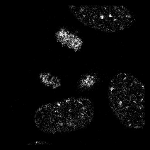
A confocal volume taken at low excitation (source) and the STED volume (target) of one field of fixed mouse embryonic fibroblasts stained with SiR-DNA, on one microscope (3D-RCAN, Chen et al., Nature Methods 2021).
Read before comparing (9)
- Pairs. On a Leica SP8 3X STED microscope, the confocal volume was taken at 0.1% of the laser's power, to match the live-cell SNR the network was meant for, and the STED volume right after it. The nuclei are in a variety of mitotic stages. The paper counts 35 such pairs; the archive holds 33.
- Values. Both volumes come as 'Processed' float32 on a 0 to 1 scale, the processing unsaid, and are kept as delivered. The confocal holds five values only (0, 0.25, 0.5, 0.75, 1), a few photons a voxel, with 0.7 to 5% of its voxels not 0; the STED about 200 to 300 values. truth.dynamic_range is 1, the scale's width.
- Alignment. No registration of these pairs is described. Checked on the twenty cases, the shift in plane between the two is 0.9 pixel in the median (45 nm) and at most 1.9 pixels (95 nm), mostly along y; along z a confocal slice matches its own STED slice or the one below, within a slice. That pixel is not registered and counts as error for every method.
- Pixel size. The files carry none. The paper gives slices 0.16 µm apart and, for the nuclei, 2048 x 2048 pixels of 25 nm, but the files hold 1024 x 1024: the corpus infers pixels of 50 nm, under which the nuclei measure 4 to 26 µm across, where 25 nm would make them half that. The headers hold micrometres as if they were millimetres.
- Mask. The nuclei, from the STED volume's brightest value along z, smoothed, above Otsu's threshold and grown by 1 µm: 5 to 41% of the field on the test cases. On the whole field an empty image would score an SSIM of 0.71 and an MAE of 0.025, a hair from the confocal's 0.024. Made from the target, the mask tells where the nuclei are, not what they look like.
- Baseline. Inside the mask on the ten test cases, with KonfAI's criteria at a range of 1, a constant at the target's median scores an MAE of 0.109 (0.060 to 0.155), a PSNR of 16.3 dB and an SSIM of 0.12; the confocal volume itself 0.121 (0.061 to 0.192), 15.1 dB and 0.09. The source, darker than its target, loses to the constant on eight of the ten.
- Target. The STED volume is a measurement with its own photon noise, not a clean truth: no method reaches an error of 0.
- Outside numbers. None: the paper reports resolution and example images for these nuclei, no error against the STED volume. All 33 pairs are its training data, so the 3D-RCAN models, and any network trained on this record, have seen the test cases.
- Overlap. No other dataset of the corpus holds images of this record, and a later one built on it, its denoising or expansion pairs, shares no file with these. Its microtubule and nuclear pore pairs, 6 slices deep, under the 7 voxels the benchmark's SSIM needs, are not cases.
No method has a result on every case of this dataset yet.
| Accuracy | Cost | ||||||||
|---|---|---|---|---|---|---|---|---|---|
| # | Method | MAE | Change | PSNR µm | SSIM | Time s | RAM GB | GPU GB | Images |
| Source as given | 0.1 ±0.0 | 15.08 ±1.71 | 0.092 ±0.024 | ||||||
MAE: mean absolute error of a synthetic image against its target, inside the body, in the image's units. PSNR: the same by signal-to-noise ratio, higher is better. SSIM: the same by structural similarity, 1 is perfect.
Case by case
Click a score to look at that result: the target image, and over it the method's synthetic image. Underlined scores have their image; for the others you see the case without the result.
About the data
- Test cases
- 10 test fields drawn with seed 2026 among the archive's 33 SiR-DNA pairs, dna_01 to dna_33, the provider's training folder (it gives these pairs no test set), then 10 for development. A case is one field, with one to six nuclei.
- Truth
- target image as released the STED volume of the field as delivered, on the provider's 0 to 1 scale, on the confocal volume's own grid: the two were scanned one after the other on one microscope, and the provider's files give them voxel for voxel, so the corpus registers and resamples nothing
- Source
- zenodo.org/records/4624364
doi.org/10.5281/zenodo.4624364
doi.org/10.1038/s41592-021-01155-x
doi.org/10.1101/2020.08.27.270439
github.com/AiviaCommunity/3D-RCAN - Licence
- CC BY 4.0 (the Zenodo record's licence field); the record's Readme.txt says BSD 3-Clause
10 cases for tuning, never tested
dna_02, dna_24, dna_09, dna_05, dna_12, dna_29, dna_08, dna_03, dna_06, dna_07