 fidopt
Mouse whole-body light-sheet (AIMOS)
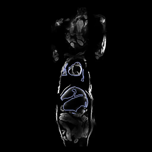
Light-sheet fluorescence scans of two cleared mice, cropped from the head to the hips, showing the tissue's auto-fluorescence, with six organs drawn by hand on both (AIMOS, Schoppe et al. 2020), paired by us.
Read before comparing (10)
- Imaging. Light-sheet microscopy at 6 µm of mice made transparent by tissue clearing. The images are the tissue's auto-fluorescence, which shows the organs by their texture, in 16-bit counts without unit; the propidium iodide images are not used.
- Mice. 15 mice of three strains, both sexes, 1 to 4 months old; which mouse is which is not given, so a pair may join two strains, and which carry tumours is not said.
- Voxel size. The files give none; prepare.py sets 0.12 mm, the one grid of the paper's series on which a scan, 30 x 60 mm, covers a mouse from the head to the hips. Were the size another, every distance would scale with it and the Dice would not change.
- Truth. Experts drew the organs by hand on regularly spaced slices, interpolated between them and refined them in ITK-SNAP, so the outlines step between the drawn slices. Both lungs are one label and both kidneys another, and every scan holds all six. No second annotation of these scans is described.
- Images. Brightness varies between mice and within a scan, with straight steps along the image axes in some. On mouse_13 the field of view cuts the heart, part of which lies outside the image and its label. No mask is given.
- Alignment. As delivered the organs of a pair overlap at a Dice of 0.18 (0.12 to 0.26). The moving image's header carries one translation, at most 4 mm, from elastix mutual information inside each body, no annotation used. A rigid fit turns the mice 1.6 to 10.6 degrees, a matter of posture, so only the translation is kept.
- Baseline. With no registration the seven pairs start at a Dice of 0.21 (0.04 to 0.37) and an HD95 of 4.0 mm (3.0 to 7.0). The spleen scores 0.00, its two outlines apart, and mouse_4_mouse_12 starts lowest, its organs 6 mm apart along the body.
- SyN. ANTs' SyN, the corpus's baseline, run from an earlier affine start that reached a Dice of 0.33 but scaled the mice by 0.83 to 1.22, ends at 0.45 (0.33 to 0.56), better on every pair, with an HD95 of 2.6 mm (1.8 to 3.7) and no folding.
- Ranked apart. Seven test pairs miss the main board's ten, and 15 mice allow no more. Experts drew the six organs on both images, and the start leaves a Dice of 0.21, far under 0.8.
- Outside numbers. None: the dataset was published for segmentation and the pairing is ours.
| Accuracy | Smoothness | Cost | ||||||||
|---|---|---|---|---|---|---|---|---|---|---|
| # | Method | Dice | Change | HD95 mm | SDlogJ | Folds % | Time s | RAM GB | GPU GB | Images |
| 1 | ConvexAdam (MIND)GPUapp | 0.443 ±0.150 | +0.231 | 2.82 ±1.05 | 0.099 | 0.22 | 15 | 3.1 | 5.0 | |
| 2 | anatomix + ConvexAdamGPUapp | 0.320 ±0.123 | +0.108 | 3.66 ±1.15 | 0.096 | 0.20 | 46 | 3.9 | 20.0 | |
| No registration | 0.212 ±0.097 | 4.03 ±1.25 | 0.000 | 0.00 | ||||||
The order averages each method's ranks on Dice, HD95 mm, SDlogJ, so the first one is not always the best in every column. ± is the 95% interval of the mean over the 7 pairs. Two neighbours are tied when a paired Wilcoxon test on the shared pairs does not find the upper one better on Dice (5%): the places a method could hold are then shown under its own. The best value of each column is in colour. Click a row to see its images, a column title to sort by it. Time and memory measured on Laptop.
Dice: how much the organ contours overlap, 1 is perfect. HD95: distance between the contours' surfaces. SDlogJ: how irregular the deformation is, 0 is perfectly smooth. Folds: share of the image where the deformation folds over itself.
Pair by pair
Click a score to look at that result: the fixed image, and over it the moving image after registration, with the annotations. Underlined scores have their image; for the others you see the pair without the result. A green cell gained against no registration on that pair, a red one lost.
About the data
- Test pairs
- All seven pairs are test pairs and none is left for development. The 15 mice are put in the order of sha256("2026:AIMOSLightSheet:<mouse>") and paired two by two, the first fixed; mouse_9, the last, is left out.
- Truth
- contours by experts the dataset's six organs on each scan, drawn by hand by experts: brain (1), heart (2), lungs (3, both in one label), liver (4), kidneys (5, both in one label), spleen (6)
- Source
- doi.org/10.7910/DVN/LL3C1R
dataverse.harvard.edu/api/access/datafile/3796610
doi.org/10.1038/s41467-020-19449-7
doi.org/10.5281/zenodo.4048770
doi.org/10.1016/j.cell.2019.11.013
media.springernature.com/original/springer-static/esm/art%3A10.1038%2Fs41467-020-19449-7/MediaObjects/41467_2020_19449_MOESM3_ESM.pdf
media.springernature.com/original/springer-static/esm/art%3A10.1038%2Fs41467-020-19449-7/MediaObjects/41467_2020_19449_MOESM4_ESM.xlsx - Licence
- CC0 1.0