 fidopt
Advanced-MRI-Breast-Lesions
Fat-suppressed T1-weighted MR of both breasts at 1.5 T, the second phase after contrast, every suspicious enhancing lesion of the report outlined by a radiologist, thresholded and called tumour or benign (Advanced-MRI-Breast-Lesions, TCIA).
10 test casesbreastimage MR
Read before comparing (9)
- Pool. 632 sessions of 632 patients from one institution, 2018 to 2021, on a GE Signa HDxt. 99 carry a DICOM SEG, one segment a lesion (82 tumour, 83 benign); the provider does not say why the others have none.
- Image. The second phase after contrast of the provider's registered dynamic series, on which the SEG lies: axial, prone, about 200 s after the pre-contrast phase. The other phases, the T2 series and the late images are not given. The values have no unit, with a few negative values the provider's registration left and thin lines of zeros across some slices.
- Phase. The provider thresholds each outline on the first or the second subtraction without saying which. On the 20 cases built, 89% to 99% of a case's labelled voxels lie above 75 on the second, against 29% to 95% on the first, never 100%. A method sees the enhanced image, not the subtraction the truth is a threshold of.
- Truth. A certified radiologist with 23 years of experience outlined every enhancing or suspicious position the report mentions; each outline was then cut to the voxels above 75 on a subtraction. One reader, the boundary the threshold's: graded semi-automatic. Small pieces beside a test label's main body, at most 4% of its voxels, count as truth. Enhancement the report does not mention is not labelled.
- Classes. Tumour covers carcinomas and high-risk lesions, from the pathology of a biopsy or of surgery; benign covers lesions benign at pathology or unchanged after at least a year. The image alone often cannot tell them apart: a benign lesion written as breast_tumour is a false positive, and the three test sessions with benign lesions only hold no tumour.
- Size. On the test cases breast_tumour is in seven, 0.52 to 5.72 ml a case, and breast_benign_lesion in five, 0.28 to 2.07 ml. Over the 99 SEGs a lesion has a median of 0.7 ml, and 21 of the 165 are under 0.1 ml. AMBL-049 and AMBL-626 have breast implants.
- Checks. On the 20 cases each of the 32 lesions carries the clinical file's label, on its side and within a slice of its position; the outlines of five test cases sit on enhancing lesions.
- Outside numbers. No inter-reader error and no published score on these labels were found, so the room left is unknown. No network checked the labels, and the surface Dice is at 1 mm, the provider giving no tolerance.
- Overlap. No descriptor of the corpus names this collection. BreastNACT, ISPY1BreastMR and ISPY2_Syn are earlier cohorts from the United States, while these sessions date from 2018 to 2021 at one institution, so no patient scan is shared.
No method has a result on every case of this dataset yet.
Case by case
Click a score to look at that result: the image, the truth in green and the method's map in red. Underlined scores have their image; for the others you see the case without the result.
About the data
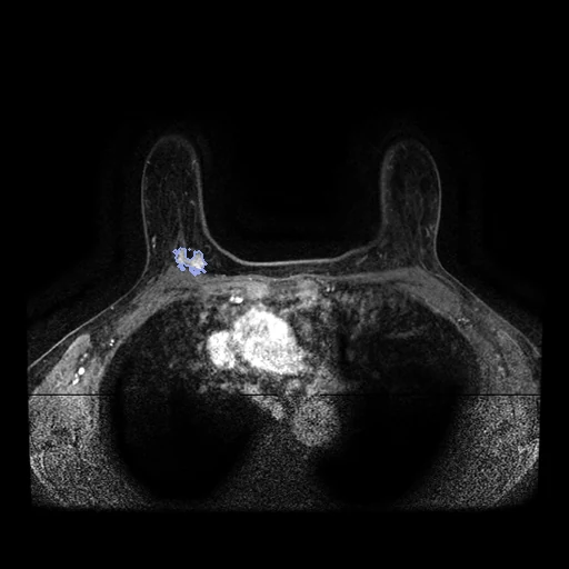
The first test caseClick to look at every case.
- Test cases
- 10 test sessions drawn with seed 2026 by what their SEG holds, among the 89 with a SEG outside development, one session a patient: 5 with tumours only, 3 with benign lesions only, 2 with both. 10 development sessions are the last of the same seeded order, each group in proportion: 5 with tumours only, 3 with benign lesions only, 2 with both. The 533 sessions without a SEG are not cases.
- Truth
- contours by a program, verified by people every enhancing or suspicious position the radiology report mentions, outlined by hand by a certified radiologist with 23 years of experience, the outline then cut automatically to the voxels above 75 on the subtraction of the pre-contrast image from the first or second image after contrast; the provider calls each lesion tumour (carcinomas and high-risk lesions, from the pathology of a biopsy or of surgery) or benign (from pathology, or from a follow-up of at least one year)
- Source
- www.cancerimagingarchive.net/collection/advanced-mri-breast-lesions
doi.org/10.7937/C7X1-YN57
www.cancerimagingarchive.net/wp-content/uploads/Advanced-MRI-Breast-Lesions-DA-Clinical-Sep2024.xlsx - Licence
- CC BY 4.0
10 cases for tuning, never tested
AMBL-507, AMBL-559, AMBL-565, AMBL-571, AMBL-563, AMBL-579, AMBL-599, AMBL-593, AMBL-034, AMBL-596