 fidopt
Aphasia Recovery Cohort stroke lesions
T2-weighted brain MR of a person with aphasia, months to years after a left-hemisphere stroke, with the chronic lesion drawn by an expert (Aphasia Recovery Cohort, University of South Carolina, OpenNeuro ds004884).
22 test casesbrainimage MR
Read before comparing (7)
- Cohort. People with aphasia after a left-hemisphere stroke, recruited at the University of South Carolina at least 6 months after it, aged 21 to 80, without other neurological disease. Version 1.0.2 holds 230 of them; a second release is kept hidden. All were scanned at 3 T, on a Siemens Trio and, after an upgrade, a Prisma fit.
- Images. The T2w the lesion was drawn on, as delivered and defaced: sagittal 3D at 1 mm, a turbo spin echo on the Trio and a SPACE on the Prisma, values without unit. It is the first session with a T2w, the second on sub-M2281. No T1, FLAIR, diffusion or functional image and no mask is given: a score here comes from the T2w alone.
- Truth. 'An expert drew a lesion map on the T2w scan from the first visit' (the paper): who, with which tool or rules, and whether anyone checked is not said, and no agreement between readers is published. On the first ten test cases each map covers the bright tissue of the old infarct, leaves the enlarged ventricle out, and often steps from one axial slice to the next.
- Lesions. 0.2 to 364 ml over the 228 maps, median 69.2 ml; test case sub-M2149's is a few white-matter specks. A Dice here is mostly the main lesion's: the first ten test maps, in 2 to 6 pieces each, keep a Dice of 0.84 to 0.98 (mean 0.93) when moved by one voxel along one axis.
- Checks. Every map built holds 0 and 1 on its T2w's grid and is written under its T2w's header. On the first 20 built each lies on brighter signal than the rest of the head, as an old infarct does. On all 228 maps the lesion lies left of its image's centre, so the headers' left is the patient's left.
- Outside numbers. None: no segmentation score is published on this cohort. TotalSegmentator makes no stroke lesion, so the side check stands in for a network check of the label.
- Overlap. No other dataset of the corpus holds these subjects. SOOPStroke, the authors' acute cohort, shares no identifiers. Its 20 patients built first were compared with all 228 subjects, and with the 22 drawn last on sex, race, age and the lesion's side and volume: the eight pairs left differ in scan date or lesion place, so none is the same person, as far as the files tell. Its 40 later candidates were checked against the 42 subjects used here, and the twelve that stayed possible are left out of its pool.
No method has a result on every case of this dataset yet.
Case by case
Click a score to look at that result: the image, the truth in green and the method's map in red. Underlined scores have their image; for the others you see the case without the result.
About the data
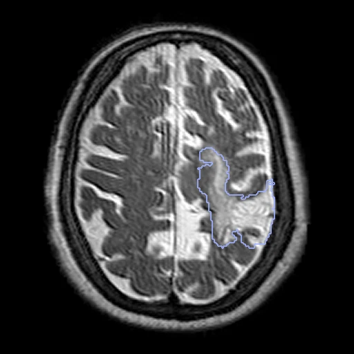
The first test caseClick to look at every case.
- Test cases
- 22 test subjects drawn with seed 2026 among the 228 with a lesion map, the two scanners in proportion: 11 on the Trio, 11 on the Prisma. 20 development subjects, the last of the same seeded order, the two scanners in proportion: 10 on each. sub-M2003 and sub-M2039 have no lesion map.
- Truth
- contours by experts the dataset's lesion map of the chronic stroke, drawn by an expert on the T2-weighted image of the subject's first visit; one reader, no review or agreement published
- Source
- openneuro.org/datasets/ds004884
doi.org/10.18112/openneuro.ds004884.v1.0.2
s3.amazonaws.com/openneuro.org/ds004884
doi.org/10.1038/s41597-024-03819-7
github.com/neurolabusc/AphasiaRecoveryCohortDemo - Licence
- CC0
20 cases for tuning, never tested
sub-M2139, sub-M2002, sub-M2117, sub-M2106, sub-M2270, sub-M2017, sub-M2179, sub-M2048, sub-M2305, sub-M2115, sub-M2234, sub-M2022, sub-M2306, sub-M2104, sub-M2168, sub-M2041, sub-M2214, sub-M2086, sub-M2123, sub-M2152