 fidopt
Bonn focal cortical dysplasia (FLAIR)
FLAIR MR of the brain of a person with epilepsy due to a focal cortical dysplasia type II, the dysplastic cortex outlined by a neurologist and reviewed by another (University Hospital Bonn, OpenNeuro ds004199).
28 test casesbrainimage MR
Read before comparing (8)
- Cohort. People with epilepsy due to a focal cortical dysplasia type II treated at the University Hospital Bonn, the dysplasia verified by histology or suspected on imaging, scanned on one 3 T Siemens Trio with an 8-channel head coil before its 2014 update and a 32-channel one after. Five were read as MRI-negative at first, test cases sub-00100 and sub-00107 among them.
- Images. The FLAIR as delivered, defaced, read as floats without unit: on 78 patients a sagittal 3D FLAIR at 1 mm isotropic, on 7 a 2D FLAIR with slices of 3.7 to 5.2 mm. Test case sub-00074 and development cases sub-00027, sub-00112 and sub-00120 are 2D; the other test cases are 3D. No T1 and no mask are given: a score comes from the FLAIR alone.
- Truth. One of two neurologists experienced in epilepsy imaging outlined the dysplastic cortex by hand on the FLAIR, one lesion a patient, and the other reviewed it in all three planes. They settled disagreements with the patient's other findings, such as PET, invasive EEG and the postoperative MRI, which is how a mask exists on an image read as normal. No second tracing and no agreement are published.
- Mask shape. On the ten test cases looked at, each mask covers the cortex and the white matter beneath it as a region around the dysplasia, several with straight sides, rather than a tight outline: this caps the Dice a method can reach.
- Lesions. Small and subtle: 0.19 to 52 ml over the 85 masks, median 2.3 ml, 81 in one piece. Inside the 28 test masks the FLAIR's median is 0.97 to 1.53 times that of the rest of the head; sub-00107's is no brighter. Moved by one voxel along one axis, those masks keep a Dice of 0.35 to 0.93, mean 0.83.
- Checks. prepare.py writes each mask under its FLAIR's header, the origins differing by up to 0.001 mm. On all 38 cases each mask holds 0 and 1 on its FLAIR's grid and lies on the hemisphere participants.tsv names. sub-00048's mask, named acq-32ch10, lies on its only FLAIR, acq-T2sel.
- Outside numbers. None: the paper publishes no segmentation score. TotalSegmentator makes no dysplasia, so the hemisphere check stands in for a network check of the label.
- Overlap. No other dataset of the corpus holds these patients: none comes from Bonn, and ISLES2022 comes from Munich and Bern.
No method has a result on every case of this dataset yet.
Case by case
Click a score to look at that result: the image, the truth in green and the method's map in red. Underlined scores have their image; for the others you see the case without the result.
About the data
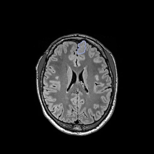
The first test caseClick to look at every case.
- Test cases
- The provider's test split taken whole: its 28 patients, 12 scanned before the scanner's 2014 update and 16 after. 10 development patients drawn in the seeded order among the 57 of its training split. The 85 controls, all in the training split, have no lesion and are left out.
- Truth
- contours by experts the dataset's mask of the dysplastic cortex, drawn by hand in FSL on the FLAIR by one of two neurologists experienced in epilepsy imaging and reviewed by the other in all three planes, a disagreement settled with all the patient's other findings (morphometric analysis, SPECT, PET, invasive EEG, postoperative MRI) until both agreed; one lesion a patient, its location set beforehand by two neuroradiologists
- Source
- openneuro.org/datasets/ds004199
doi.org/10.18112/openneuro.ds004199.v1.0.6
s3.amazonaws.com/openneuro.org/ds004199
doi.org/10.1038/s41597-023-02386-7 - Licence
- CC0
10 cases for tuning, never tested
sub-00090, sub-00139, sub-00146, sub-00120, sub-00112, sub-00078, sub-00097, sub-00044, sub-00027, sub-00080