 fidopt
UCSD-VS-Longitudinal, vestibular schwannoma
Contrast-enhanced T1 MR of the internal auditory canals at a vestibular schwannoma patient's first examination, the tumour outlined by a network, corrected by hand and approved by a neuroradiologist (UCSD-VS-Longitudinal, TCIA).
14 test casesbrainimage MR
Read before comparing (7)
- Cohort. Patients with a sporadic vestibular schwannoma at UC San Diego Health, 2 to 4 examinations each, under observation, after radiotherapy or after surgery, on mostly GE scanners at 1.5 T and 3 T.
- Case. The post-contrast T1 of the internal auditory canals (t1postIAC) at the first examination, with the provider's mask; the other sequences and examinations are left out. The test cases are 2D axial slabs of 13 to 24 slices 2.5 to 3.0 mm apart, 0.14 to 0.35 mm in the plane, defaced, in arbitrary units. No mask is given.
- Truth. An nnU-Net trained on Vestibular-Schwannoma-SEG (the crossMoDA family) proposed each mask in 3D; it was corrected by hand in ITK-SNAP and approved by a board-certified neuroradiologist. The number of readers, their agreement and how many proposals changed are not given.
- Treatment. Nine test cases are before any treatment and five after: VS_0056, 2 days after surgery, and VS_0128, 107 days after, where the mask is what enhances in and around the operated tumour, and VS_0098, VS_0126 and VS_0202 after radiotherapy. Every test case holds tumour.
- Room. The test tumours hold 0.014 to 16.0 ml, median 0.52 ml. VS_0019 and VS_0034 are under 50 voxels: a prediction one slice off scores 0. A test mask moved one voxel in the plane keeps a Dice of 0.80 to 0.98, moved one slice 0.00 to 0.85. The surface Dice is at 1 mm, as the provider sets no tolerance.
- Checks. On the 26 cases every mask holds 0 and 1 on its image's grid and follows an enhancing mass in the canal or the cerebellopontine angle; VS_0138 is outlined with its cyst. TotalSegmentator makes no vestibular schwannoma, so no network checks the label.
- Overlap. No other descriptor names this collection, and its patients are not those of VestibularSchwannoma, VestibularSchwannomaMCRC or VestibularSchwannomaMCRC2.
No method has a result on every case of this dataset yet.
Case by case
Click a score to look at that result: the image, the truth in green and the method's map in red. Underlined scores have their image; for the others you see the case without the result.
About the data
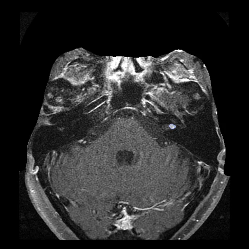
The first test caseClick to look at every case.
- Test cases
- 14 test patients drawn with seed 2026 by scanner maker among 141 (121 GE, 13 Siemens, 6 Canon, 1 Time Medical), under the name VestibularSchwannomaUCSD, and 12 development patients. Left out of the 191: 34 whose first mask is on the whole-head T1, and 16 without a whole-head T1 at one of the first two examinations, the pool of a registration set planned on the same patients.
- Truth
- contours by experts the dataset's mask of the enhancing tumour on the post-contrast T1 of the internal auditory canals: proposed by an nnU-Net trained on Vestibular-Schwannoma-SEG, reviewed and corrected by hand in ITK-SNAP and approved by a board-certified neuroradiologist
- Source
- www.cancerimagingarchive.net/collection/ucsd-vs-longitudinal
doi.org/10.7937/wefa-cp23
www.cancerimagingarchive.net/wp-content/uploads/UCSD-VS-Longitudinal_clinical_information-2.tsv - Licence
- CC BY 4.0
12 cases for tuning, never tested
VS_0119, VS_0134, VS_0143, VS_0240, VS_0120, VS_0088, VS_0238, VS_0071, VS_0160, VS_0081, VS_0005, VS_0091