 fidopt
CPTAC-CCRCC-Tumor-Annotations
An abdominal CT of a patient with clear cell renal cell carcinoma before treatment, the kidney tumours outlined by radiologists and reviewed by board-certified radiologists (CPTAC-CCRCC-Tumor-Annotations on CPTAC-CCRCC, TCIA).
10 test casesabdomenimage CT
Read before comparing (9)
- Annotations. Version 2 of 2025-10-20 of TCIA's annotations. Radiologists holding MBBS degrees or higher drew each lesion by hand in the axial plane and US board-certified radiologists reviewed it, as RECIST 1.1 target lesions: five a scan and two an organ at most, the primary lesion whatever its size, other lesions above 1 cm.
- Cases. A case is a patient's CT series with a contour named after a kidney, before treatment: the earliest such examination, then the series with the most images, then the highest series number. 52 patients have one: the pool. The other phases carry their own contours and are not used; the 7 patients annotated on MR are too few for a set of their own.
- Labels. Only the kidney tumours are written. The other target lesions, on 25 of the 52 series and 3 of the 10 test cases (liver, adrenal glands, nodes, the renal vein and vena cava, lung), are not. No set of all target lesions was built: the caps leave metastases and nodes undrawn, which would charge a method that found them.
- Truth. It passes the bar of a main leaderboard, with limits. A kidney with more than two tumours, or a second one under 1 cm, has tumour left undrawn. Two contours may be one mass, as the two halves on test case C3N-01646. Benign cysts are not drawn, where KiTS labels them apart. One annotator and one reviewer a scan, no agreement published.
- Checks. On the 19 cases with one side drawn, the label's centre lies on the side its name gives, and the ten test outlines sit on a renal mass, not on the kidney around it. TotalSegmentator makes no tumour: on C3N-00244 its left kidney holds every voxel of the 'LEFT KIDNEY' tumour. The filled volumes are 0.6 to 1.5 times the report's.
- Images. 512 by 512 pixels of 0.62 to 1.0 mm, slices 0.625 to 5 mm apart, in Hounsfield units, the scanner's padding left outside the reconstructed circle. The series are clinical venous, nephrographic and delayed phases; test case C3N-01989 shows no enhancement of the kidneys. Where a series holds two passes (C3N-00305, C3N-00313, C3N-03018), the image is the stack with the most contours.
- Room. The ten test tumours hold 6.4 to 518 ml (median 52; 1.2 to 620 ml in development). Moved by one voxel in plane, the test masks keep a Dice of 0.95 to 0.99 (mean 0.97); by one slice, 0.82 to 0.98 (0.92). The surface Dice is at 1 mm, the provider giving no tolerance.
- Overlap. KiTSCT and AorticTreeCTA hold C4KC-KiTS, other patients. TotalSegMR's test image s0042 and LiverSegmentsMR's QqIPw7GVi8YiXjfX are MR series of C3L-00800 and C3L-00609, hence their exclusion. The other 38 images those two sets then held match no MR series of CPTAC-PDA, CPTAC-UCEC or CPTAC-CCRCC by slice count, rows, columns and pixel size; a series holding several volumes was not tried.
- Public. The annotations are public since 2023: a network trained on them may have seen the test cases.
No method has a result on every case of this dataset yet.
Case by case
Click a score to look at that result: the image, the truth in green and the method's map in red. Underlined scores have their image; for the others you see the case without the result.
About the data
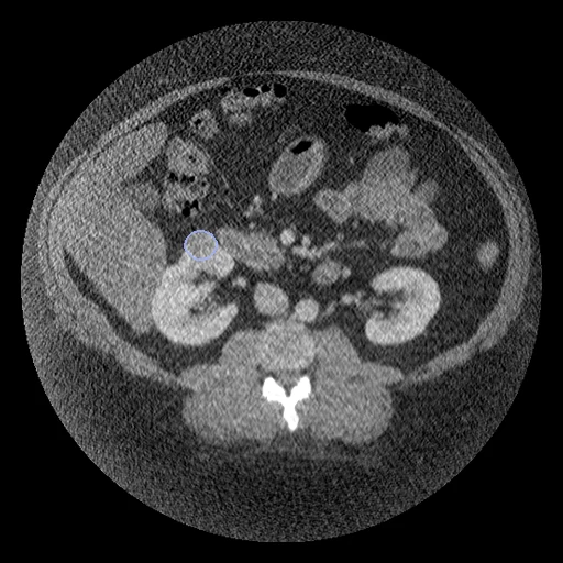
The first test caseClick to look at every case.
- Test cases
- 10 test patients drawn with seed 2026 by the maker of the annotated CT among the 42 outside development (4 GE, 4 Siemens, 1 Philips, 1 Toshiba), and 10 development patients, the last of the same seeded order; one CT each. C3L-00800 and C3L-00609, whose MR TotalSegMR and LiverSegmentsMR hold, are left out; neither has a kidney contour on CT.
- Truth
- contours by experts the target lesions named after the right or the left kidney, two a kidney at most: the renal tumour, drawn by hand in the axial plane by radiologists (MBBS or higher) and reviewed by US board-certified radiologists, as RECIST target lesions
- Source
- www.cancerimagingarchive.net/analysis-result/cptac-ccrcc-tumor-annotations
doi.org/10.7937/SKQ4-QX48
www.cancerimagingarchive.net/wp-content/uploads/Metadata_Report_CPTAC_CCRCC_2025_10_20-1.csv
www.cancerimagingarchive.net/collection/cptac-ccrcc
doi.org/10.7937/K9/TCIA.2018.OBLAMN27
services.cancerimagingarchive.net/nbia-api/services/v1/getSeries?Collection=CPTAC-CCRCC - Licence
- CC BY 4.0
10 cases for tuning, never tested
C3N-00313, C3N-01654, C3L-01352, C3N-01361, C3N-03018, C3N-00492, C3L-01459, C3N-02439, C3L-01976, C3N-00194