 fidopt
PSMA-PET-CT-Lesions (autoPET III and IV), PET lesions
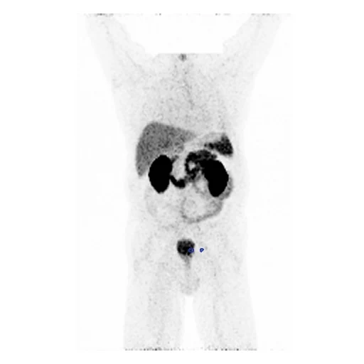
Whole-body PSMA-PET in SUV of a man with suspected or diagnosed prostate cancer, every PSMA-avid tumour lesion segmented on the PET by one reader and validated by board-certified experts (PSMA-PET-CT-Lesions, autoPET III and IV).
Read before comparing (11)
- Pool. 597 PSMA-PET/CT studies of 378 men at LMU University Hospital Munich, 2014 to 2022, on three scanners, with 18F- or 68Ga-PSMA; 539 hold a lesion. A case is each patient's first positive study, so no patient is in two cases.
- Left out. The 58 studies without a lesion, as AutoPET_Seg does for FDG: a method could only lose on them. PSMA_e3c4639102fe5c51, whose header starts the PET 7.2 minutes after injection while the image shows an hour's uptake, so no SUV can be computed; only the studies drawn were checked for this.
- Image. The attenuation-corrected PET in body-weight SUV on its own grid: 4.07 mm pixels and 2 mm slices on the Biograph mCT Flow 20, 2.73 and 3.27 mm on the Discovery 690, 4.07 and 5 mm on the Biograph 64. The SUV agrees within 0.003 with the provider's NIfTI release on one study of each scanner.
- PET alone. A method gets one image, and the truth was drawn on the PET's grid, so the CT is neither given nor downloaded. The autoPET III and IV entries read PET and CT together: their numbers are not these.
- Truth. One reader with 3 years in hybrid imaging pre-segmented each lesion by a threshold in circular VOIs and corrected it slice by slice; board-certified experts with 4 and more than 10 years validated every lesion. No second segmentation: graded expert.
- Ignore region. TCIA's defaced PET blanks a box over the skull base and the neck, where the SEGs keep their lesions. Each case's exact PET zeros, that box and most of the air, are taken out of both maps before scoring. On the test cases this removes 220 of 8,587 lesion voxels from PSMA_120974a1c4058f4a_2000-11-21 and 20 of 13,010 from PSMA_14c16d0ce2b3229d_2002-09-11, nothing elsewhere.
- Lesions. 1.05 to 791 ml a test case (median 18 ml), in 1 to 146 pieces. A case's Dice is that of its largest lesions, so a method that misses the small ones loses little; autoPET also scores the volumes of missed and false lesions, not computed here.
- Room. The first ten test masks moved by one voxel keep a Dice of 0.55 to 0.84 a case (mean 0.71). The surface Dice is at 1 mm, under every scanner's voxel, so a boundary one voxel off misses: the moves keep 0.74 of it.
- Checks. On 17 of the cases every map equals the provider's release voxel for voxel, and on a coronal maximum intensity projection every outlined lesion lies on focal uptake. The 34 others hold lesions of median SUV 1.7 or more; the 1.7 is development case PSMA_2740fa8c813cd29c's single 23-voxel lesion, peaking at 3.3 where its surroundings read 1.0.
- Overlap. AutoPSMA, PSMAFollowUp and AutoPSMA_CTPET_Syn hold images of the same collection; their patients are left out. The autoPET challenges (segmentation screening rows 222 to 226) hold this PSMA set: a dataset built from them should leave out the patients of cases and development.
- Outside numbers. None as scored here, from the PET alone with the blanked box taken out. The masks are autoPET III and IV's public training data: a network trained on them has seen these patients.
No method has a result on every case of this dataset yet.
Case by case
Click a score to look at that result: the image, the truth in green and the method's map in red. Underlined scores have their image; for the others you see the case without the result.
About the data
- Test cases
- 27 test studies drawn with seed 2026 by scanner among the 246 outside development, and 24 development studies, the last of the same seeded order, each scanner in proportion: 9 Biograph mCT Flow 20, 10 Discovery 690 and 5 Biograph 64-4R TruePoint, none a patient of another dataset. The pool is 270 patients' first study with lesions, less the 98 patients of AutoPSMA, PSMAFollowUp and AutoPSMA_CTPET_Syn, and PSMA_e3c4639102fe5c51, whose header gives no SUV.
- Truth
- contours by experts every PSMA-avid tumour lesion, primary tumour and/or metastases, segmented on the PET by one reader with 3 years of experience in hybrid imaging (in circular VOIs, the voxels above a threshold the reader set pre-segmented, then corrected slice by slice) and validated by board-certified experts with 4 and more than 10 years; where the defaced PET holds no data (its exact zeros: the box blanked over the skull base and the neck, and the air) both maps are taken out of the scoring
- Source
- www.cancerimagingarchive.net/collection/psma-pet-ct-lesions
doi.org/10.7937/r7ep-3x37
www.cancerimagingarchive.net/wp-content/uploads/PSMA-PET-CT-Lesions-CLINICAL_v01_20251118.tsv
services.cancerimagingarchive.net/nbia-api/services/v1/getSeries?Collection=PSMA-PET-CT-Lesions
fdat.uni-tuebingen.de/records/rdkqd-wdh87 - Licence
- CC BY 4.0
24 cases for tuning, never tested
PSMA_c80781b803041d12_1998-01-05, PSMA_7a522636008cf840_2003-06-24, PSMA_99278c16213e2429_1998-11-11, PSMA_6ef5e5c442e4ce63_2000-12-20, PSMA_c67186371fb31240_2002-07-23, PSMA_35852148926fc57d_1997-03-26, PSMA_3c928684b9d209d2_1999-06-04, PSMA_dc8b0fa3643a6878_2001-12-19, PSMA_5813d6984082108d_1999-09-16, PSMA_b5e009b02fb0a848_2003-10-10, PSMA_560e93923a626c54_1998-05-06, PSMA_0bf6ac34fa0c97c0_1999-11-16, PSMA_5b644577e75785bc_2001-07-03, PSMA_0cd58730e7446445_1998-07-24, PSMA_9baf4ca6321e66ad_1999-11-09, PSMA_81b53d3911e09543_2003-09-09, PSMA_a6509098ad09ada5_1999-04-21, PSMA_ad7bc1aa5b586751_2000-12-28, PSMA_bed76b3b7c2b172e_2003-08-22, PSMA_2740fa8c813cd29c_1997-03-17, PSMA_08a881912350c103_1997-08-20, PSMA_53d9705e1ddc8d81_2003-11-19, PSMA_ede705184679888a_2000-01-20, PSMA_90092e3de48644f1_2001-08-03