 fidopt
Adrenal-ACC-Ki67-Seg
Contrast-enhanced abdominal CT of a patient with an adrenocortical carcinoma before its resection, the tumour outlined semi-automatically and refined by hand by a radiologist (Adrenal-ACC-Ki67-Seg, MD Anderson Cancer Center, TCIA).
10 test casesabdomenimage CT
Read before comparing (7)
- Cases. The collection holds 53 operated patients with an adrenocortical carcinoma proven by pathology, from 2006 to 2018. Each has one tumour, on one side, and one DICOM SEG on one post-contrast CT series: a case is that CT. The 71 other CT series carry no outline and are not used.
- Truth. The tumour was segmented semi-automatically in AMIRA, then refined by hand by an experienced radiologist, for radiomics. One reader and no agreement between readers published: graded semi-automatic, its own error unknown. The label is the tumour mass alone, not the gland. No network checked it, and the surface Dice uses 1 mm, as the provider gives no tolerance.
- Tumours. On the ten test CTs they hold 9.7 to 1326 ml (median 38 ml), each in one piece but Adrenal_Ki67_Seg_042, and every centre lies on the side the clinical file names. On Adrenal_Ki67_Seg_016 and Adrenal_Ki67_Seg_014 the outline is about 4 cm long for a resected tumour of 11.6 and 9 cm; CT and surgery lie 5 to 237 days apart.
- Images. 512 by 512 pixels of 0.68 to 0.98 mm, slices 1 to 5 mm apart, of the abdomen alone or of the chest, abdomen and pelvis, in Hounsfield units as delivered, with -3023, -2048 or -1024 outside the reconstructed circle. The outlined series of 11 of the 53 patients, test cases Adrenal_Ki67_Seg_018 and Adrenal_Ki67_Seg_042 among them, names no maker.
- Adrenal_Ki67_Seg_014. This test case joins two acquisitions in one series that overlap by one slice. The corpus stacks its 58 slices at 5 mm, as the provider's SEG does, so one level appears twice and the slices above it lie about 5 mm off; image and labels agree slice for slice.
- Public models. The collection is public since May 2023 and listed in SAT-DS, the CVPR 2025 BiomedSegFM challenge and VISTA3D: a network trained on them may have seen the test cases. No published score on these cases was found.
- Overlap. No other descriptor of the corpus names this collection. HCCTACE, also from MD Anderson, is another disease and other scans; TCIA de-identifies each collection on its own, so a patient in both could not be told.
No method has a result on every case of this dataset yet.
Case by case
Click a score to look at that result: the image, the truth in green and the method's map in red. Underlined scores have their image; for the others you see the case without the result.
About the data
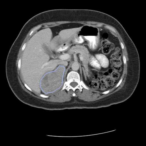
The first test caseClick to look at every case.
- Test cases
- 10 test patients drawn with seed 2026 by the maker of the outlined CT among the 43 outside development: 4 GE, 2 Siemens, 2 whose series names no maker, 1 Philips and 1 Toshiba. 10 development patients, the last of the same seeded order, each maker in proportion: 4 GE, 3 Siemens, 2 without a maker and 1 Philips. One CT a patient.
- Truth
- contours by a program, verified by people the adrenocortical carcinoma, proven by pathology after its resection, in one label: segmented semi-automatically in AMIRA, then refined by hand by an experienced radiologist, for the extraction of radiomic features
- Source
- www.cancerimagingarchive.net/collection/adrenal-acc-ki67-seg
doi.org/10.7937/1FPG-VM46
doi.org/10.1016/j.crad.2020.01.012
www.cancerimagingarchive.net/wp-content/uploads/Adrenal-ACC-Ki67-Seg_SupportingData_20230522.xlsx - Licence
- CC BY 4.0
10 cases for tuning, never tested
Adrenal_Ki67_Seg_015, Adrenal_Ki67_Seg_001, Adrenal_Ki67_Seg_030, Adrenal_Ki67_Seg_048, Adrenal_Ki67_Seg_050, Adrenal_Ki67_Seg_053, Adrenal_Ki67_Seg_009, Adrenal_Ki67_Seg_046, Adrenal_Ki67_Seg_034, Adrenal_Ki67_Seg_004