 fidopt
ReMIND, brain tumour before resection
Pre-operative head MR of an adult about to have a brain tumour resected at Brigham and Women's Hospital: the one defaced series the surgical team outlined the tumour on, with that whole-tumour outline (ReMIND, TCIA).
10 test casesbrainimage MR
Read before comparing (12)
- Cohort. 114 consecutive patients operated on with image guidance: gliomas, 11 metastases and other lesions, a few found by pathology not to be tumours. 51 had a craniotomy before, 43 of the 97 in the pool.
- Cases. The series the outline names: a contrast-enhanced T1 for 54 pool patients, a T2 for 27, a FLAIR for 12 and a T1 without contrast for 4, the draw's strata. The test cases are six contrast-enhanced T1, three T2 SPACE and one 2D FLAIR. A method is told mr, not the contrast.
- One series. The provider gives no patient's sequences on one grid, and only 7 of the 114 have all four before surgery: the dataset joins the single-image lesion sets, not the brain tumour track.
- Images. The DICOM series as TCIA serves them, values untouched, defaced by the provider, skull and scalp kept; no mask. Nine test cases are 3D at about 1 mm; ReMIND-111 is a 2D FLAIR of 55 slices 3 mm apart. On all 20 cases the outline lies on the side the clinical spreadsheet gives.
- Outline grid. On 8 of the 20 cases (test cases ReMIND-014, 070, 072, 086 and 111, development cases 010, 027 and 100) the provider's outline lies on another lattice; prepare.py brings it onto the image's grid by nearest neighbour in world coordinates, its edge within half a voxel.
- Truth. For surgical planning a neurosurgical fellow outlined the tumour by hand and the attending neurosurgeon refined it if needed: the enhancing region with any necrotic core, or the solid part within the T2 and FLAIR hyperintensity, without oedema. It is a surgeon's target: on ReMIND-106 it follows straight lines, and on non-enhancing tumours its edge is a judgement.
- Agreement. Two neurosurgical research fellows outlined ten cases again: a Dice of 0.922 (SD 0.028) and a mean surface distance of 0.68 mm on five enhancing tumours, 0.864 (SD 0.037) and 1.24 mm on five non-enhancing ones.
- Room. A test outline moved by one voxel keeps a Dice of 0.89 to 0.96: above about 0.9 methods differ within the truth's spread. The surface Dice, at 1 mm, is stricter than the observers on non-enhancing tumours.
- Name. The label, brain_tumour, is close to BraTS's tumour core for enhancing tumours and to the whole tumour for the others. Outlines of cavities, residual tumour and targets, and Brainlab's automatic structures, are left out.
- Tumours. The ten test outlines hold 4.5 to 69.3 ml (median 24.3), each in one piece, among them a metastasis (ReMIND-014) and treatment effects (072). Four were operated on before (049, 070, 075 and 106), so an older cavity lies beside the outline.
- Outside numbers. None is scored as here. The outlines are public: a network trained on ReMIND may have seen these patients.
- Overlap. No other dataset of the corpus holds these patients. Learn2Reg's ReMIND2Reg and the screening's ReMIND rows hold the same patients: a dataset built later from them keeps these test patients in its test cases and these development patients out of them.
No method has a result on every case of this dataset yet.
Case by case
Click a score to look at that result: the image, the truth in green and the method's map in red. Underlined scores have their image; for the others you see the case without the result.
About the data
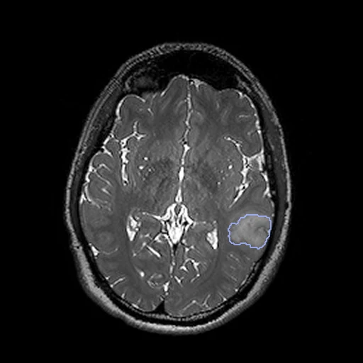
The first test caseClick to look at every case.
- Test cases
- 10 test patients drawn with seed 2026 by the contrast of the outlined series among the 87 outside development; 10 development patients, the last of the same seeded order, each contrast in proportion (six contrast-enhanced T1, three T2, one FLAIR). The pool is the 97 of 114 patients with one pre-operative outline: 16 have two, ReMIND-032 none.
- Truth
- contours by experts the dataset's pre-operative whole tumour, outlined by hand for surgical planning in Brainlab Elements by a neurosurgical fellow and refined where needed by the attending neurosurgeon: the enhancing region with any necrotic core for an enhancing tumour, the solid or nodular part within the T2 and FLAIR hyperintensity for one that does not enhance
- Source
- www.cancerimagingarchive.net/collection/remind
doi.org/10.7937/3RAG-D070
doi.org/10.1038/s41597-024-03295-z
www.cancerimagingarchive.net/wp-content/uploads/ReMIND-Dataset-Clinical-Data-September-2023.xlsx
services.cancerimagingarchive.net/nbia-api/services/v1/getSeries?Collection=ReMIND - Licence
- CC BY 4.0
10 cases for tuning, never tested
ReMIND-050, ReMIND-054, ReMIND-013, ReMIND-094, ReMIND-029, ReMIND-027, ReMIND-105, ReMIND-112, ReMIND-100, ReMIND-010