 fidopt
Soft-tissue-Sarcoma
T2-weighted fat-saturated or STIR MR of a soft-tissue sarcoma of the extremities before treatment, the tumour and any visible oedema around it drawn slice by slice by an expert radiation oncologist (Soft-tissue-Sarcoma, TCIA).
10 test caseslimbsimage MR
Read before comparing (10)
- Cohort. 51 patients with a soft-tissue sarcoma of the extremities, none metastatic or recurrent, on clinical protocols that differ, 12 at the McGill University Health Centre and 39 at an outside institution; most tumours lie in the thigh. The contours were drawn on a T2-weighted fat-saturated series (26 patients) or a STIR (25).
- Images. The series the expert drew on, as read: scanner values on scales that differ (maxima of 406 to 5091 on the 20 built), its own grid and plane, slices 4 to 7 mm thick with gaps of 1 to 2 mm. Most are axial; STS_011 is sagittal and STS_023 coronal. The T1 MR and the PET/CT are not given. No mask.
- Truth. One expert radiation oncologist drew GTV_Mass on all 51 patients and, on the 32 with visible oedema, GTV_Edema, the tumour with its oedema. One reader, no measure of agreement: graded expert.
- Labels. 1 is GTV_Mass, 2 what GTV_Edema adds to it, absent on the 19 patients without visible oedema (test cases STS_008, STS_035, STS_043). The oedema is scored as a structure of its own, with the tumour's weight. A label a case lacks is not scored unless a method writes it, a false positive at 0.
- Sizes. On the ten test cases the tumour holds 27 to 751 ml (median 262) over 7 to 29 slices, and the oedema, on the seven that have one, 2.4 to 254 ml (median 119).
- Room. Test masks moved by one voxel in plane keep a mean Dice of 0.98 for the tumour and 0.94 for the oedema, by one slice 0.87 and 0.54, the thick slices weighing on the oedema. The surface Dice is at 1 mm, the provider setting no tolerance.
- Outside numbers. None on these cases as scored here: Neubauer et al. 2020 cropped and resampled the leg tumours and do not say which contour they scored. The contours are public: a network trained on this collection may have seen these patients.
- Overlap. No other dataset of the corpus holds these patients. The collection's T1 MR and PET/CT are screened for registration and synthesis, and SAROS labels some of its CTs: a dataset built from them leaves out these 20 test and development patients.
- Side. For STS_036 (development) the spreadsheet's 'right groin' contradicts the MR header and the CT, where the tumour lies on the patient's left. No label has a side, so no score depends on it.
- Checks. On the 20 built, each label map holds only 0 to 2 on its image's grid, and the contours lie on the mass and on the bright tissue around it.
No method has a result on every case of this dataset yet.
Case by case
Click a score to look at that result: the image, the truth in green and the method's map in red. Underlined scores have their image; for the others you see the case without the result.
About the data
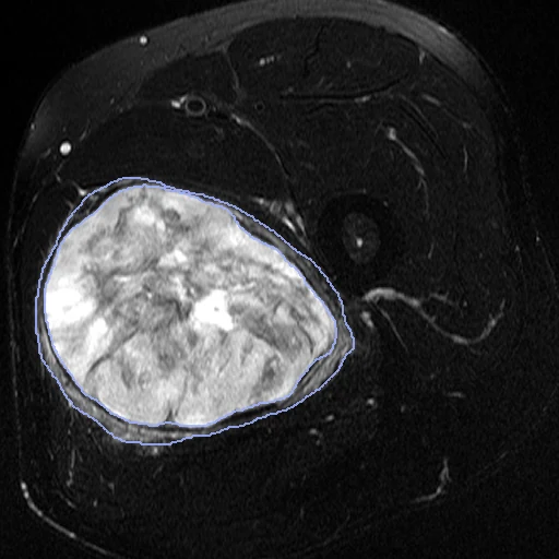
The first test caseClick to look at every case.
- Test cases
- 10 test patients drawn with seed 2026 among the 41 outside development, by sequence in proportion: five T2-weighted fat-saturated, five STIR. 10 development patients, the last of the same seeded order. All 51 patients have the expert's contours.
- Truth
- contours by experts the dataset's two contours, drawn slice by slice on the T2-weighted fat-saturated or STIR MR by an expert radiation oncologist: 1 the tumour without the visible oedema around it (GTV_Mass), 2 the oedema, what the contour of the tumour with that oedema (GTV_Edema, drawn for the 32 patients with visible oedema) adds to it
- Source
- www.cancerimagingarchive.net/collection/soft-tissue-sarcoma
doi.org/10.7937/K9/TCIA.2015.7GO2GSKS
doi.org/10.1088/0031-9155/60/14/5471
www.cancerimagingarchive.net/wp-content/uploads/INFOclinical_STS.xlsx
services.cancerimagingarchive.net/nbia-api/services/v1/getSeries?Collection=Soft-tissue-Sarcoma
arxiv.org/abs/2008.12544 - Licence
- CC BY 3.0
10 cases for tuning, never tested
STS_011, STS_018, STS_003, STS_038, STS_004, STS_036, STS_040, STS_028, STS_023, STS_033