 fidopt
Mediastinal-Lymph-Node-SEG
Contrast-enhanced chest CT of a patient at the baseline of an oncology trial, every abnormal mediastinal lymph node delineated by hand (Mediastinal-Lymph-Node-SEG, the fully annotated scans of LNQ2023).
12 test caseslungimage CT
Read before comparing (7)
- Pool. 513 patients with enlarged lymph nodes, one CT each, read by the imaging core lab of Massachusetts General Hospital, Dana-Farber and Brigham and Women's. The 120 fully annotated are LNQ2023's 20 validation and 100 test cases; the partially annotated are its training cases.
- Truth. One trained radiologist drew every node judged abnormal, an estimated short axis over 1 cm, freehand on axial slices in 3D Slicer, with an instructor and a senior specialist consulted. One reading, no agreement reported. Nodes near 1 cm may have been missed; smaller nodes are not labelled, so a method that segments them is scored wrong there. All nodes share one label.
- Images. The CTs as delivered, in Hounsfield units, slices 3 to 5 mm apart on the test CTs. Outside the reconstructed circle they hold -3024, -2048 or -1024, and case_0501 reaches 12323 on Toshiba's extended scale; all left so.
- Nodes. On the ten test CTs drawn first the nodes hold 3.0 to 71.3 ml in 4 to 17 pieces, from the thoracic inlet to the oesophageal hiatus, soft tissue with no voxel of lung or bone. The two added later are larger: case_0142 holds 133.5 ml, case_0201 116.9 ml.
- Room. On the challenge's 100 test CTs, among these 120, the best entry reached a median Dice of 0.712, and entries trained on the partial labels alone about 0.61; the bulky nodes of Hodgkin's lymphoma were the hardest. The challenge set no tolerance, so the surface Dice is at 1 mm.
- Exposure. The full labels have been public since the collection's release in August 2024: a model trained on the collection may have seen these test cases.
- Overlap. No other descriptor names the collection or the challenge. SpineMetsCT, from Brigham and Women's, holds other scans, though TCIA's separate de-identification means a shared patient could not be told. The screening's LNQ2023 row is these images again; its CT-Lymph-Nodes row is another cohort.
No method has a result on every case of this dataset yet.
Case by case
Click a score to look at that result: the image, the truth in green and the method's map in red. Underlined scores have their image; for the others you see the case without the result.
About the data
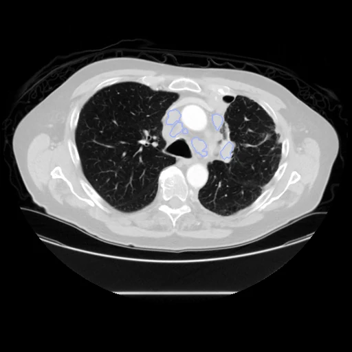
The first test caseClick to look at every case.
- Test cases
- 12 test CTs drawn by scanner maker with seed 2026 among the 110 fully annotated patients outside development: 9 GE, 2 Toshiba, 1 Siemens, none of the one Philips. 10 development CTs, the last of the same seeded order, each maker in proportion: 7 GE, 2 Toshiba, 1 Siemens. The 393 partially annotated patients are not cases: only the nodes a trial followed were drawn.
- Truth
- contours by experts every lymph node of the mediastinum judged abnormal, an estimated short axis over 1 cm, in one label: drawn by hand on axial slices in 3D Slicer by a trained radiologist, with the support of an instructor in radiology and in consultation with a senior imaging specialist; smaller nodes are not labelled
- Source
- www.cancerimagingarchive.net/collection/mediastinal-lymph-node-seg
doi.org/10.7937/QVAZ-JA09
arxiv.org/abs/2408.10069
melba-journal.org/2025:001
lnq2023.grand-challenge.org
www.cancerimagingarchive.net/wp-content/uploads/Mediastinal-Lymph-Node-SEG-VER1-CLINICAL-1.csv - Licence
- CC BY 4.0
10 cases for tuning, never tested
case_0089, case_0666, case_0098, case_0808, case_0228, case_0230, case_0771, case_1021, case_0319, case_0256