 fidopt
NLSTseg
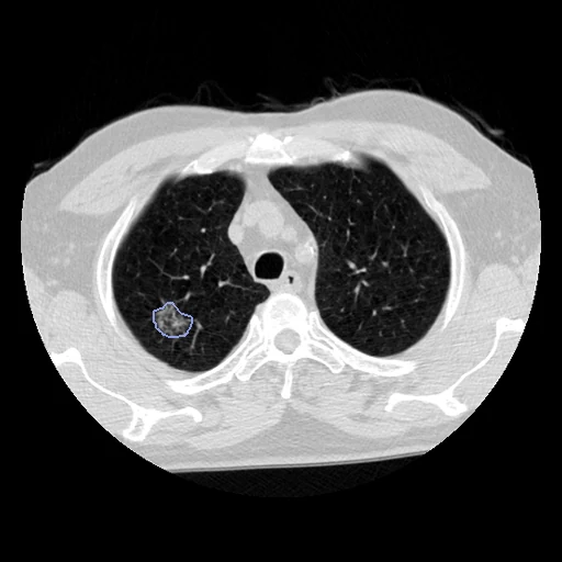
Low-dose CT of a National Lung Screening Trial participant diagnosed with lung cancer, from the screening year of the diagnosis, with the cancer and any suspicious nodule beside it painted by hand (NLSTseg).
Read before comparing (10)
- Source. One CT a participant: of the screening year of the diagnosis, the series with the most slices, ties broken by kernel.
- Truth. A researcher with two years of imaging research and an oncology radiologist with five years painted each lesion along its outline in 3D Slicer, from the lobe NLST's records give; a radiation oncologist and a radiologist, ten years each, cross-verified every lesion. With no contrast, a central tumour's edge against the hilum, mediastinum or heart was drawn where a changed window showed it.
- Labels. Tumours and the suspicious nodules beside them are all label 1, lung_nodule, as in NLSTNodules and LNDbNodules; the lesion type is not kept. Of a diffuse cancer of more than five lesions, the five most prominent are painted. 124505's only lesion, a nodule, is listed twice in the table, once in its mask.
- Left out. Every other nodule: a screening CT holds more, and a method that outlines them is charged with their voxels. A method gets the CT alone, with no mask, finds the cancer and outlines it.
- Lesions. The ten test CTs drawn first hold 13 tumours of 110 to 8,789 mm³ (median 1,362 mm³), ground-glass to solid, each in the lobe the table names but on 206543 (76% in the left lower lobe, not the left upper). On three, bits of 1 to 11 voxels of paint lie 1 to 7 mm from their lesion.
- Images. The NLST DICOM series as delivered, in Hounsfield units. Each mask lies on the voxels it was painted on, checked on the twenty cases built first and by prepare.py on the forty others; on 211328 that is the kernel C reconstruction, not the kernel D one Image.xlsx names.
- TotalSegmentator. Version 2.18's lung_nodules task scores a Dice of 0.28 and an HD95 of 197 mm on test case 100570, outlining part of its tumour and three far spots, and 0.37 and 57 mm on 209095, finding two of its three tumours. HD95 here mostly counts what is found away from the cancer.
- NLSTNodules. 147 of NLSTseg's participants are among LIDC-annot-NLST501's 501, NLSTNodules' source, among them test participants 102658, 120289 and 209095 and development participant 212522, none in NLSTNodules' cases. On the five CTs both datasets hold, left out here, NLSTseg's lesions are NLSTNodules' nodules or masses drawn smaller (Dice 0.60 to 0.81); NLSTNodules also marks the other nodules.
- Outside numbers. None: the authors' 2D U-Net is scored on its own split. NLSTseg is public with its masks, so a model trained on it has seen these test CTs, and one trained on LIDC-annot-NLST501 three of them.
- Overlap. The pool leaves out the 128 participants prepare.py lists for the corpus's other NLST datasets (Lung250M and L2R_NLST, LUNA25, NLSTNodules, NLSTKernel_Syn), 19 of them NLSTseg's. The sixty here, checked by ID against every descriptor, are in no other dataset, and NLSTKernel_Syn leaves them out. Lung250M's 22 NLST training pairs and Learn2Reg NLST's training pairs publish no participant ID and cannot be checked.
| Accuracy | Cost | |||||||
|---|---|---|---|---|---|---|---|---|
| # | Method | Dice | Surface Dice | HD95 mm | Time s | RAM GB | GPU GB | Images |
| 1 | KonfAI segmentation app · TotalSegmentator (lung_nodules), the authors' packageGPUapp | 0.557 ±0.102 | 0.538 ±0.102 | 81.76 ±29.81 | 41 | 19.4 | 5.3 | |
The order averages each method's ranks on Dice, Surface Dice, HD95 mm, so the first one is not always the best in every column. ± is the 95% interval of the mean over the 30 cases. Two neighbours are tied when a paired Wilcoxon test on the shared cases does not find the upper one better on Dice (5%): the places a method could hold are then shown under its own. The best value of each column is in colour. Click a row to see its images, a column title to sort by it. Time and memory measured on Workstation.
Dice: how much the organ contours overlap, 1 is perfect. Surface Dice: the share of the contours' surfaces within a tolerance of each other, 1 is perfect. HD95: distance between the contours' surfaces.
Case by case
Click a score to look at that result: the image, the truth in green and the method's map in red. Underlined scores have their image; for the others you see the case without the result.
About the data
- Test cases
- 30 test CTs drawn with seed 2026 among the 555 outside development, then 30 development CTs, the last of the same seeded order; the draw is not stratified. The pool is the 585 of NLSTseg's 605 participants left once the corpus's other NLST participants and one broken series are out. The provider publishes no test set.
- Truth
- contours by experts the lung cancer NLST diagnosed, and any nodule the readers found suspicious beside it, each painted along its outline in 3D Slicer by a researcher and an oncology radiologist, from the lobe NLST's records give, and cross-verified by a radiation oncologist and a radiologist of ten years each: tumours and nodules alike label 1, lung_nodule, the corpus's name for a lung nodule or mass
- Source
- doi.org/10.5281/zenodo.14838349
doi.org/10.1038/s41597-025-05742-x
pmc.ncbi.nlm.nih.gov/articles/PMC12375103
github.com/irene2023study/NLSTseg
doi.org/10.5281/zenodo.17362624
www.cancerimagingarchive.net/collection/nlst
doi.org/10.7937/TCIA.HMQ8-J677
www.cancerimagingarchive.net/analysis-result/lidc-annot-nlst501
raw.githubusercontent.com/multimodallearning/Lung250M-4B/main/data_curation/nlst_test_nbia.csv - Licence
- CC BY 4.0 (the Zenodo record of the masks and tables, and NLST's CT images on TCIA)
30 cases for tuning, never tested
212522, 109189, 213630, 106553, 126967, 209512, 211328, 211893, 107561, 104815, 102660, 217501, 114746, 111454, 115524, 115571, 210090, 102388, 134082, 121099, 124505, 213442, 201018, 104363, 121893, 132606, 119597, 104999, 214812, 131611