 fidopt
ISPY1-Tumor-SEG-Radiomics
First post-contrast phase of the pre-treatment breast DCE MR of a woman with locally advanced breast cancer, the whole primary tumour outlined by hand and approved by a breast radiologist (ISPY1-Tumor-SEG-Radiomics, TCIA).
16 test casesbreastimage MR
Read before comparing (9)
- Pool. 163 women of the I-SPY 1 TRIAL (ACRIN 6657), with stage 2 or 3 breast cancer of at least 3 cm, treated by neoadjuvant chemotherapy at several centres from May 2002 to March 2006. The analysis result keeps those whose pre-treatment DCE has at least two post-contrast phases and whose histopathology and recurrence-free survival are known.
- Image. A unilateral sagittal fat-suppressed T1-weighted 3D gradient echo at 1.5 T. The case is the provider's first post-contrast phase, copied as is: bias-corrected (N3), resampled to 1 mm and z-scored over the patient's three phases with CaPTk 1.8.1, the grid the outline lies on. The tumour's median z-score is 3.0 to 29.0 on the test cases, the whole image's -0.3 to -1.3.
- One image. A method gets only this phase, the one the outline was drawn on and where the tumour enhances. The pre-contrast and second post-contrast phases are not given, nor the phase on its acquired grid, which the outline does not lie on.
- Truth. The manual structural tumour volume (masks_stv_manual): the whole primary tumour, enhancing and non-enhancing, satellite lesions left out. A breast radiologist located it from the provider's functional tumour volume and delineated it in ITK-SNAP; a fellowship-trained breast radiologist reviewed, edited and approved every outline. It meets the main leaderboard's bar, but with no second reader its own error is unknown.
- Pieces. Five of the ten test outlines drawn first carry 1 to 41 small pieces apart from the main body, at most 1.3% of their voxels, such as 41 around ISPY1_1145's 198 ml body; they count in the Dice and the HD95. The tumours measure 1.9 to 200 ml on the test cases. The surface Dice is at 1 mm, on the 1 mm grid.
- Room. The provider's 3D residual U-Net reached a median Dice of 0.74 over the held-out folds of its nested cross-validation. Its shipped outlines (masks_stv_resunet, not used) score 0.65 on the ten test cases drawn first (0.01 to 0.91; 0.01 on ISPY1_1062, a dense breast enhancing strongly around the tumour); the paper does not say whether they come from held-out folds.
- Checks. TotalSegmentator makes no breast tumour, so the labels were checked by eye: on the 20 cases drawn first, and ISPY1_1094 and 1158, every outline lies on an enhancing mass. The bright artefacts the paper reports outside the breast, on ISPY1_1183 (development) and ISPY1_1187, lie away from the outline.
- Overlap. No other dataset of the corpus holds I-SPY 1 images. BreastNACT, UCSF's pilot study, enrolled from 1995 to 2002, and neither collection names a patient of the other; ISPY2_Syn is a later trial, 2010 to 2016. MAMA-MIA (screening segmentation 119) annotates I-SPY 1 patients again: a build of it should leave out the 30 patients used here.
- Outside numbers. A network trained on this analysis result or on MAMA-MIA may have seen the test cases.
No method has a result on every case of this dataset yet.
Case by case
Click a score to look at that result: the image, the truth in green and the method's map in red. Underlined scores have their image; for the others you see the case without the result.
About the data
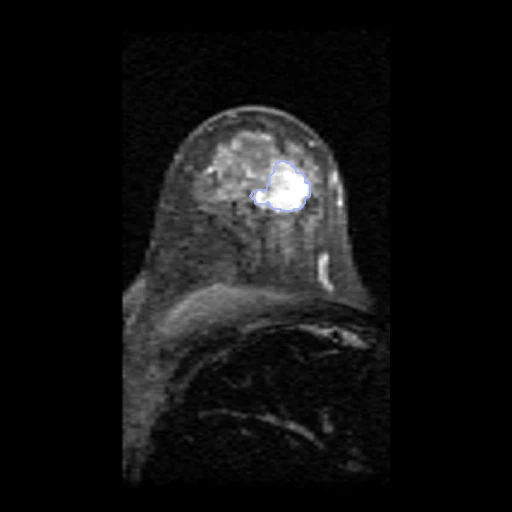
The first test caseClick to look at every case.
- Test cases
- 16 test patients drawn with seed 2026 by scanner maker among the 149 of the analysis result outside development (11 GE, 4 Siemens, 1 Philips), and 14 development patients, the last of the same seeded order, each maker in proportion (10 GE, 3 Siemens, 1 Philips); ISPY1_1237 is kept out of development, its outline lying off its image's grid. One examination a patient, the pre-treatment DCE.
- Truth
- contours by experts the analysis result's structural tumour volume: the whole primary tumour, its enhancing and non-enhancing parts, delineated by hand in ITK-SNAP on the first post-contrast phase by a board-certified breast radiologist, from the location the provider's tumour box and functional tumour volume gave, then reviewed, edited and approved by a board-certified, fellowship-trained breast radiologist; satellite lesions left out
- Source
- www.cancerimagingarchive.net/analysis-result/ispy1-tumor-seg-radiomics
doi.org/10.7937/TCIA.XC7A-QT20
doi.org/10.1038/s41597-022-01555-4
www.cancerimagingarchive.net/wp-content/uploads/ispy1_README.txt
www.cancerimagingarchive.net/collection/ispy1
doi.org/10.7937/K9/TCIA.2016.HdHpgJLK - Licence
- CC BY 3.0
14 cases for tuning, never tested
ISPY1_1161, ISPY1_1183, ISPY1_1177, ISPY1_1146, ISPY1_1189, ISPY1_1181, ISPY1_1037, ISPY1_1125, ISPY1_1132, ISPY1_1032, ISPY1_1090, ISPY1_1196, ISPY1_1222, ISPY1_1107