 fidopt
FDG-PET-CT-Lesions (autoPET), PET lesions
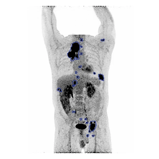
Whole-body FDG-PET in SUV, from the neck down, of a patient with melanoma, lymphoma or non-small cell lung cancer, every FDG-avid lesion judged malignant drawn by an experienced radiologist (FDG-PET-CT-Lesions, the autoPET data).
Read before comparing (10)
- Pool. 1,014 whole-body FDG-PET/CT studies of 900 patients at University Hospital Tübingen, on one Siemens Biograph mCT, 501 of them positive. A case is a patient's first study with at least one FDG-avid lesion, so no patient is in two cases.
- Held out. PETCT_76ebd5c736_2005-12-29 has its three lesions, 1.6 ml, all in the box the defacing blanked. PETCT_1f65acff65_2007-05-06's header puts the PET 5.6 minutes after the injection, against the protocol's 60, so no SUV can be computed. PETCT_aa27cb9156_2003-12-14's SEG names another PET series than its study's. Only the studies drawn were checked.
- Image. The attenuation-corrected PET in body-weight SUV on its own grid, 2.04 mm in plane and 3 mm between slices, computed with AutoPET_CTPET_Syn's function, which that dataset checked against the provider's NIfTI release. No mask is given.
- PET alone. The segmentation contract hands a method one image, and the truth was drawn on the PET's grid from its uptake, the CT only shown beside it: the CT is not given. The provider's nnU-Net and the autoPET entries read the PET and the CT together, so their numbers are not these.
- Truth. From the clinical report of a radiologist and a nuclear medicine physician, one experienced radiologist drew every FDG-avid lesion judged malignant slice by slice on the PET, the uncertain studies reviewed in consensus. One reader and no measure of agreement: graded expert. Physiological uptake is in no label.
- Ignore region. The public PETs are defaced: a box from the base of the neck up reads 0. Wherever the PET reads exactly 0, that box and the air, both maps are zeroed before scoring. On the test cases this takes 6,899 of 14,121 lesion voxels out of PETCT_3d6f425a76_2002-01-28 and 7,268 of 11,137 out of PETCT_f144b214af_2001-02-23, in the neck.
- Lesions. Outside the ignore region the 30 test cases hold 0.40 to 2,465 ml of lesion (median 87 ml) in 1 to 126 pieces, 205 of the 544 under 0.5 ml. A case's Dice is that of its largest lesions, so missing small ones costs little; autoPET also scores the volumes missed and falsely found, not computed here.
- Room. Moved by one voxel, the 30 test masks keep a Dice of 0.62 to 0.93 (mean 0.84, six moves); the ten drawn first keep 0.75 of the surface Dice, taken at 1 mm, under the PET's voxel. The provider's nnU-Net, reading the SUV and the CT, reached a mean Dice of 0.73 in five-fold cross-validation over the collection. No outside number is scored as here.
- Checks. On all 30 test cases each ignore region is exactly its PET's zeros. On the 20 cases built first, in coronal projections every outlined lesion lies on focal uptake, none on the heart, the kidneys or the bladder.
- Overlap. AutoPETCT and AutoPET_CTPET_Syn come from the same collection; their patients are left out. The autoPET challenges hold this FDG set: a dataset built from them should leave out this dataset's patients, listed in datasets/partitions.json. The masks are autoPET's public training data, so a network trained on it has seen these patients.
No method has a result on every case of this dataset yet.
Case by case
Click a score to look at that result: the image, the truth in green and the method's map in red. Underlined scores have their image; for the others you see the case without the result.
About the data
- Test cases
- 30 test studies drawn with seed 2026 by diagnosis in proportion (11 melanoma, 9 lymphoma, 10 lung cancer), then 30 development studies, the last of the same seeded order, in proportion too (11 melanoma, 9 lymphoma, 10 lung cancer). The pool, 456 studies, is each patient's first study with lesions, less the patients of AutoPETCT and AutoPET_CTPET_Syn and three studies without usable truth or SUV. draw.held_out lists the strata's studies so left out; ten more of those datasets' patients are kept out of development.
- Truth
- contours by experts every FDG-avid lesion judged malignant from the clinical PET/CT report, primary tumour and metastases, drawn slice by slice on the PET in NORA by one experienced radiologist with the CT beside it or over it, the uncertain studies reviewed in consensus with the radiologist and the nuclear medicine physician of the report; where the defaced PET holds no data (its exact zeros: the box blanked from the base of the neck up, and the air) both maps are taken out of the scoring
- Source
- www.cancerimagingarchive.net/collection/fdg-pet-ct-lesions
doi.org/10.7937/gkr0-xv29
doi.org/10.1038/s41597-022-01718-3
www.cancerimagingarchive.net/wp-content/uploads/Clinical-Metadata-FDG-PET_CT-Lesions.csv
services.cancerimagingarchive.net/nbia-api/services/v1/getSeries?Collection=FDG-PET-CT-Lesions - Licence
- CC BY 4.0
30 cases for tuning, never tested
PETCT_f01e83894d_2005-03-24, PETCT_42e9f16c09_2003-01-05, PETCT_c3cc732d95_2005-07-21, PETCT_4b688f46b0_2005-02-18, PETCT_98c6af8b90_2003-05-26, PETCT_249dd35d0c_2005-05-20, PETCT_7fbb39290e_2002-08-15, PETCT_275fb00a66_2001-05-04, PETCT_7147385005_2002-05-09, PETCT_1fcfa34d10_2003-09-19, PETCT_760c77b289_2002-01-20, PETCT_d51664a9e4_2004-11-05, PETCT_af73b85f2c_2003-06-19, PETCT_5c55b3087d_2000-12-11, PETCT_4404466919_2005-09-10, PETCT_b04be2846e_2004-11-15, PETCT_ec6b934720_2002-10-25, PETCT_09ee00bdc6_2005-09-30, PETCT_8b74860ce6_2005-03-10, PETCT_5255c79083_2001-10-20, PETCT_06a46414eb_2006-03-12, PETCT_a9d7a14ba1_2000-08-14, PETCT_61348439bf_2005-07-24, PETCT_6c660140ca_2006-08-06, PETCT_64909533c5_2003-04-14, PETCT_74bbceaeeb_2005-06-03, PETCT_9c8842d12e_2004-01-25, PETCT_84e7715ed9_2002-01-04, PETCT_1b1bdfc35b_2000-10-29, PETCT_11afab3485_2003-05-25