 fidopt
LIDC-annot-NLST501
Low-dose chest CT of a National Lung Screening Trial participant, with the lung nodules and masses that at least two of four independent radiologists segmented (LIDC-annot-NLST501, a TCIA analysis result of 2026).
Read before comparing (10)
- Cohort. 501 low-dose NLST CTs, one a participant, chosen by Median Technologies: 152 with an NLST lung cancer diagnosis and 349 without. A cancer participant's CT is at diagnosis or the latest within a year before it, the others' their first; slices are under 5 mm and cover the whole lung.
- Reads. Four of 20 board-certified radiologists read each CT independently, blind to clinical data, after LIDC-IDRI's protocol: nodules of 4 to 30 mm and masses above 30 mm reported, nothing under 4 mm, and up to six of the most suspicious segmented in 3D, ground-glass parts included. Vessels, airways and the pleura were left to each reader.
- Pool. The CTs where every read segmented every nodule and mass it reported, so no reader stopped at six (240 of the 501), and where two reads segmented one same nodule (208 of the 240). 116385's corrected series still skips a slice position and is left out. Test case 204367's three slices filled with copies by the provider hold no truth: it is kept.
- Truth. Label 1 where at least two of the four reads put a nodule or a mass. The ten test CTs drawn first hold 1 to 7 nodules each, of 7 to 3,090 mm³. On 120570 two reads outlined a mass and two a nodule of about 1,000 mm³: the truth keeps 3,090 mm³ there.
- Left out. Nodules under 4 mm, other focal findings and nodules one read alone segmented are not in the truth: on the first ten test CTs, 25 such parts (0 to 4 a CT). A method that outlines one is charged with its voxels.
- Spread. Each read scored against the voxels two of the other three hold: on the first ten test CTs the 40 reads reach a median Dice of 0.70 (0 to 0.88), seven at 0; on the development CTs 0.16 to 0.90 (median 0.65). No method should be expected to beat that. A case's Dice pools its nodules, so the largest weighs most; HD95 mostly counts nodules found or missed.
- TotalSegmentator. Version 2.18's lung_nodules scores a Dice of 0.71 on 122866's nodule of 1,089 mm³, drawing it smaller than the readers, and finds none of 108433's three nodules of about 6 mm.
- Images. The NLST DICOM series as delivered, in Hounsfield units, from GE, Siemens and Philips scanners, slices 1.25 to 2.5 mm apart over the twenty test CTs; some GE CTs read -3024 or -2048 HU outside the reconstructed circle. No mask is given: a method gets the CT alone and must find the nodules.
- Outside numbers. None: the provider's paper is in submission. The CTs and reads are public, so a model trained on LIDC-annot-NLST501 has seen these test cases.
- Overlap. By NLST participant ID, none of the corpus's thirty other NLST participants (Lung250M, L2R_NLST, LUNA25) is here; NLSTKernel_Syn leaves out all 501, and NLSTSeg these 38. Lung250M's 22 NLST training pairs and Learn2Reg NLST's publish no participant ID and cannot be checked.
| Accuracy | Cost | |||||||
|---|---|---|---|---|---|---|---|---|
| # | Method | Dice | Surface Dice | HD95 mm | Time s | RAM GB | GPU GB | Images |
| 1 | KonfAI segmentation app · TotalSegmentator (lung_nodules), the authors' packageGPUapp | 0.288 ±0.125 | 0.347 ±0.151 | 125.12 ±47.86 | 40 | 19.0 | 5.3 | |
The order averages each method's ranks on Dice, Surface Dice, HD95 mm, so the first one is not always the best in every column. ± is the 95% interval of the mean over the 20 cases. Two neighbours are tied when a paired Wilcoxon test on the shared cases does not find the upper one better on Dice (5%): the places a method could hold are then shown under its own. The best value of each column is in colour. Click a row to see its images, a column title to sort by it. Time and memory measured on Workstation.
Dice: how much the organ contours overlap, 1 is perfect. Surface Dice: the share of the contours' surfaces within a tolerance of each other, 1 is perfect. HD95: distance between the contours' surfaces.
Case by case
Click a score to look at that result: the image, the truth in green and the method's map in red. Underlined scores have their image; for the others you see the case without the result.
About the data
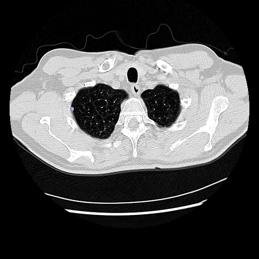
- Test cases
- 20 test CTs drawn with seed 2026 among the 207 of LIDC-annot-NLST501's 501 whose four reads segmented every nodule and mass they reported and share a nodule, outside development; 18 development CTs, the last of the same seeded order. The corpus's NLST participants and one broken series are left out. The provider publishes no test set.
- Truth
- contours by experts the voxels that at least two of the CT's four independent reads put inside a nodule of 4 to 30 mm or a mass they segmented (LIDC's 50% agreement), each read by one of 20 board-certified radiologists who outlined their up to six most suspicious lesions in 3D
- Source
- www.cancerimagingarchive.net/analysis-result/lidc-annot-nlst501
doi.org/10.7937/y2dx-e334
www.cancerimagingarchive.net/wp-content/uploads/LIDC-annot-NLST501_TCIA_documentation.pdf
www.cancerimagingarchive.net/collection/nlst
doi.org/10.7937/TCIA.HMQ8-J677
raw.githubusercontent.com/multimodallearning/Lung250M-4B/main/data_curation/nlst_test_nbia.csv - Licence
- CC BY 4.0 (the analysis result's masks and tables, and NLST's CT images, on their TCIA pages)
18 cases for tuning, never tested
106850, 130695, 205736, 122937, 118970, 212863, 124298, 211707, 123521, 212510, 207647, 206971, 201769, 211436, 129124, 123546, 118560, 104765