 fidopt
Vestibular-Schwannoma-SEG, tumour and cochlea
High-resolution T2 MR of the head of one patient with a vestibular schwannoma, with the tumour and the cochlea drawn by the treating neurosurgeon and physicist.
12 test casesbrainimage MR
Read before comparing (1)
- Cases. The fixed images of VestibularSchwannoma's pairs, as delivered, with their own labels: a test pair's image is a test case, a development pair's a development case. The moving images carry a known synthetic deformation and are left out.
No method has a result on every case of this dataset yet.
Case by case
Click a score to look at that result: the image, the truth in green and the method's map in red. Underlined scores have their image; for the others you see the case without the result.
About the data
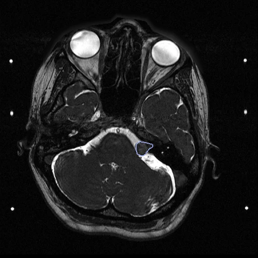
The first test caseClick to look at every case.
- Truth
- contours by experts the tumour and the cochlea, drawn by the treating neurosurgeon and physicist and exported into the T2 by the planning software
- Source
- www.cancerimagingarchive.net/collection/vestibular-schwannoma-seg
doi.org/10.7937/TCIA.9YTJ-5Q73
doi.org/10.1038/s41597-021-01064-w
github.com/KCL-BMEIS/VS_Seg - Licence
- CC BY 4.0
11 cases for tuning, never tested
VS-SEG-001/fixed, VS-SEG-002/fixed, VS-SEG-003/fixed, VS-SEG-230/fixed, VS-SEG-019/fixed, VS-SEG-149/fixed, VS-SEG-045/fixed, VS-SEG-050/fixed, VS-SEG-165/fixed, VS-SEG-093/fixed, VS-SEG-071/fixed