 fidopt
Light My Cells, transmitted light to nucleus fluorescence, 2D
One field of a cell culture in 2D: a transmitted-light image (bright field, phase contrast or DIC) as source, and the nucleus fluorescence image of the same field and focal plane as target (Light My Cells, France-BioImaging).
10 test casescellsource transmitted light, target nucleus fluorescence2D, distances in µm
Read before comparing (12)
- Case. The source is one plane of the field's transmitted-light z-stack, uint16 as delivered: the plane at the nucleus image's focus (the provider records that plane's index, TheZ, and the stage z agrees where given). The target is the provider's best-focus plane of the nucleus stain: Hoechst, DAPI, DRAQ5, SiR-DNA or a stably expressed fluorescent histone, by study. 512 x 512 to 2048 x 2048 pixels of 0.05 to 0.16 µm, written in micrometres as if millimetres.
- Modality. 'brightfield' covers the three transmitted-light contrasts, bright field, phase contrast and DIC; the draw's strata keep them apart.
- Alignment. Both images come from one microscope, objective and camera (a confocal's detectors for myoblast_genethon and fibroblast_crsa) at one stage position, on one grid; nothing is registered. prepare.py refuses a set whose two files differ in grid, plane or recorded stage position. On the built cases the nuclei fall on the outlines the DIC and bright-field images show, within a few pixels by eye; what is left counts as error for every method.
- Values. Each nucleus image is scaled as the challenge's evaluation scales it, its 2nd percentile to 0 and its 99.8th to 1, not clipped; truth.dynamic_range is 1. The challenge scaled the prediction the same way; the corpus scores it as returned, so a method must give the target's scale.
- Mask. The whole field, as the challenge scores it: a mask made from the target would give the methods the nuclei's places, which is the task.
- Baseline. On the whole field of the 10 test cases, at a range of 1, a constant at the target's median scores an MAE of 0.094 (0.034 to 0.196), a PSNR of 15.3 dB (12.0 to 19.8) and an SSIM of 0.61 (0.30 to 0.84). The background it gets right lifts its SSIM where nuclei are few: u2os_h2b_gfp 0.84.
- Focus. In urinary_bladder_carcinoma the provider's best plane for the nucleus (DRAQ5) is the last of its 27, and the image is blurred: its focus likely lies past the stack.
- Saturation. myoblast_genethon's nucleus image, from a confocal, is 12-bit with 1.7% of its pixels at 4095, so its 99.8th percentile is that ceiling and its brightest nuclei are flat at 1; its values come in steps of about 69 (98 distinct values, 76% of the field at 0).
- Samples. A case is a Biosample of the deposit, so that a culture imaged by several studies, with other objectives or modalities, never falls in both test and development. U2OS IGH (studies 29, 30) is taken as U2OS H2B GFP (19, 21, 23): one description of the cells, word for word, and one specimen named 'st21_29_30', imaged on two microscopes. HeLa, one cell line, is four samples, each another lab's culture or stable line on another microscope: hela_igh and hela_vienna in test, hela_roussy and scan_hela_h2b_gfp in development. HeLa Schaffner (study 16) labels mitochondria only and is not a case.
- Organelles. Mitochondria, tubulin and actin images are in 12, 5 and 2 of the deposit's 15 samples; a sample sits in one dataset only, so they are not cases.
- Overlap. No other dataset of the corpus holds Light My Cells images; the data descriptor calls all 56,984 previously unpublished. A registration dataset made of these pairs under a known warp would derive from this one.
- Outside numbers. The deposit is the challenge's training database, public with its targets: a model trained on it, the challenge's among them, has seen every case. The challenge scored 36 and then 322 test images from an acquisition site held out of the deposit.
No method has a result on every case of this dataset yet.
Case by case
Click a score to look at that result: the target image, and over it the method's synthetic image. Underlined scores have their image; for the others you see the case without the result.
About the data
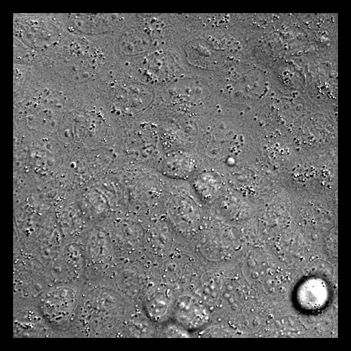
The first test caseClick to look at every case.
- Test cases
- 10 test samples drawn with seed 2026 by transmitted-light modality in proportion, and the other 4 for development, one case a sample. A sample is a cell culture of the deposit with a nucleus image, 14 of them (its 15 Biosamples with one, U2OS IGH taken as U2OS H2B GFP), that several of its 30 studies may image; its field is drawn by prepare.py among the sample's acquisition sets.
- Truth
- target image as released the deposit's nucleus fluorescence image of the field, the best-focus plane the provider chose in its fluorescence z-stack, scaled as the challenge's evaluation scales it (2nd percentile to 0, 99.8th to 1)
- Source
- www.ebi.ac.uk/biostudies/BioImages/studies/S-BIAD1047
doi.org/10.1038/s41597-026-07004-w
lightmycells.grand-challenge.org
codeberg.org/FranceBioImaging/LightMyCellsDatabase
codeberg.org/FranceBioImaging/LightMyCellsChallenge - Licence
- CC BY 4.0 (the BioImage Archive study's License field)
4 cases for tuning, never tested
hela_roussy, human_liver_carcinoma, scan_hela_h2b_gfp, fibroblast_crsa