 fidopt
I-SPY 2 DCE MR, pre-contrast to early post-contrast
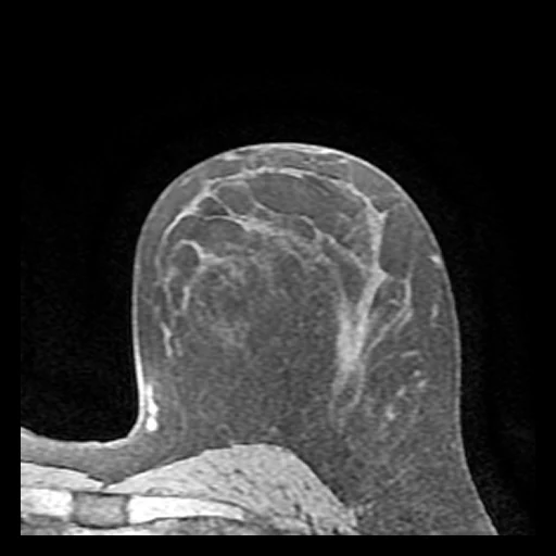
Fat-suppressed T1-weighted breast MR of a woman with locally advanced breast cancer before treatment: the pre-contrast phase (source) and the early post-contrast phase (target) of one dynamic acquisition, on one grid (ISPY2, TCIA).
Read before comparing (10)
- Pool. The I-SPY 2 trial (NCT01042379) tests new neoadjuvant agents in women with locally advanced breast cancer. The collection holds the MR of 719 patients at up to four visits; a case is a patient's pre-treatment DCE, axial and bilateral, the patient prone. GE scanned 425 of the 717 patients, Siemens 209 and Philips 83.
- Images. UCSF's Breast Imaging Research Program stored every phase in one VOLSER series, cropped to the breast with the tumour, or bilateral where the acquisition had fewer than 384 voxels a row. On the 20 cases drawn first the crop keeps whole voxels at the acquired spacing, and no registration is recorded.
- Pair. Source and target are the phases the SER map's timing indices name pre-contrast and early, phases 0 and 2 on all 60 cases drawn; the early one comes 127 to 152 s after the injection on the ten test cases drawn first. Both are slices of one series on one grid; nothing is registered.
- Motion. Motion between the phases stays in the pair and counts as error for every method: a rigid registration, not applied, moves the body's voxels by 0.1 to 2.5 mm on average on the ten test cases drawn first, up to 3.3 mm on ISPY2-518000.
- Values. The scanners' 16-bit magnitudes without unit, on scales that differ by scanner; the Philips test case ISPY2-572793 holds them in steps of 23.
- Mask. The body in the field, from the source alone by an Otsu threshold: the breast with its fat, the skin and the chest wall, the lungs mostly out, 36% to 64% of the image on the test cases. On ISPY2-705381 and ISPY2-930903, the two GE 3 T cases drawn, it takes in a band of the air's noise along the skin.
- Range. truth.dynamic_range, 2502, is the target's 99.9th percentile inside the mask pooled over the ten test cases drawn first, whose own run from 200 to 3107: the PSNR and SSIM of dim scans read high. The MAE is in each scan's units, so its mean weighs the brighter scans most.
- Baseline. Inside the mask on the 30 test cases a constant at the target's median scores an MAE of 116 (17 to 537), a PSNR of 23.9 dB (11.0 to 39.1) and an SSIM of 0.53 (0.14 to 0.89). The source itself scores 71 (11 to 397), 27.2 dB (12.3 to 42.1) and 0.77 (0.37 to 0.95), better than the constant on 28 of the 30: what a method must add is the enhancement.
- Public copies. BreastDCEDL_ISPY2 (TCIA, CC BY 4.0) holds the same voxels of 982 patients, without their geometry, and its training split holds 6 of the ten test cases drawn first. MAMA-MIA also holds I-SPY 2 patients. A network trained on either may have seen the test cases.
- Overlap. No dataset of the corpus holds I-SPY 2 or ACRIN-6698 images; BreastNACT and ISPY1BreastMR are earlier cohorts. Later builds from MAMA-MIA or other I-SPY 2 data should leave out the 60 patients used here, the test and development cases.
No method has a result on every case of this dataset yet.
| Accuracy | Cost | ||||||||
|---|---|---|---|---|---|---|---|---|---|
| # | Method | MAE | Change | PSNR dB | SSIM | Time s | RAM GB | GPU GB | Images |
| Source as given | 104.1 ±35.5 | 26.80 ±2.87 | 0.716 ±0.067 | ||||||
MAE: mean absolute error of a synthetic image against its target, inside the body, in the image's units. PSNR: the same by signal-to-noise ratio, higher is better. SSIM: the same by structural similarity, 1 is perfect.
Case by case
Click a score to look at that result: the target image, and over it the method's synthetic image. Underlined scores have their image; for the others you see the case without the result.
About the data
- Test cases
- 30 test patients drawn with seed 2026 by scanner maker in proportion (18 GE, 9 Siemens, 3 Philips) among the 717 with a pre-treatment VOLSER DCE, one examination each. Then 30 development patients, the last of the same seeded order, each maker in proportion: 18 GE, 9 Siemens, 3 Philips.
- Truth
- target image as released the patient's early post-contrast phase, the one the provider's laboratory took as early for its enhancement maps (an effective 127 to 152 s after the injection on the ten test cases drawn first), from the same series and acquisition as the source, on its grid as acquired, not registered
- Source
- www.cancerimagingarchive.net/collection/ispy2
doi.org/10.7937/TCIA.D8Z0-9T85
www.cancerimagingarchive.net/wp-content/uploads/ACRIN-6698-ISPY2-Shared-Private-Tag-Data-Dictionary_20210520.xlsx
www.cancerimagingarchive.net/analysis-result/breastdcedl_ispy2
clinicaltrials.gov/study/NCT01042379 - Licence
- CC BY 4.0
30 cases for tuning, never tested
ISPY2-255535, ISPY2-746731, ISPY2-345574, ISPY2-994987, ISPY2-246134, ISPY2-306127, ISPY2-277848, ISPY2-116165, ISPY2-676365, ISPY2-261304, ISPY2-553141, ISPY2-299364, ISPY2-357034, ISPY2-136536, ISPY2-930903, ISPY2-252748, ISPY2-802105, ISPY2-451816, ISPY2-590220, ISPY2-660915, ISPY2-333145, ISPY2-166782, ISPY2-422450, ISPY2-317131, ISPY2-794428, ISPY2-623569, ISPY2-439489, ISPY2-916645, ISPY2-115638, ISPY2-262131