 fidopt
Vestibular-Schwannoma-SEG, contrast-enhanced T1 to T2
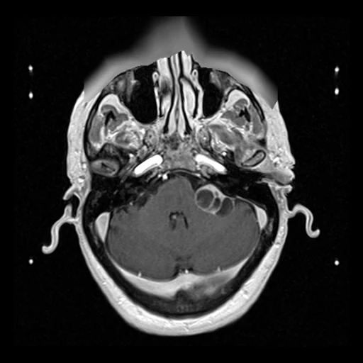
Contrast-enhanced T1 MR of the head of a patient with a vestibular schwannoma (source) and the high-resolution T2 of the same Gamma Knife session (target), the head fixed in a stereotactic frame (Vestibular-Schwannoma-SEG, TCIA).
Read before comparing (9)
- Cohort. 242 consecutive adults with a single sporadic vestibular schwannoma treated by Gamma Knife radiosurgery in London between 2012 and 2018. For planning, each had a contrast-enhanced T1 (MPRAGE) and a high-resolution T2 (3D CISS or 3D turbo spin echo) on a Siemens Avanto at 1.5 T; on the 20 cases built first the T2 starts 7 to 9 minutes after the T1.
- Alignment. The provider's: with the head in a Leksell frame, Leksell GammaPlan computes the transform between the series from the frame's fiducials (inv_T1_LPS_to_T2_LPS.tfm, rigid, rotations of 0.10 to 0.46 degrees on the 20 built first). prepare.py reads the T1 through it on the T2's grid, linearly, 0 past the T1's field. The T2 is not resampled and the corpus registers nothing.
- Residual. The provider does not state the fiducial registration's accuracy, and one rigid transform cannot hold a difference of distortion between the sequences: what it leaves counts against every method. On checkerboards of the 20 built first, the skull, brainstem, cerebellum, ventricles and vessels run on from one image into the other.
- Target. The CISS T2 is 448 x 448 voxels of 0.47 mm, 80 slices of 1.5 mm (70 on VS-SEG-115); the turbo spin echo T2 384 x 384 of 0.55 mm, 40 slices of 1.5 mm around the canals. The T1 covers 95 to 100% of it. The two give different targets, so the draw takes them in proportion.
- Scanners. Two test cases were scanned at 3 T (VS-SEG-115 on a TrioTim, VS-SEG-200 on a Prisma fit), and their values run lower.
- Mask. The head inside both fields: the T1 on the T2's grid above a twentieth of its 99th percentile, opened, its largest connected part, holes filled slice by slice, kept where the T1 has data; 1.4 to 3.1 litres on the test cases. It keeps some noise beside the ears on a few cases; the frame's fiducials lie outside it.
- Baseline. PSNR and SSIM take a range of 4095, the T2's 12-bit storage. Inside the mask on the 21 test cases, a constant at the target's whole-image median scores an MAE of 247 (107 to 308), one at its median inside the mask 168 (88 to 223), and the source 376 (211 to 412): the T1 is brighter than the T2 over most of the head.
- Overlap. The corpus's VestibularSchwannoma holds 23 of these patients as registration pairs, the T1 deformed by a known warp, so they are left out (VestibularSchwannoma_Seg takes their T2s): of the 242, 234 have both series on NBIA, which leaves 211. VestibularSchwannomaMCRC leaves out the 58 patients the provider's multi-centre collection shares with this one.
- Public. The collection is the source of the crossMoDA challenges, unpaired contrast-enhanced T1 to T2 domain adaptation: a network trained on crossMoDA data has seen these patients' images. The collection's tumour and cochlea contours are not part of this dataset.
No method has a result on every case of this dataset yet.
| Accuracy | Cost | ||||||||
|---|---|---|---|---|---|---|---|---|---|
| # | Method | MAE | Change | PSNR dB | SSIM | Time s | RAM GB | GPU GB | Images |
| Source as given | 375.5 ±20.9 | 19.23 ±0.57 | 0.371 ±0.016 | ||||||
MAE: mean absolute error of a synthetic image against its target, inside the body, in the image's units. PSNR: the same by signal-to-noise ratio, higher is better. SSIM: the same by structural similarity, 1 is perfect.
Case by case
Click a score to look at that result: the target image, and over it the method's synthetic image. Underlined scores have their image; for the others you see the case without the result.
About the data
- Test cases
- 21 test patients drawn with seed 2026 by T2 sequence (17 CISS, 4 turbo spin echo), then 19 development patients, the last of the same seeded order, each sequence in proportion (15 CISS, 4 turbo spin echo). The pool is the 234 patients with both series on NBIA, less the 23 of the corpus's VestibularSchwannoma (draw.held_out), whose T1 is this dataset's source and whose labels name both images.
- Truth
- target image as released the patient's high-resolution T2 of the same session, as delivered, on its own grid: the source is brought onto it through the provider's frame transform
- Source
- www.cancerimagingarchive.net/collection/vestibular-schwannoma-seg
doi.org/10.7937/TCIA.9YTJ-5Q73
doi.org/10.1038/s41597-021-01064-w
www.cancerimagingarchive.net/wp-content/uploads/Vestibular-Schwannoma-SEG_matrices-Mar-2021.zip
github.com/KCL-BMEIS/VS_Seg - Licence
- CC BY 4.0
19 cases for tuning, never tested
VS-SEG-173, VS-SEG-212, VS-SEG-107, VS-SEG-081, VS-SEG-194, VS-SEG-157, VS-SEG-094, VS-SEG-150, VS-SEG-116, VS-SEG-148, VS-SEG-078, VS-SEG-213, VS-SEG-129, VS-SEG-189, VS-SEG-156, VS-SEG-020, VS-SEG-223, VS-SEG-142, VS-SEG-169