 fidopt
CSXA cervical radiographs
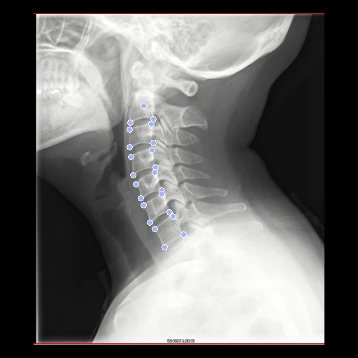
Lateral radiographs of the cervical spine of two people, with 23 keypoints on the vertebral bodies of C2 to C7 placed and checked by spine surgeons on each (Cervical Spine X-ray Atlas, Dongzhimen Hospital, Beijing).
Read before comparing (9)
- People. 4963 people seen at one hospital, 3202 women and 1761 men aged 18 to 87: 4782 with neck pain or cervical spondylosis and 181 without symptoms. No dates, devices or settings are given. Every test pair joins two different curvatures of the dataset's five.
- Images. Each is a capture of the hospital viewer's screen, not the radiograph's own file, with the viewer's marks fixed on the screen: window and level, a side marker, coloured lines. prepare.py keeps red, green and blue as delivered; the grey radiograph's bands agree within 8 levels on over 98.9% of the pixels. No mask.
- Negatives. Four test images show bone dark on a light ground: the fixed images of 1681048_1178032, 3702146_0679150 and 4951037_1925068 and the moving image of 4328170_1914066, as does the moving image of development pair 1935069_4489026. On the first ten test pairs greedy (NCC) did worse than no registration on the two inverted ones, while ANTs' SyN improved all ten.
- Unit. The millimetre of the viewer's ruler, from the provider's pixel equivalent: 0.17 to 0.50 mm a pixel across the archive. Scaled endplates measure 18.2 to 19.4 mm whatever the pixel size, so the scale holds, but whether the ruler is calibrated at the detector or the patient is not said: a millimetre may hold the magnification.
- Truth. 23 points an image: the centre of C2, the corners of its lower endplate and the four corners of C3 to C7; C1 and C2's upper endplate are left out as unclear. One spine surgeon placed them all, three others reviewed them in turn, all four agreed the changes, and a senior surgeon reviewed last.
- Truth error. The paper gives no agreement between readers. On the same corners, three radiologists placing them independently came within 1 mm of each other for 79 to 86% of the points (Yan et al. 2022). The centre of C2, with no edge to mark it, is the loosest point.
- Alignment. Each image lies on its own grid at the origin. The moving image's header carries one translation from elastix mutual information inside each body, the best of several starts, no annotation used. A rigid fit turns the necks 0 to 11 degrees, a matter of posture, so only the translation is kept; size and curvature are left to the methods.
- Baseline. With no registration the 30 test pairs start at a TRE of 13.9 mm (3.3 to 28.5), rTRE 0.041. The points lie on six vertebral bodies: the board does not measure the skull, the soft tissue or the posterior elements.
- Outside numbers. None: the paper derives measurements from the keypoints and registers nothing.
| Accuracy | Smoothness | Cost | ||||||||
|---|---|---|---|---|---|---|---|---|---|---|
| # | Method | TRE mm | Change | rTRE | SDlogJ | Folds % | Time s | RAM GB | GPU GB | Images |
| 11–3 | vismatch matchers · RoMaGPU | 8.28 ±2.87 | −5.65 | 0.0249 ±0.0087 | 0.061 | 0.00 | 12 | 3.2 | 6.9 | |
| 21–3 | vismatch matchers · DISK + LightGlueGPU3 failed | 8.27 ±3.55 | −5.66 | 0.0246 ±0.0105 | 0.361 | 0.28 | 3 | 2.1 | 2.9 | |
| 31–3 | vismatch matchers · SIFT + LightGlueGPU3 failed | 13.38 ±12.12 | −0.55 | 0.0397 ±0.0358 | 0.225 | 3.24 | 3 | 2.0 | 0.5 | |
| 44–6 | vismatch matchers · LoFTRGPU | 24.98 ±24.47 | +11.05 | 0.0741 ±0.0739 | 0.322 | 1.04 | 3 | 2.0 | 2.4 | |
| 44–6 | vismatch matchers · XFeatGPU | 12.82 ±6.98 | −1.11 | 0.0378 ±0.0205 | 0.668 | 1.23 | 3 | 2.2 | 0.4 | |
| 64–6 | vismatch matchers · ALIKED + LightGlueGPU | 32.15 ±32.51 | +18.22 | 0.0926 ±0.0925 | 1.021 | 3.50 | 3 | 2.3 | 1.8 | |
| No registration | 13.93 ±2.58 | 0.0413 ±0.0075 | 0.000 | 0.00 | ||||||
The order averages each method's ranks on TRE mm, SDlogJ, so the first one is not always the best in every column. ± is the 95% interval of the mean over the 30 pairs. Two neighbours are tied when a paired Wilcoxon test on the shared pairs does not find the upper one better on TRE mm (5%): the places a method could hold are then shown under its own. The best value of each column is in colour. Click a row to see its images, a column title to sort by it. Time and memory measured on Laptop.
TRE: distance between matching landmarks. rTRE: the TRE over the length of the image's diagonal, to compare datasets of other sizes and units. SDlogJ: how irregular the deformation is, 0 is perfectly smooth. Folds: share of the image where the deformation folds over itself.
Pair by pair
Click a score to look at that result: the fixed image, and over it the moving image after registration, with the annotations. Underlined scores have their image; for the others you see the pair without the result. A green cell gained against no registration on that pair, a red one lost.
About the data
- Test pairs
- 30 test pairs drawn with seed 2026 among the 2451 outside development, and 30 development pairs. The 4963 images, one a person, are put in the order of sha256("2026:CSXA:<image>") and paired two by two, the first fixed, one image left over. One hospital and one source: the draw is not stratified.
- Truth
- landmarks by experts the dataset's 23 keypoints on each image: the centre of C2, the two corners of its lower endplate and the four corners of the vertebral bodies of C3 to C7, placed by one spine surgeon, reviewed in turn by three others and the changes agreed by all four (Ran et al. 2024)
- Source
- doi.org/10.57760/sciencedb.15391
doi.org/10.1038/s41597-024-03383-0
github.com/yran888/CSXA-dataset
doi.org/10.1186/s12891-022-05927-0 - Licence
- CC BY 4.0
30 pairs for tuning, never tested
3575143_4441024, 2590130_3790148, 3515142_2094124, 4376179_1338035, 1424038_0366132, 2926134_4196162, 4251165_2541130, 0861165_3843150, 4329170_0583140, 4112159_3505142, 2176125_0206127, 1935069_4489026, 1577043_0972025, 0473138_2809133, 0611145_2330127, 1895064_1177032, 4292168_4378179, 1838060_2599131, 4893035_2660131, 4101159_3789148, 4107159_3076136, 4237164_2762133, 0919022_1602044, 2940135_1389037, 3762148_2625131, 1152030_0983025, 0380132_2115124, 2582130_3487141, 0601144_1861062, 4871034_3981154