 fidopt
CT follow-up of liver metastases, liver
Abdominal CT of one patient followed for liver metastases, with the liver and its metastases as one mask.
Read before comparing (3)
- Cases. The fixed images of LiverMetsCT's pairs, each on its pair's side of the split. The moving images are left out: their headers carry the pair's start, so they are not the images as delivered.
- Values. Not Hounsfield units: the provider clipped each CT to a soft-tissue window (level 40, width 450 HU) and scaled it to [0, 1], so a value v stands for about 450 v - 185 HU; air reads 0.004, the liver about 0.6. They are kept as delivered, float32 (float64 for Patient-49 and Patient-50).
- Methods. A method that clips or normalises with fixed Hounsfield values, thresholds air in HU, or runs a network trained on Hounsfield units sees an almost flat image unless it rescales. Air, lung, gas, bone and dense contrast are flat.
| Accuracy | Cost | |||||||
|---|---|---|---|---|---|---|---|---|
| # | Method | Dice | Surface Dice | HD95 mm | Time s | RAM GB | GPU GB | Images |
| 11–2 | KonfAI segmentation app · MRSegmentator, KonfAI appGPUapp | 0.771 ±0.291 | 0.641 ±0.245 | 27.39 ±42.78 | 2.5 | 6.9 | ||
| 21–2 | MONAI wholeBody CTGPUapp | 0.623 ±0.222 | 0.383 ±0.157 | 43.23 ±21.97 | 14 | 3.6 | 3.8 | |
| 33–5 | CT-FM whole-body segmentationGPUapp | 0.000 ±0.000 | 0.000 ±0.000 | 178.65 ±26.87 | 8 | 2.5 | 5.8 | |
| 43–5 | KonfAI segmentation app · TotalSegmentator (total), the authors' packageGPUapp | 0.000 ±0.000 | 0.000 ±0.000 | 14.7 | 2.3 | |||
| 53–5 | KonfAI segmentation app · TotalSegmentator (total), KonfAI appGPUapp | 0.000 ±0.000 | 0.000 ±0.000 | 14 | 2.7 | 4.0 | ||
The order averages each method's ranks on Dice, Surface Dice, HD95 mm, so the first one is not always the best in every column. ± is the 95% interval of the mean over the 10 cases. Two neighbours are tied when a paired Wilcoxon test on the shared cases does not find the upper one better on Dice (5%): the places a method could hold are then shown under its own. The best value of each column is in colour. Click a row to see its images, a column title to sort by it. Time and memory measured on Workstation.
1 result not ranked: an older run, cases still missing, or a failure on every case
| Accuracy | Cost | ||||||||
|---|---|---|---|---|---|---|---|---|---|
| # | Method | Dice | Surface Dice | HD95 mm | Time s | RAM GB | GPU GB | Machine | Images |
| MONAI wholeBody CTGPUappolder run | 0.623 ±0.222 | 0.384 ±0.157 | 43.23 ±21.97 | 8 | 2.3 | 3.7 | Laptop | ||
Dice: how much the organ contours overlap, 1 is perfect. Surface Dice: the share of the contours' surfaces within a tolerance of each other, 1 is perfect. HD95: distance between the contours' surfaces.
Case by case
Click a score to look at that result: the image, the truth in green and the method's map in red. Underlined scores have their image; for the others you see the case without the result.
About the data
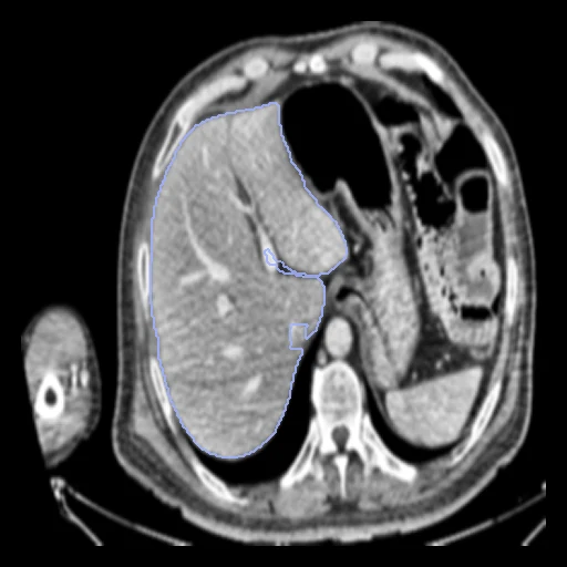
- Truth
- contours by experts the liver (label 1), metastases included: the dataset's liver and metastasis masks together, drawn by a deep-learning network, corrected in ITK-SNAP by trained annotators and verified by radiologists
- Licence
- CC0 1.0
10 cases for tuning, never tested
Patient-01/fixed, Patient-02/fixed, Patient-03/fixed, Patient-20/fixed, Patient-26/fixed, Patient-07/fixed, Patient-05/fixed, Patient-29/fixed, Patient-13/fixed, Patient-22/fixed