 fidopt
RCC-AID
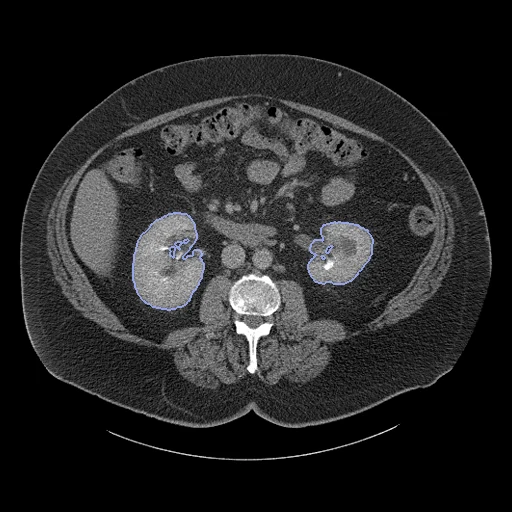
Contrast-enhanced abdominal CT of a patient with a clear cell, papillary or chromophobe renal cell carcinoma (TCGA-KIRC, TCGA-KIRP, TCGA-KICH), kidneys, masses and cysts segmented by a model and corrected by students (RCC-AID).
Read before comparing (8)
- Pool. RCC-AID kept 129 contrast-enhanced CTs of 91 patients from TCGA's 1,915 renal CT scans, by metadata and a reader study; 38 of the 129 hold a cyst. A preprint counts 142 CTs of 101 patients in an earlier record; the files used are IDC release 25's 129, read as DICOM label maps.
- Cases. A case is a patient's earliest study, then the venous, arterial or delayed phase in that order, then the file name, cut to the slices the segmentation covers. The 10 test CTs are 5 venous, 3 arterial and 2 delayed, slices 1 to 5 mm apart, in Hounsfield units with the scanner's padding at -3024 or -3027.
- Truth. A model's masks corrected by two trained students, a radiologist seeing only the cases they marked uncertain; no agreement between readers is published. It passes the bar of a main leaderboard with these limits. The kidney label stops where the annotated slices stop and is one class, its sides left to the scorer's merge of kidney_left and kidney_right.
- Scans. They are what the clinics acquired, not always before surgery: development case TCGA-CW-6088 follows a right nephrectomy. The test masses hold 7.4 to 440 ml (median 56); one test case holds a cyst (TCGA-KM-8442, 1.2 ml).
- Checks. Seven cases looked at one by one show the labels on the kidneys, the masses on renal masses and the cysts on fluid-dense renal lesions. TotalSegmentator 2.18.0 on TCGA-B9-4617 scores a kidney Dice of 0.94, and on it and TCGA-BP-4352 puts the mass in the kidney on the side the label gives.
- Room. Moved by one voxel in plane, the test masks keep a Dice of 0.95 to 0.99 for the masses and 0.96 to 0.98 for the kidneys; moved by one slice, 0.70 to 0.98 and 0.87 to 0.97. The surface Dice is at 1 mm, the provider giving no tolerance.
- Overlap. Checked by the patient IDs of every descriptor and prepare.py: no other of the 91 appears elsewhere. The 9 with an IDC MR are left out because TotalSegMR and LiverSegmentsMR hold IDC MR whose patients are not all identified. KiTSCT and AorticTreeCTA (C4KC-KiTS) and CPTACCCRCC_Seg (CPTAC-CCRCC) are other collections with other patient IDs; SAROSBodyCT draws no TCGA kidney collection.
- Public labels. The annotations are public since 2026: a network trained on them may have seen the test cases.
| Accuracy | Cost | |||||||
|---|---|---|---|---|---|---|---|---|
| # | Method | Dice | Surface Dice | HD95 mm | Time s | RAM GB | GPU GB | Images |
| 11–4 | KonfAI segmentation app · MRSegmentator, KonfAI appGPUapp1 of 3 structures | 0.851 ±0.067 | 0.759 ±0.063 | 14.88 ±10.88 | 7.0 | 6.1 | ||
| 21–4 | MONAI wholeBody CTGPUapp1 of 3 structures2 failed | 0.678 ±0.261 | 0.600 ±0.232 | 14.45 ±11.33 | 16 | 3.7 | 7.7 | |
| 31–4 | KonfAI segmentation app · TotalSegmentator (total), KonfAI appGPUapp2 of 3 structures | 0.599 ±0.170 | 0.523 ±0.140 | 16.38 ±11.76 | 3.2 | 6.4 | ||
| 41–4 | KonfAI segmentation app · TotalSegmentator (total), the authors' packageGPUapp2 of 3 structures | 0.554 ±0.162 | 0.485 ±0.135 | 16.91 ±12.92 | 14.5 | 3.9 | ||
| 5 | KonfAI segmentation app · TotalSegmentator (kidney_cysts), the authors' packageGPUapp1 of 3 structures | 0.005 ±0.014 | 0.020 ±0.050 | 71.39 | 24 | 16.6 | 2.5 | |
The order averages each method's ranks on Dice, Surface Dice, HD95 mm, so the first one is not always the best in every column. ± is the 95% interval of the mean over the 10 cases. Two neighbours are tied when a paired Wilcoxon test on the shared cases does not find the upper one better on Dice (5%): the places a method could hold are then shown under its own. The best value of each column is in colour. Click a row to see its images, a column title to sort by it. Time and memory measured on Workstation.
1 result not ranked: an older run, cases still missing, or a failure on every case
| Accuracy | Cost | ||||||||
|---|---|---|---|---|---|---|---|---|---|
| # | Method | Dice | Surface Dice | HD95 mm | Time s | RAM GB | GPU GB | Machine | Images |
| MONAI wholeBody CTGPUapp1 of 3 structuresolder run | 0.847 ±0.054 | 0.727 ±0.065 | 17.96 ±12.59 | 9 | 2.4 | 8.3 | Laptop | ||
Dice: how much the organ contours overlap, 1 is perfect. Surface Dice: the share of the contours' surfaces within a tolerance of each other, 1 is perfect. HD95: distance between the contours' surfaces.
Case by case
Click a score to look at that result: the image, the truth in green and the method's map in red. Underlined scores have their image; for the others you see the case without the result.
About the data
- Test cases
- 10 test patients drawn with seed 2026 by collection (8 TCGA-KIRC, 1 TCGA-KIRP, 1 TCGA-KICH) among the 78 of the 91 annotated patients the corpus cannot hold elsewhere, one CT each, then 10 development patients, the last of the same seeded order. Left out: the 4 patients whose CT and MR L2R_AbdomenMRCT holds and 9 others with an MR series in the Imaging Data Commons. The provider gives no split.
- Truth
- contours by a program, verified by people RCC-AID's label map, one class each: 1 the kidneys without the masses and cysts, 2 the renal masses, 3 the renal cysts. A segmentation model made the first kidney and lesion masks; two trained students checked and corrected them and annotated the tumours and cysts, a board-certified radiologist checking the uncertain cases (the paper's abstract). IDC converted the NIfTI masks to DICOM label map segmentations, every segment marked SEMIAUTOMATIC.
- Source
- doi.org/10.5281/zenodo.22284133
doi.org/10.5281/zenodo.20719257
doi.org/10.59275/j.melba.2026-2dg2
www.citedrive.com/en/discovery/rcc-aid-renal-cell-carcinoma-ai-dataset-for-medical-imaging-research
doi.org/10.64898/2026.04.22.26351451
portal.imaging.datacommons.cancer.gov
pypi.org/project/idc-index-data/25.0.0
doi.org/10.7937/K9/TCIA.2016.V6PBVTDR
doi.org/10.7937/K9/TCIA.2016.ACWOGBEF
doi.org/10.7937/K9/TCIA.2016.YU3RBCZN
services.cancerimagingarchive.net/nbia-api/services/v1/getSeries?Collection=TCGA-KIRC - Licence
- CC BY 3.0 (TCIA TCGA CTs) and CC BY 4.0 (RCC-AID annotations, IDC's DICOM conversion)
10 cases for tuning, never tested
TCGA-CZ-5451, TCGA-BP-4789, TCGA-B9-4117, TCGA-BP-4965, TCGA-BP-4971, TCGA-KN-8424, TCGA-B0-5097, TCGA-BP-4995, TCGA-CW-6088, TCGA-BP-4768