 fidopt
HCC-TACE-Seg
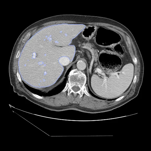
Contrast-enhanced liver CT of a patient with hepatocellular carcinoma, 1 to 12 weeks before a first transarterial chemoembolisation at MD Anderson Cancer Center, with the liver, tumour and intrahepatic vessels segmented (HCC-TACE-Seg).
Read before comparing (10)
- Truth. Three radiology residents drew the masks with AMIRA's semi-automatic tools, a body imaging radiologist with 20 years of experience reviewed them, and STAPLE fused the three readings; no agreement between readers is reported. Kept: liver, liver_tumor (viable and necrotic parts together) and liver_vessels. The SEG's fourth segment, the abdominal aorta, covers 6 to 18 slices on 19 of the 20 cases built, a region of interest, and is left out.
- Liver. Each voxel is in one segment at most, so the liver label is the parenchyma alone. A method that writes the whole liver, as TotalSegmentator does, puts the tumour and the vessels in it: they are 4 to 80% of the liver on the test cases (median 6%), and a perfect whole liver would score a liver Dice of 0.33 to 0.98 (median 0.97).
- Structures. On the test cases the vessels come in 58 to 1,483 pieces and hold 8 to 189 ml; the tumours hold 20 to 2,923 ml, in one piece on 9 of the 10.
- Outlines. Drawn slice by slice, they jump between neighbouring slices on some cases: the tumour's boundary by several centimetres on HCC_056, part of the left lobe on HCC_102, and on HCC_043 and HCC_100 slices of a lobe are left out of the liver. Outside the liver label's largest piece lie under 0.3% of its voxels on 8 test cases, 1.7% on HCC_099 and 4% on HCC_043.
- CT. The series the SEG refers to, in Hounsfield units as delivered. Where a series holds two phases, prepare.py takes the one under the SEG's frames or, where both lie there (HCC_100, HCC_007, HCC_030), the one whose vessels the vessel mask finds. On HCC_043 the masks fit another series of the examination, which is used; on HCC_019 they are placed by their position in the world.
- Phase. Under the vessel mask the 20 CTs built read 97 to 217 HU (median 186), the portal veins opacified, as on the portal venous phase the data paper says the masks were drawn on.
- Cut liver. On HCC_001 the slice under the SEG's lowest frame, 749 voxels of the liver's tip, is missing from the collection, so that frame is left out. On 6 of the 20 cases built the liver reaches the first or the last slice and may be cut.
- TotalSegmentator. Version 2.18 on test case HCC_034 scores a liver Dice of 0.943 and an HD95 of 24 mm against the parenchyma, 0.973 and 2.5 mm against the whole liver. Its liver_vessels task scores a tumour Dice of 0.888 and a vessel Dice of 0.485, 45% of its vessels lying outside the liver, where the dataset draws none.
- Outside numbers. None: the collection has no challenge and no official split.
- Overlap. No other dataset of the corpus holds these patients: LiverPhasesCT is a cohort from Chongqing; MSD_HepaticVessel, MSD_Spleen and CRLMCT come from Memorial Sloan Kettering. AMPLIFAI, listed in the screening as re-annotating this collection among others, is not in the corpus.
| Accuracy | Cost | |||||||
|---|---|---|---|---|---|---|---|---|
| # | Method | Dice | Surface Dice | HD95 mm | Time s | RAM GB | GPU GB | Images |
| 11–4 | KonfAI segmentation app · MRSegmentator, KonfAI appGPUapp1 of 3 structures | 0.838 ±0.080 | 0.595 ±0.106 | 21.99 ±4.00 | 3.8 | 6.8 | ||
| 21–4 | MONAI wholeBody CTGPUapp1 of 3 structures | 0.841 ±0.069 | 0.570 ±0.098 | 21.97 ±5.58 | 16 | 3.8 | 6.4 | |
| 31–4 | KonfAI segmentation app · TotalSegmentator (total), KonfAI appGPUapp1 of 3 structures | 0.827 ±0.088 | 0.579 ±0.104 | 23.25 ±6.47 | 18 | 3.2 | 5.6 | |
| 41–4 | KonfAI segmentation app · TotalSegmentator (total), the authors' packageGPUapp1 of 3 structures | 0.825 ±0.093 | 0.588 ±0.109 | 23.31 ±5.77 | 17.2 | 3.4 | ||
| 5 | KonfAI segmentation app · TotalSegmentator (liver_vessels), the authors' packageGPUapp2 of 3 structures | 0.514 ±0.120 | 0.402 ±0.103 | 23.08 ±6.83 | 25 | 14.1 | 2.6 | |
The order averages each method's ranks on Dice, Surface Dice, HD95 mm, so the first one is not always the best in every column. ± is the 95% interval of the mean over the 10 cases. Two neighbours are tied when a paired Wilcoxon test on the shared cases does not find the upper one better on Dice (5%): the places a method could hold are then shown under its own. The best value of each column is in colour. Click a row to see its images, a column title to sort by it. Time and memory measured on Workstation.
1 result not ranked: an older run, cases still missing, or a failure on every case
| Accuracy | Cost | ||||||||
|---|---|---|---|---|---|---|---|---|---|
| # | Method | Dice | Surface Dice | HD95 mm | Time s | RAM GB | GPU GB | Machine | Images |
| MONAI wholeBody CTGPUapp1 of 3 structuresolder run | 0.841 ±0.069 | 0.570 ±0.098 | 21.97 ±5.58 | 13 | 2.5 | 6.2 | Laptop | ||
Dice: how much the organ contours overlap, 1 is perfect. Surface Dice: the share of the contours' surfaces within a tolerance of each other, 1 is perfect. HD95: distance between the contours' surfaces.
Case by case
Click a score to look at that result: the image, the truth in green and the method's map in red. Underlined scores have their image; for the others you see the case without the result.
About the data
- Test cases
- 10 test cases drawn with seed 2026 among the 95 patients outside development, out of the collection's 105, and 10 development cases. A case is a patient's CT before treatment, the one the masks were drawn on; the other phases and the CT after treatment are left out.
- Truth
- contours by experts 3 structures, the dataset's masks on the CT before treatment: 1 the liver's parenchyma, 2 the tumour (one a patient, its viable and necrotic parts together), 3 the vessels inside the liver; drawn with AMIRA's semi-automatic tools by three radiology residents, reviewed by a body imaging radiologist of 20 years' experience, the three readings fused by STAPLE
- Source
- www.cancerimagingarchive.net/collection/hcc-tace-seg
doi.org/10.7937/TCIA.5FNA-0924
doi.org/10.1038/s41597-023-01928-3 - Licence
- CC BY 4.0
10 cases for tuning, never tested
HCC_070, HCC_007, HCC_004, HCC_038, HCC_030, HCC_087, HCC_102, HCC_094, HCC_022, HCC_040