 fidopt
Cardiac CT of growing pigs (A heart for pigs)
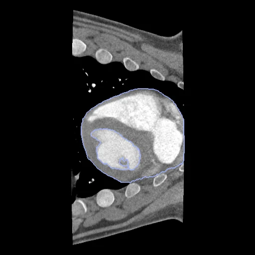
Contrast-enhanced ECG-gated cardiac CT of one growing pig at two consecutive scanning rounds, the first phase of the later (fixed) and the earlier (moving), the heart and left ventricle drawn on both (Norsvin, NMBU, NTNU).
Read before comparing (9)
- Case. One pig at two consecutive rounds, the later as fixed so that the smaller heart of the earlier round lies inside its field. Between the two the heart grows, its labelled volume by 1.08 to 2.75 times on the test pairs, and moves in the chest. The first and fifth phases of one scan were set aside: unregistered they overlap at a Dice of 0.81.
- Truth. Drawn by hand on the contrast images; the page does not say by whom, with which tool or how far two readers differ. Label 2, the contrast-filled left ventricular cavity, is cut out of label 1, which holds the myocardium and the other chambers: the board says nothing of single chambers, valves or vessels.
- Pairs. The files carry no orientation, and the scans lie one way up or turned over by round and position, so pairs form within each group: rounds 1-2 and 2-3 on the back, rounds 4-5 and 5-6 with the stomach scans of rounds 5 and 6, 36 pairs from the ten pigs.
- Alignment. Each pair gets one elastix translation on mutual information inside each body, from the intensities alone, written in the moving headers; both scans keep their voxels. With no registration from it the 10 test pairs score a Dice of 0.71 (0.40 to 0.84) and an HD95 of 11.3 mm (5.2 to 28.5), against 0.73 after a fit on the centres of label 1.
- Room. It comes mostly from the first interval: from round 2 on, consecutive rounds start about as close as two phases of one scan. The draw takes the four intervals in proportion (3, 3, 2 and 2 pairs), but after the replacements the stratum of rounds 1 and 2 holds fewer test pairs than its share: there elastix could not place the grown heart (Dice 0.10 to 0.18).
- Field of view. A scan covers only 120 to 160 mm along the scanner's axis, which from round 2 on the heart nearly fills. In the fixed image of Stent_2_7596_rounds_5_6 the heart touches an end of the scan: what lies beyond is not scored.
- Images. ECG-gated 4D CT of 10 or 11 phases on two Canon scanners; the first phase typically corresponds to diastole. Hounsfield units as delivered, -2048 outside the reconstruction circle. The non-contrast scans, whose masks were carried by the provider's transform rather than drawn, are not used.
- Animals. Ten pigs, scanned in six rounds from about 15 to 130 kg, their folders naming five Control and five Stent animals unexplained. The test pairs come from eight pigs, the development pairs from the two others (Control_1_7569 and Stent_1_7547). Several test pairs share a pig, and Control_4_7570's two share its round-2 scan: they are not ten independent animals.
- Outside numbers. None: no published registration result uses this dataset.
| Accuracy | Smoothness | Cost | ||||||||
|---|---|---|---|---|---|---|---|---|---|---|
| # | Method | Dice | Change | HD95 mm | SDlogJ | Folds % | Time s | RAM GB | GPU GB | Images |
| 1 | NiftyRegCPUapp | 0.875 ±0.061 | +0.161 | 15.54 ±10.13 | 2.151 | 3.78 | 750 | 14.8 | ||
| No registration | 0.714 ±0.101 | 11.32 ±5.00 | 0.000 | 0.00 | ||||||
The order averages each method's ranks on Dice, HD95 mm, SDlogJ, so the first one is not always the best in every column. ± is the 95% interval of the mean over the 10 pairs. Two neighbours are tied when a paired Wilcoxon test on the shared pairs does not find the upper one better on Dice (5%): the places a method could hold are then shown under its own. The best value of each column is in colour. Click a row to see its images, a column title to sort by it. Time and memory measured on Laptop.
Dice: how much the organ contours overlap, 1 is perfect. HD95: distance between the contours' surfaces. SDlogJ: how irregular the deformation is, 0 is perfectly smooth. Folds: share of the image where the deformation folds over itself.
Pair by pair
Click a score to look at that result: the fixed image, and over it the moving image after registration, with the annotations. Underlined scores have their image; for the others you see the pair without the result. A green cell gained against no registration on that pair, a red one lost.
About the data
- Test pairs
- 10 test pairs drawn with seed 2026 among the 29 outside development, from each round interval in proportion, and 7 development pairs. Three pairs of rounds 1 and 2 (Control_3_7602, Control_4_7570, Stent_5_7607) were replaced by the next pairs of the seeded order not of rounds 1 and 2 nor of a development animal, since no start from the intensities places their hearts.
- Truth
- contours by experts the provider's masks of the first phase of each scan, drawn by hand on the contrast-enhanced images: 1 the heart outside the left ventricle, 2 the left ventricle (its contrast-filled cavity)
- Source
- doi.org/10.25452/figshare.plus.30398977.v2
plus.figshare.com/articles/dataset/A_spatially_and_temporally_aligned_contrast-non-contrast_cardiac_CT_dataset_of_pigs/30398977 - Licence
- CC BY 4.0
7 pairs for tuning, never tested
Control_1_7569_rounds_1_2, Control_1_7569_rounds_2_3, Control_1_7569_rounds_4_5, Stent_1_7547_rounds_1_2, Stent_1_7547_rounds_2_3, Stent_1_7547_rounds_4_5, Control_1_7569_rounds_5_6