 fidopt
M-CRIB neonatal brain, 94 regions
T2 MR of the brain of one term neonate with the manual M-CRIB 2.0 parcellation of 94 regions.
Read before comparing (3)
- Cases. The fixed images of MCRIB's pairs: a test pair's image is a test case, a development pair's a development case. M-CRIB has ten neonates, so the test set holds three images and the development set two. The moving images are left out.
- Images. The authors' preprocessed T2, on which the labels were drawn: bias corrected (N4ITK), skull-stripped (FSL's BET), aligned on the AC-PC line in 3D Slicer and resampled with FLIRT from 0.5 x 0.5 x 1 mm to 0.63 mm. The values are as delivered, 32-bit floats without unit, with 300 to 1,700 negative voxels around each brain, outside its labels. No mask is given.
- Contrast. The white matter, not yet myelinated, is brighter than the cortex on these T2, the reverse of an adult T2, and the CSF brighter still (measured on three brains): a method trained on adult brains meets another contrast.
| Accuracy | Cost | ||||||||
|---|---|---|---|---|---|---|---|---|---|
| # | Method | Dice | Surface Dice | HD95 mm | Time s | RAM GB | GPU GB | Machine | Images |
| 11–2 | SynthSeg 1.0CPUapp23 of 94 structures | 0.480 ±0.178 | 0.513 ±0.195 | 7.79 ±1.02 | 26 | 5.0 | Laptop | ||
| 21–2 | FastSurferVINNGPUapp86 of 94 structures | 0.106 ±0.072 | 0.187 ±0.090 | 14.50 ±1.55 | 34 | 4.3 | 6.3 | Workstation | |
The order averages each method's ranks on Dice, Surface Dice, HD95 mm, so the first one is not always the best in every column. ± is the 95% interval of the mean over the 3 cases. Two neighbours are tied when a paired Wilcoxon test on the shared cases does not find the upper one better on Dice (5%): the places a method could hold are then shown under its own. The best value of each column is in colour. Click a row to see its images, a column title to sort by it.
1 result not ranked: an older run, cases still missing, or a failure on every case
| Accuracy | Cost | ||||||||
|---|---|---|---|---|---|---|---|---|---|
| # | Method | Dice | Surface Dice | HD95 mm | Time s | RAM GB | GPU GB | Machine | Images |
| FastSurferVINNGPUapp86 of 94 structuresolder run | 0.106 ±0.072 | 0.187 ±0.091 | 14.39 ±2.02 | 44 | 3.3 | 6.0 | Laptop | ||
Dice: how much the organ contours overlap, 1 is perfect. Surface Dice: the share of the contours' surfaces within a tolerance of each other, 1 is perfect. HD95: distance between the contours' surfaces.
Case by case
Click a score to look at that result: the image, the truth in green and the method's map in red. Underlined scores have their image; for the others you see the case without the result.
About the data
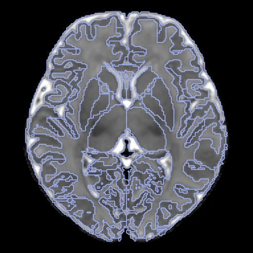
- Truth
- contours by experts the dataset's manual M-CRIB 2.0 parcellation of each brain, 94 regions: the 62 cortical regions of the Desikan-Killiany-Tourville protocol, an 'unknown' label on each side for unassigned voxels in front of the amygdala, and 30 others (cerebral white matter, ventricles, CSF, basal ganglia, thalami, hippocampi, amygdalae, ventral diencephalon, cerebellum, brainstem, corpus callosum)
- Source
- osf.io/4vthr
osf.io/mnwv9
osf.io/2yx8u
doi.org/10.3389/fnins.2019.00034
doi.org/10.1002/hbm.24948
doi.org/10.1016/j.neuroimage.2016.09.068 - Licence
- CC BY 4.0
2 cases for tuning, never tested
P05_P07/fixed, P06_P03/fixed