 fidopt
BraTS-PEDs paediatric high-grade glioma
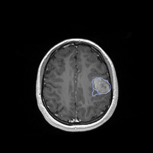
Pre-treatment head MR of a child with a high-grade glioma: T1, T1Gd, T2 and FLAIR as one four-channel image on the SRI24 atlas at 1 mm, with four tumour sub-regions labelled (BraTS-PEDs, TCIA).
Read before comparing (10)
- Pool. Children with a histologically confirmed high-grade glioma, diffuse midline gliomas among them, imaged before surgery, radiation or chemotherapy at 1.5 or 3 T on several vendors' scanners: CBTN 116, the DMG/DIPG Registry 80, DFCI-BCH-BWH-PEDs-HGG 61. The provider left out examinations with marked motion or artefacts, and removed subject 00023 as a duplicate of 00119.
- Images. The provider co-registered the four sequences on SRI24 at 1 mm, 240 x 240 x 155 voxels, normalised the intensities and defaced the heads, leaving the skull; no mask is given. The channels come in the order T1, T1Gd, T2, FLAIR, and a method that reads one value a voxel picks its channel in its adapter.
- Slices. CBTN's metadata give slices of 0.9 to 5 mm, and thick slices stay blurred once resampled, as on test cases 00054, 00065, 00226 and 00227. The collection page lists 00024 and 00098 as skull-stripped, wholly or on the T1Gd; 00024 is not drawn, 00098 is a development case.
- Truth. A paediatric automatic segmentation following the RAPNO recommendations, revised in ITK-SNAP by volunteer neuroradiologists, reviewed by board-certified attending neuroradiologists until consensus and approved by paediatric neuroradiologists. One map a subject, no second reader and no measure of agreement: graded semi-automatic.
- Labels. 1 enhancing tumour, on the T1Gd against the T1; 2 non-enhancing tumour, abnormal signal neither enhancing nor cystic; 3 cyst, fluid bright on the T2; 4 peritumoral oedema, the FLAIR hyperintensity around the tumour. Adult BraTS's oedema holds the infiltrative non-enhancing tumour, so a method trained on adult labels may call label 2 oedema.
- Scoring. Each label is scored on its own; a label a case lacks is not scored unless a method writes it, a false positive at 0. The 25 test tumours hold 6.7 to 241.7 ml. Six test maps (00003, 00065, 00096, 00176, 00179, 00211) hold stray pieces of 1 to 20 voxels away from the tumour, kept as delivered.
- Checks. On the 20 cases built first every channel and label map equals the package's file, the labels lie on the tumour, and the channel contrasts match them. The 28 added later hold only labels 0 to 4 on their image's grid; two were looked at in three planes.
- Room. Test labels moved by one voxel keep a Dice of 0.82 for the enhancing tumour (0.69 to 0.94, seven cases), 0.91 for the non-enhancing tumour, 0.79 for the cyst and 0.86 for the oedema. The surface Dice, at 1 mm since BraTS sets no tolerance, forgives such a move.
- Outside numbers. BraTS-PEDs scores nested regions, and its 2023 edition merged the non-enhancing tumour, the cyst and necrosis into one label, so no published number is scored as here. The training labels are public: a network trained on BraTS data may have seen these subjects.
- Overlap. No other descriptor names BraTS-PEDs, CBTN, the DMG/DIPG Registry or DFCI-BCH-BWH-PEDs-HGG; BraTS2021 holds adults from other sites. A dataset built later from BraTS 2023 to 2025's paediatric data or from DFCI-BCH-BWH-PEDs-HGG must leave out these 48 subjects; test cases 00176 and 00179 are that collection's id_86 and id_89.
| Accuracy | Cost | |||||||
|---|---|---|---|---|---|---|---|---|
| # | Method | Dice | Surface Dice | HD95 mm | Time s | RAM GB | GPU GB | Images |
| 1 | MONAI BraTS SegResNetGPUapp2 of 4 structures | 0.039 ±0.036 | 0.027 ±0.027 | 84.89 ±7.76 | 3.5 | 3.4 | ||
The order averages each method's ranks on Dice, Surface Dice, HD95 mm, so the first one is not always the best in every column. ± is the 95% interval of the mean over the 25 cases. Two neighbours are tied when a paired Wilcoxon test on the shared cases does not find the upper one better on Dice (5%): the places a method could hold are then shown under its own. The best value of each column is in colour. Click a row to see its images, a column title to sort by it. Time and memory measured on Laptop.
Dice: how much the organ contours overlap, 1 is perfect. Surface Dice: the share of the contours' surfaces within a tolerance of each other, 1 is perfect. HD95: distance between the contours' surfaces.
Case by case
Click a score to look at that result: the image, the truth in green and the method's map in red. Underlined scores have their image; for the others you see the case without the result.
About the data
- Test cases
- 25 test subjects drawn by source with seed 2026 among the 234 training subjects outside development: 11 from CBTN, 8 from the DMG/DIPG Registry, 6 from DFCI-BCH-BWH. 23 development subjects, the last of the same seeded order, each source in proportion: 11 from CBTN, 7 from the DMG/DIPG Registry, 5 from DFCI-BCH-BWH. The pool is the 257 training subjects with a label map; the validation subjects have none, and the test set is given on request only.
- Truth
- contours by a program, verified by people the dataset's four labels, 1 the enhancing tumour, 2 the non-enhancing tumour, 3 the cystic component, 4 the peritumoral oedema, by the RAPNO recommendations: a paediatric automatic segmentation revised in ITK-SNAP by volunteer neuroradiologists given the four sequences, reviewed by board-certified attending neuroradiologists and returned for refinement until consensus
- Source
- www.cancerimagingarchive.net/collection/brats-peds
doi.org/10.7937/dx5c-tj86
www.cancerimagingarchive.net/wp-content/uploads/BraTS-PEDs_metadata.tsv
www.cancerimagingarchive.net/wp-content/uploads/BraTS-PEDs_metadata_glossary.tsv
www.cancerimagingarchive.net/wp-content/uploads/BraTS-PEDs_Imaging_Info.tsv
doi.org/10.48550/arXiv.2404.15009
doi.org/10.48550/arXiv.2305.17033
doi.org/10.59275/j.melba.2025-f6fg
arxiv.org/abs/2407.08855
www.cancerimagingarchive.net/collection/dfci-bch-bwh-peds-hgg - Licence
- CC BY 4.0
23 cases for tuning, never tested
BraTS-PED-00005-000, BraTS-PED-00192-000, BraTS-PED-00167-000, BraTS-PED-00044-000, BraTS-PED-00240-000, BraTS-PED-00034-000, BraTS-PED-00147-000, BraTS-PED-00025-000, BraTS-PED-00188-000, BraTS-PED-00036-000, BraTS-PED-00155-000, BraTS-PED-00243-000, BraTS-PED-00089-000, BraTS-PED-00038-000, BraTS-PED-00228-000, BraTS-PED-00126-000, BraTS-PED-00098-000, BraTS-PED-00264-000, BraTS-PED-00086-000, BraTS-PED-00136-000, BraTS-PED-00196-000, BraTS-PED-00047-000, BraTS-PED-00112-000