 fidopt
Myocardial perfusion SPECT
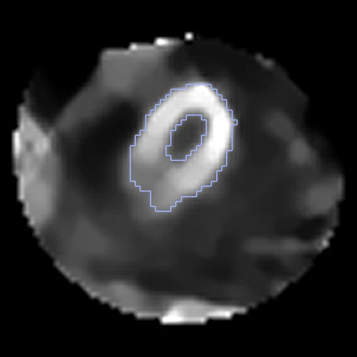
Rest myocardial perfusion SPECT (99mTc-MIBI) of two patients, each with the dataset's mask of the left ventricular wall (Myocardial perfusion scintigraphy image database, PhysioNet), paired by us.
Read before comparing (9)
- Truth. One coarse label on a 4 mm grid: the wall is 14 to 22 mm thick, 3.5 to 5.5 voxels, blurred by the camera. It says where the left ventricle sits, its size and direction, little of its shape.
- Masks. Two nuclear medicine experts selected in 3D Slicer the voxels as bright as the left ventricle and erased the rest, other specialists checking each mask: graded semi-automatic. The outline is an intensity threshold of the image, so a method that matches intensities follows it. 8 of the 40 masks keep 1 to 4 isolated voxels.
- Doubt. The paper says both that 40 and that all 83 of its rest examinations were segmented by hand; the release holds 101 masks, on stress and prone scans too, and its README calls them 'predicted masks (if included)'. Whether some are a network's output is not said; the corpus uses rest scans only, the kind the paper describes.
- Pairs. Only 8 patients have a rest and a stress scan with a mask on each, too few for ten test pairs, so a pair joins two patients.
- Alignment. Each scan is in the camera's frame: as delivered the walls overlap at 0.32 (0.00 to 0.64) on the 20 pairs, centres 12 to 80 mm apart. The corpus gives one elastix translation on the intensities inside each body, no annotation used, in the moving header; a rigid fit turns the hearts 1.6 to 16.5 degrees, left to the methods.
- Baseline. With no registration the 10 test pairs start at a Dice of 0.72 (0.55 to 0.85), against 0.78 after a translation that put the centres of the two walls together, and an HD95 of 12.5 mm (6.9 to 20.8).
- Values. 16-bit integers without unit, each scan scaled to its own maximum, which lies outside the wall on 36 of the 40 scans; the wall's median runs from 699 to 17372, so raw intensities compare two scales.
- Cohort. Patients referred to Hospital Israelita Albert Einstein, São Paulo, in February and March 2023, on a GE Discovery NM 530c CZT camera; the field is 70 x 70 x 50 voxels of 4 mm around the heart. No diagnosis, defect or artefact is labelled; the paper found artefacts on 47 of its 83 patients.
- Outside numbers. None: the pairing is ours.
| Accuracy | Smoothness | Cost | ||||||||
|---|---|---|---|---|---|---|---|---|---|---|
| # | Method | Dice | Change | HD95 mm | SDlogJ | Folds % | Time s | RAM GB | GPU GB | Images |
| 1 | ConvexAdam (MIND)GPUapp | 0.828 ±0.053 | +0.108 | 8.04 ±3.11 | 0.067 | 0.04 | 5 | 2.0 | 0.4 | |
| No registration | 0.720 ±0.079 | 12.52 ±3.32 | 0.000 | 0.00 | ||||||
The order averages each method's ranks on Dice, HD95 mm, SDlogJ, so the first one is not always the best in every column. ± is the 95% interval of the mean over the 10 pairs. Two neighbours are tied when a paired Wilcoxon test on the shared pairs does not find the upper one better on Dice (5%): the places a method could hold are then shown under its own. The best value of each column is in colour. Click a row to see its images, a column title to sort by it. Time and memory measured on Laptop.
Dice: how much the organ contours overlap, 1 is perfect. HD95: distance between the contours' surfaces. SDlogJ: how irregular the deformation is, 0 is perfectly smooth. Folds: share of the image where the deformation folds over itself.
Pair by pair
Click a score to look at that result: the fixed image, and over it the moving image after registration, with the annotations. Underlined scores have their image; for the others you see the pair without the result. A green cell gained against no registration on that pair, a red one lost.
About the data
- Test pairs
- 10 test pairs drawn with seed 2026 among the 30 outside development, and 10 development pairs. The pool is each patient's supine rest scan with a mask, 80 patients, the 81st's only scan having none; in the order of sha256("2026:MyocardialSPECT:<scan>"), two by two, each patient in one pair.
- Truth
- contours by a program, verified by people the left ventricular wall on each scan (1), the dataset's mask
- Source
- physionet.org/content/myocardial-perfusion-spect/1.0.0
doi.org/10.13026/ce2z-dw74
doi.org/10.1371/journal.pone.0312257 - Licence
- ODbL 1.0
10 pairs for tuning, never tested
47178689_11174108, 99648767_28771481, 67754110_34187527, 57935602_05758527, 39876800_75500164, 08804386_47600173, 64609613_87000848, 61762032_85230764, 32067012_94893933, 34357317_85471860