 fidopt
Spine-Mets-CT-SEG
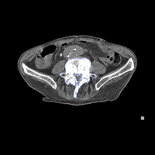
Radiotherapy simulation CT of a patient with spinal metastases at Brigham and Women's Hospital, each vertebra from T1 to L5 segmented in the annotated slices (Spine-Mets-CT-SEG, TCIA).
Read before comparing (11)
- Pool. 55 patients with spinal metastases, one CT each: the simulation scan of a spine radiotherapy, stereotactic or not, on a Siemens SOMATOM Confidence (36 CTs) or a GE LightSpeed RT16 (19), without gating or breath hold.
- Images. The CTs as delivered, in Hounsfield units; 13055 is on the scanner's extended scale, up to 30710 in metal. 13653's series holds each slice three times, and prepare.py keeps each once.
- Truth. A spine biomechanics expert (R. N. Alkalay) segmented each level with 3D Slicer's Grow from Seeds, and an expert radiologist (D. B. Hackney) confirmed anatomy and labelling. One segmentation a CT, no agreement between readers: graded semi-automatic. Masks follow the bone, leaving holes where a lytic lesion has eaten it (15001's L4).
- Coverage. T1 to L5 as the field of view allows, 11 to 17 vertebrae a test CT; no cervical vertebra, sacrum or rib is labelled or scored. A vertebra the scan's end cuts is annotated in the part the scan holds (15041's T5, 13977's T7), though the preprint says such vertebrae were not.
- Ignore region. On 13055, a test CT, and 13683, a development CT, the labels stop on a plane inside the scan, through T1 and T2 on one and T8 and T9 on the other. The slices beyond are taken out of both maps before scoring and never shown, so a method that segments the whole vertebra loses nothing.
- Metastases. Not segmented: a lesion is part of its vertebra's label. 183 of the 787 vertebrae annotated hold one, 58 blastic, 54 lytic and 71 mixed. Eight of the ten test CTs hold such a vertebra.
- Held out. 15094 and 14659 skip a level, so part of each spine is named a level off; 13627 and 13800 leave a vertebra of the image unlabelled (T1 on 13627, T2 on 13800), so a method that segments it scores a false positive; 14655's T1 to L5 are a level off its ribs. The draw took the next CTs of its order.
- TotalSegmentator. Version 2.18's fast model gives every vertebra the truth's name on 19 of the 20 CTs built, with a mean Dice of 0.80 to 0.89 on nine test CTs. On 13055, a spine of compression fractures, it names T1 to T4 and T11 to L2 a level higher; its full model scores a mean Dice of 0.47 there.
- Masks. Each label lies on bone (234 to 626 HU on the test CTs). Small pieces apart from their vertebra, on the test CTs at most 0.66 ml of 15041's T12, are scored as delivered.
- Room. The provider's nnU-Net, trained on these 55 CTs, reached a mean Dice of 0.943 (± 0.022) in three-fold cross-validation; its weights are announced, not released. Methods part on the right level and the last points of the Dice; the surface Dice is at 1 mm, as the provider sets no tolerance.
- Overlap. No descriptor names the collection, so no other dataset of the corpus holds these CTs. No outside number compares: no challenge uses it.
| Accuracy | Cost | |||||||
|---|---|---|---|---|---|---|---|---|
| # | Method | Dice | Surface Dice | HD95 mm | Time s | RAM GB | GPU GB | Images |
| 11–2 | KonfAI segmentation app · TotalSegmentator (total), the authors' packageGPUapp | 0.858 ±0.099 | 0.840 ±0.109 | 3.94 ±3.79 | 37.6 | 6.3 | ||
| 21–2 | KonfAI segmentation app · TotalSegmentator (total), KonfAI appGPUapp | 0.844 ±0.121 | 0.827 ±0.129 | 4.71 ±4.30 | 3.0 | 10.2 | ||
| 3 | MONAI wholeBody CTGPUapp | 0.715 ±0.109 | 0.634 ±0.135 | 10.50 ±5.02 | 24 | 4.1 | 19.8 | |
The order averages each method's ranks on Dice, Surface Dice, HD95 mm, so the first one is not always the best in every column. ± is the 95% interval of the mean over the 10 cases. Two neighbours are tied when a paired Wilcoxon test on the shared cases does not find the upper one better on Dice (5%): the places a method could hold are then shown under its own. The best value of each column is in colour. Click a row to see its images, a column title to sort by it. Time and memory measured on Laptop.
1 result not ranked: an older run, cases still missing, or a failure on every case
| Accuracy | Cost | ||||||||
|---|---|---|---|---|---|---|---|---|---|
| # | Method | Dice | Surface Dice | HD95 mm | Time s | RAM GB | GPU GB | Machine | Images |
| MONAI wholeBody CTGPUappfailed on every case | 0.000 ±0.000 | 0.000 ±0.000 | Workstation | ||||||
Dice: how much the organ contours overlap, 1 is perfect. Surface Dice: the share of the contours' surfaces within a tolerance of each other, 1 is perfect. HD95: distance between the contours' surfaces.
Case by case
Click a score to look at that result: the image, the truth in green and the method's map in red. Underlined scores have their image; for the others you see the case without the result.
About the data
- Test cases
- 10 test CTs drawn with seed 2026 by scanner among the 40 outside development: 7 Siemens SOMATOM Confidence and 3 GE LightSpeed RT16. 10 development CTs, the last of the same seeded order (7 Siemens, 3 GE). Five CTs whose levels are not whole or not those of their spine are left out.
- Truth
- contours by a program, verified by people each vertebra from T1 to L5 in the slices the provider annotated, one label a vertebra in its numbering (T1 to L5 as 1 to 17): grown from seeds in 3D Slicer by a spine biomechanics expert, the segmentation and the levels confirmed by a radiologist; a vertebra the scan's end cuts is labelled in the part the scan holds, and the slices beyond a plane where the annotation stops inside the scan are the ignore region
- Source
- www.cancerimagingarchive.net/collection/spine-mets-ct-seg
doi.org/10.7937/kh36-ds04
doi.org/10.1101/2024.10.14.24314447
github.com/rouge1616/Spine-Mets-CT-SEG
www.cancerimagingarchive.net/wp-content/uploads/Spine-Mets-CT-SEG_Clinical.xlsx - Licence
- CC BY 4.0
10 cases for tuning, never tested
12196, 12459, 14313, 14874, 13683, 10456, 14316, 15027, 15001, 11471