 fidopt
TotalSegmentator hip_implant, CT
A CT of one hip carrying an implant, at the resolution it was reconstructed at, the implant segmented, from the training set of TotalSegmentator's task hip_implant.
10 test casespelvisimage CT
Read before comparing (9)
- Source. The deposit, by Jakob Wasserthal of the University Hospital Basel, is the training set of TotalSegmentator's task hip_implant: 71 CTs, 'CT images + manual segmentations'. No paper describes it, and nothing says who drew the implants, with what tool, where the CTs were made or how they were chosen. Graded expert on the page's word alone.
- Images. The deposit's files byte for byte, in Hounsfield units: one hip, 512 pixels of 0.29 to 0.49 mm across, slices 0.6 to 1.2 mm apart. The ten test cases and seven development cases use an extended scale, in steps of 10 HU from -10,240 up to 26,780, so the metal does not saturate; 9omM4mNH603Adztl, Gxw74fszCfYMXKhb and eJTLOt6J4iRnfNRQ stop at 3,071 HU. Metal streaks cross every image.
- Truth. The implant as an object, not its bright voxels: the outline follows the stem, head and cup, takes in the dark liner between head and cup, and joins the cup's screws with the bone between them. On the test cases 0.1 to 15% of its voxels read under 0 HU and 0.2 to 33% under 1,000 HU. How the liner and the gaps between screws were decided is not written anywhere.
- Baseline. The best of thresholds at 1,500, 2,000, 2,500 and 3,000 HU reaches a Dice of 0.68 to 0.96 on the test cases (median 0.82) and 0.60 to 0.86 on development, leaving 0.2 to 4.6 mL of bright voxels outside the truth (bone cortex, cement, streaks). That is the floor to beat.
- Cases. The test implants hold 52.6 to 124.1 mL (median 92 mL), one piece on seven and one piece with crumbs of a few voxels on the others. The development implants hold 70.5 to 106.9 mL.
- Patients. With no patient identifier, two CTs of one patient, or a patient's two hips, could be a test and a development case; this cannot be checked.
- TotalSegmentator. Version 2.18's hip_implant model was trained on this deposit and has seen every case: a Dice of 0.977 on test case FKHhft7vc8dinYZC and 0.972 on byHK8aqbFHcbv5Px, against 0.96 and 0.68 for the best threshold. That is a model on its training set, not a fair line.
- Overlap. No dataset of the corpus holds these CTs. None of the 1,228 CTs of TotalSegmentator's CT dataset, TotalSegCT's source, has its corners within 10 mm of a hip CT's, and none of the 60 images built from TotalSegCT, LiverLesionsCT and PericardialEffusionCT, the same team's deposits, contains a hip CT's box or covers 90% of it. No deposit names its patients, so a shared patient cannot be ruled out.
- Outside numbers. None: the deposit has no paper, no challenge and no split. The provider sets no surface tolerance.
No method has a result on every case of this dataset yet.
Case by case
Click a score to look at that result: the image, the truth in green and the method's map in red. Underlined scores have their image; for the others you see the case without the result.
About the data
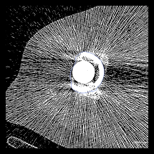
The first test caseClick to look at every case.
- Test cases
- 10 test CTs drawn with seed 2026 among the 60 outside development, and 10 development CTs, out of the deposit's 70 CTs whose label map holds an implant (prepare.py --pool); RO8vqzeBW3UBpF6j's map is empty. The provider publishes no test set and no patient identifier.
- Truth
- contours by experts the hip implant as one object, its stem, head, cup, liner and screws under one label, which the deposit calls manual segmentations and says nothing more of
- Source
- zenodo.org/records/20272031
doi.org/10.5281/zenodo.20272030
github.com/wasserth/TotalSegmentator - Licence
- CC BY 4.0
10 cases for tuning, never tested
78tN3D8on98qbsCw, hGPRZrDKWKYFxNdP, DiQGSILr77tkYpmC, 8CXodkYTIqDx0pfq, eJTLOt6J4iRnfNRQ, kk1kvge5De9PcZwh, Gxw74fszCfYMXKhb, 9omM4mNH603Adztl, TulUb9J85WT8GuwH, 82eviQZILd1czyrr