 fidopt
Spinal-Multiple-Myeloma-SEG, vertebrae and lytic lesions
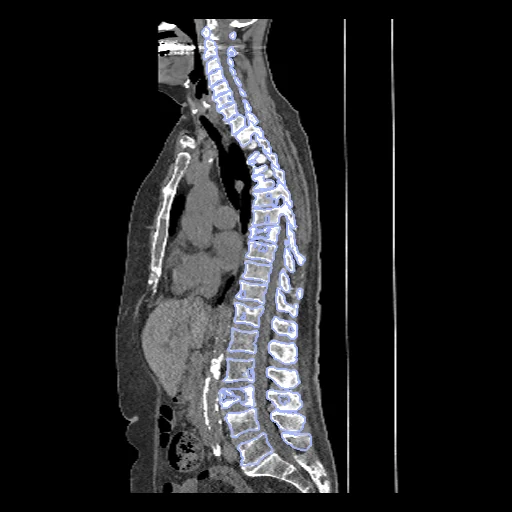
The conventional CT of a dual-layer low-dose scan of a patient with multiple myeloma, cropped to the spine, each vertebra from C1 to L5 and the spine's lytic lesions segmented (Spinal-Multiple-Myeloma-SEG, TCIA).
Read before comparing (8)
- Pool. 72 whole-body low-dose scans without contrast agent of 67 patients with multiple myeloma at the University Hospital Brno, on a Philips IQon Spectral CT, the slices of the spine kept. Patients without a lytic lesion of the spine, without spectral data or with heavy artefacts from spinal metal were excluded, so every case holds lesions.
- Images. 512 x 512 pixels of 0.88 to 0.98 mm over the full field of view, arms and table included, slices every 0.6 mm, in Hounsfield units from -1024 to 3071. They are noisy, as low-dose images are. The acquisition table gives 66 to 134 mA and the kernels B and C; the collection page's text says 10 mA and a sharp kernel.
- Anatomy. The provider's table notes a metal implant in the spine of test patients 018, 040 and 061, an L6 in 021, 055 and 060, and eleven thoracic vertebrae in 030 and in development patient 077, whose labels have no T12.
- Image. The image is the conventional CT, not the 40 keV image of the same acquisition the lesions were corrected on: it is what a CT method is trained for, and the 40 keV image is MyelomaSpineDECT_Syn's hidden target. The labels lie on both; a lesion may be fainter here than where it was drawn.
- Truth. An nnU-Net trained on VerSe 2020 drew the vertebrae and a network in a loop the lesions; both were corrected by hand, and one of two board-certified radiologists verified the lesion corrections. The page does not say how many vertebrae or lesions changed: a method close to nnU-Net on VerSe may be favoured where the proposal stayed.
- Lesions. The provider gives one segment a lesion (12 to 94 on the test cases, 3.5 to 115 mL a patient), all put under one label: the score is the Dice of all the lesions together, not detection. A lesion voxel carries the lesion's label, so a vertebra is scored without its lesions; lesions are 0.4 to 11.5% of a test case's labelled voxels.
- TotalSegmentator. Its task total gives a mean Dice of 0.914 over the 24 vertebrae of test case Myel_004 (0.85 to 0.95), 0.930 with the lesion voxels left out of both maps: a method that segments whole vertebrae loses about 0.02 to the lesions cut out of the truth.
- Overlap. No patient of this collection is in another dataset of the corpus: VerSe is from Munich and other centres, Spine-Mets-CT-SEG from Boston, this one from Brno.
| Accuracy | Cost | |||||||
|---|---|---|---|---|---|---|---|---|
| # | Method | Dice | Surface Dice | HD95 mm | Time s | RAM GB | GPU GB | Images |
| 11–2 | KonfAI segmentation app · TotalSegmentator (total), KonfAI appGPUapp24 of 26 structures | 0.890 ±0.040 | 0.905 ±0.046 | 4.17 ±1.46 | 41 | 5.7 | 6.2 | |
| 21–2 | KonfAI segmentation app · TotalSegmentator (total), the authors' packageGPUapp24 of 26 structures1 failed | 0.822 ±0.207 | 0.839 ±0.212 | 2.66 ±0.84 | 33.1 | 8.1 | ||
| 3 | CT-FM whole-body segmentationGPUapp24 of 26 structures | 0.355 ±0.089 | 0.343 ±0.074 | 82.47 ±29.21 | 398 | 15.7 | 5.6 | |
The order averages each method's ranks on Dice, Surface Dice, HD95 mm, so the first one is not always the best in every column. ± is the 95% interval of the mean over the 10 cases. Two neighbours are tied when a paired Wilcoxon test on the shared cases does not find the upper one better on Dice (5%): the places a method could hold are then shown under its own. The best value of each column is in colour. Click a row to see its images, a column title to sort by it. Time and memory measured on Workstation.
2 results not ranked: an older run, cases still missing, or a failure on every case
| Accuracy | Cost | ||||||||
|---|---|---|---|---|---|---|---|---|---|
| # | Method | Dice | Surface Dice | HD95 mm | Time s | RAM GB | GPU GB | Machine | Images |
| MONAI wholeBody CTGPUapp24 of 26 structuresfailed on every case | 0.000 ±0.000 | 0.000 ±0.000 | Laptop | ||||||
| MONAI wholeBody CTGPUapp24 of 26 structuresfailed on every case | 0.000 ±0.000 | 0.000 ±0.000 | Workstation | ||||||
Dice: how much the organ contours overlap, 1 is perfect. Surface Dice: the share of the contours' surfaces within a tolerance of each other, 1 is perfect. HD95: distance between the contours' surfaces.
Case by case
Click a score to look at that result: the image, the truth in green and the method's map in red. Underlined scores have their image; for the others you see the case without the result.
About the data
- Test cases
- 10 test patients drawn with seed 2026 among the collection's 67, one scan each, then 10 development patients, the last of the same seeded order. Of the patients scanned twice, 012, 018, 023, 043 and 052, the earlier scan is used. MyelomaSpineDECT_Syn draws its patients outside this partition, and a later dataset of this collection keeps each of these patients on the same side.
- Truth
- contours by experts each vertebra in the provider's numbering (1 to 25, C1 to L6) and every lytic lesion of the spine under one label (26): the vertebrae drawn by an nnU-Net trained on VerSe 2020 and corrected by hand; the lesions (hypodense, without a sclerotic rim, over 5 mm) drawn by a network in a loop, then removed, corrected or added by hand on the 40 keV image of the same scan, the corrections verified by one of two board-certified radiologists. A lesion voxel carries the lesion's label, so a vertebra's label is the vertebra without its lesions
- Source
- www.cancerimagingarchive.net/collection/spinal-multiple-myeloma-seg
doi.org/10.7937/k4qv-hh78
services.cancerimagingarchive.net/nbia-api/services/v1/getSeries?Collection=Spinal-Multiple-Myeloma-SEG
www.cancerimagingarchive.net/wp-content/uploads/Spinal-Multiple-Myeloma-SEG_Table_of_image_data_description.tsv
www.cancerimagingarchive.net/wp-content/uploads/Spinal-Multiple-Myeloma-SEG_Table_of_acquisition_parameters.tsv
doi.org/10.59275/j.melba.2026-d741
github.com/MISAG-BUT/Spinal-Multiple-Myeloma-SEG - Licence
- CC BY 4.0
10 cases for tuning, never tested
Myel_008, Myel_036, Myel_071, Myel_046, Myel_039, Myel_064, Myel_044, Myel_042, Myel_006, Myel_077