 fidopt
TotalSegmentator MRI
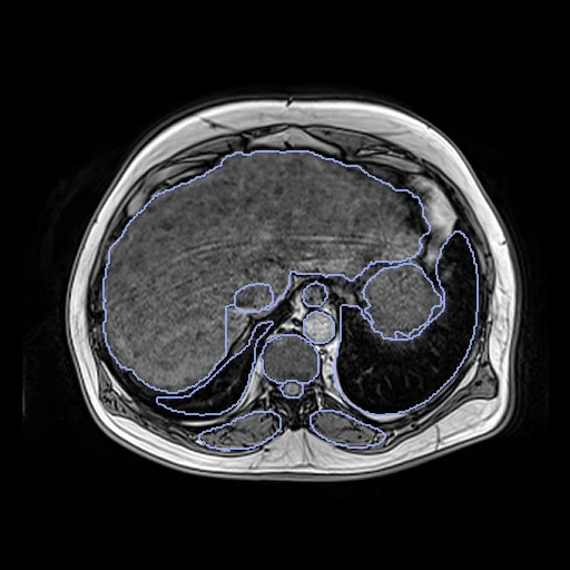
Axial T1-weighted gradient-echo MR of the upper abdomen of two adults, one kind of image on both (fat, fat-suppressed, in or opposed phase), paired by us (TotalSegmentator MRI 3.0.0, University Hospital Basel).
Read before comparing (11)
- Pool. The abdomen is the region the dataset holds most of, and its 41 axial T1-weighted gradient-echo slabs of adults the largest group of one sequence; s0163, whose slab holds the pelvis and no liver, stays out. The scans version 3.0.0 adds are not used: their annotation is not described.
- Images. Slabs of 60 to 96 slices of 3 or 3.3 mm on Siemens scanners at 1.5 or 3 T; twelve of the 19 pairs join two scanner models. Values are 16-bit integers with no common scale; no mask is given.
- Kinds. meta.csv names the sequence, not which Dixon image a slab is. The corpus read the kind off the images and checked each by eye: 20 fat images, 11 fat-suppressed after contrast, 4 in-phase and 5 opposed-phase. A pair joins two of one kind, one contrast on both sides.
- Pairs. Within each kind the slabs are put in the order of sha256("2026:TotalSegMRI:<scan>") and paired two by two, s0203 and s0253 left over: 10 fat, 5 fat-suppressed, 2 in-phase and 2 opposed-phase pairs. The pairing is ours: no outside number compares.
- Truth. Two board-certified radiologists segmented the first scans by hand or refined MRSegmentator's output; retrained nnU-Nets predicted the next, refined by hand where needed, until every examination was reviewed. Version 3.0.0 improved the labels without saying how. No agreement between readers is given; the masks are a corrected network output.
- Merging. The 50 masks overlap at their borders, 0.07 to 4.1% of a scan's labelled voxels. prepare.py gives such a voxel to the smaller structure, since the larger mask spills, as the liver's over the gallbladder on s0201, s0272 and s0278. The rule fails on s0242 (development), where the small bowel's mask takes some of the cysts of its polycystic kidneys.
- Seen. TotalSegmentator MRI was trained on these labels: 37 of the 38 scans are in its training split, s0293 in its test split. A method using that network or its features has seen this truth.
- Alignment. The slabs keep their native voxels; the moving header carries one elastix translation on mutual information, no annotation used. A rigid fit turns the bodies 1.0 to 8.5 degrees, a difference of posture, so only the translation is kept. From head to feet the start leaves shared labels' centres 17 to 67 mm apart on 7 of the 10 test pairs.
- Baseline. With no registration the 10 test pairs score a Dice of 0.18 (0.02 to 0.32), against 0.04 as delivered and 0.27 after a fit on the label centres, and an HD95 of 52 mm (30 to 97); the liver overlaps 0.48.
- Field. A test pair's images share 22 to 26 labels; a structure one slab cuts shorter overlaps only along their common length.
- Overlap. Every scan comes from Basel, none from the Imaging Data Commons, so no other dataset of the corpus holds them. meta.csv holds no patient ID; the two scans of a pair differ in sex or age, and at a glance where both are close.
| Accuracy | Smoothness | Cost | ||||||||
|---|---|---|---|---|---|---|---|---|---|---|
| # | Method | Dice | Change | HD95 mm | SDlogJ | Folds % | Time s | RAM GB | GPU GB | Images |
| 11–4 | ConvexAdam (MIND)GPUapp | 0.331 ±0.108 | +0.146 | 41.87 ±14.53 | 0.123 | 0.85 | 8 | 2.4 | 1.8 | |
| 21–4 | greedyCPUapp | 0.295 ±0.122 | +0.111 | 47.80 ±15.14 | 0.096 | 0.00 | 14 | 1.3 | ||
| 31–4 | DemonsCPUapp | 0.271 ±0.107 | +0.087 | 47.40 ±14.98 | 0.109 | 1.46 | 6 | 0.9 | ||
| 41–4 | NiftyRegCPUapp | 0.268 ±0.123 | +0.084 | 48.24 ±15.90 | 0.247 | 0.61 | 16 | 0.7 | ||
| No registration | 0.185 ±0.075 | 51.97 ±14.02 | 0.000 | 0.00 | ||||||
The order averages each method's ranks on Dice, HD95 mm, SDlogJ, so the first one is not always the best in every column. ± is the 95% interval of the mean over the 10 pairs. Two neighbours are tied when a paired Wilcoxon test on the shared pairs does not find the upper one better on Dice (5%): the places a method could hold are then shown under its own. The best value of each column is in colour. Click a row to see its images, a column title to sort by it. Time and memory measured on Laptop.
Dice: how much the organ contours overlap, 1 is perfect. HD95: distance between the contours' surfaces. SDlogJ: how irregular the deformation is, 0 is perfectly smooth. Folds: share of the image where the deformation folds over itself.
Pair by pair
Click a score to look at that result: the fixed image, and over it the moving image after registration, with the annotations. Underlined scores have their image; for the others you see the pair without the result. A green cell gained against no registration on that pair, a red one lost.
About the data
- Test pairs
- 10 test pairs drawn with seed 2026 among the 19, by image kind in proportion: 5 fat, 3 fat-suppressed, 1 in-phase and 1 opposed-phase. The other 9 are the development pairs.
- Truth
- contours by a program, verified by people the dataset's masks of the 50 structures of TotalSegmentator's task total_mr on each image, merged into one map (a voxel two masks claim goes to the smaller structure); the two images of a test pair hold 22 to 26 of them in common
- Source
- zenodo.org/records/22688334
zenodo.org/records/14710732
arxiv.org/abs/2405.19492
doi.org/10.1148/radiol.241613
github.com/wasserth/TotalSegmentator - Licence
- CC BY 4.0
9 pairs for tuning, never tested
s0181_s0222, s0252_s0171, s0201_s0241, s0242_s0158, s0166_s0159, s0254_s0197, s0293_s0199, s0155_s0207, s0289_s0270