 fidopt
CT Colonography (ACRIN 6664)
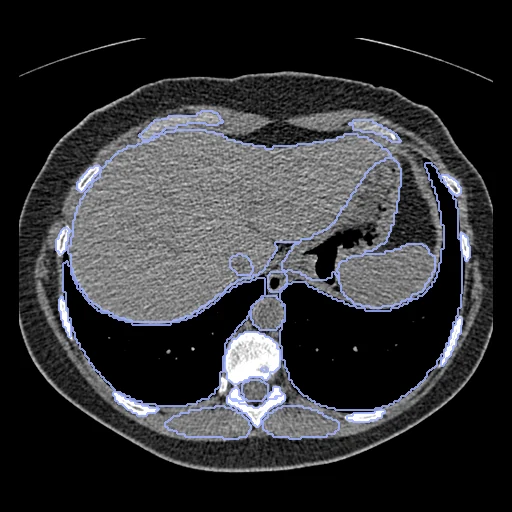
CT colonography of one patient, prone (fixed) and supine (moving) in one exam, the colon distended with gas (ACRIN 6664, the National CT Colonography Trial).
Read before comparing (7)
- Motion. The patient turns over between the two scans: the colon moves and changes shape, the gas moves to the side now up and the fluid left in the colon to the side now down, and the abdominal organs shift. A large deformation, and content that differs between the two images.
- Truth. A segmentation network's labels, the only truth, so the dataset is ranked apart from those people annotated; a method that registers on that network's features is scored against its own view of the anatomy. Both images of a pair hold 62 to 74 structures: the colon, the small bowel, the abdominal organs, vertebrae, ribs, pelvis and muscles.
- Colon. One label from the rectum to the caecum, its gas included: its overlap says whether the colon lands on the colon, not whether each segment lands on its own.
- Polyps. The trial's spreadsheets give, for 55 patients, a slice on which a polyp was seen in each position, with its segment and size. They are not turned into landmarks: a slice is all they give, the colon crosses it several times, some rows list two to eleven slices a position with nothing to pair them, and NCI cannot assure the location data.
- Alignment. The scans come in each position's scanner frame, the bones 25 to 335 mm apart. Each pair starts from an elastix rigid fit on mutual information inside each body, no label used, written in the moving image's and labels' headers. Rigid, since a translation left the pelvis visibly turned: the starts turn the moving scan by 3.8 to 30.5 degrees over the 20 pairs.
- Baseline. From that start the 10 test pairs have a label Dice of 0.48 (0.39 to 0.55) and an HD95 of 19.1 mm (11.1 to 30.5). The rigid fit on the label centres used before left 0.46, but handed the labels to the start.
- Images. Siemens, GE and Philips scanners; slices 1 to 1.25 mm thick and 0.8 mm apart, pixels of 0.59 to 0.92 mm. Prone and supine are told apart by the DICOM Patient Position; decubitus series are not used, and of two scans in one position the lower series number is taken. prepare.py sets the corners outside the reconstructed circle to air (-1024).
No method has a result on every pair of this dataset yet.
| Accuracy | Smoothness | Cost | ||||||||
|---|---|---|---|---|---|---|---|---|---|---|
| # | Method | Dice | Change | HD95 mm | SDlogJ | Folds % | Time s | RAM GB | GPU GB | Images |
| No registration | 0.478 ±0.041 | 19.10 ±4.02 | 0.000 | 0.00 | ||||||
1 result not ranked: an older run, pairs still missing, or a failure on every pair
| Accuracy | Smoothness | Cost | |||||||||
|---|---|---|---|---|---|---|---|---|---|---|---|
| # | Method | Dice | Change | HD95 mm | SDlogJ | Folds % | Time s | RAM GB | GPU GB | Machine | Images |
| ConvexAdam (MIND)GPUapp8/10 pairs | 0.713 ±0.057 | +0.235 | 13.18 ±3.92 | 0.133 | 1.65 | 77 | 9.6 | 22.5 | Laptop | ||
Dice: how much the organ contours overlap, 1 is perfect. HD95: distance between the contours' surfaces. SDlogJ: how irregular the deformation is, 0 is perfectly smooth. Folds: share of the image where the deformation folds over itself.
Pair by pair
Click a score to look at that result: the fixed image, and over it the moving image after registration, with the annotations. Underlined scores have their image; for the others you see the pair without the result. A green cell gained against no registration on that pair, a red one lost.
About the data
- Test pairs
- 10 test pairs drawn with seed 2026 among the 44 outside development, and 10 development pairs, out of the 54 patients who have a prone and a supine series and whose polyp spreadsheets give a polyp slice in both positions, for whoever marks the polyps later; 1.3.6.1.4.1.9328.50.4.0345, the 55th with such slices, has a prone series only. 1.3.6.1.4.1.9328.50.4.0518 stays in the pool, so that the draw is unchanged, but may never become development: its only supine series misses 86 of its 570 slices, and prepare.py refuses it.
- Truth
- contours by a program TotalSegmentator (task total) run by us on each image: the colon and the other structures it finds
- Source
- www.cancerimagingarchive.net/collection/ct-colonography
doi.org/10.7937/K9/TCIA.2015.NWTESAY1
doi.org/10.1056/NEJMoa0800996 - Licence
- CC BY 3.0
10 pairs for tuning, never tested
1.3.6.1.4.1.9328.50.4.0011, 1.3.6.1.4.1.9328.50.4.0040, 1.3.6.1.4.1.9328.50.4.0080, 1.3.6.1.4.1.9328.50.4.0104, 1.3.6.1.4.1.9328.50.4.0490, 1.3.6.1.4.1.9328.50.4.0347, CTC-4720202687, CTC-3174825007, CTC-3184713093, CTC-2414824407