 fidopt
Multimodal-HC, whole-body MR/CT
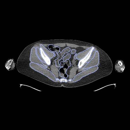
Low-dose CT (fixed) and Dixon in-phase MR (moving) of the whole body, vertex to mid-femur, of one healthy adult on two scanners in two rooms (Multimodal-HC, Rigshospitalet).
Read before comparing (10)
- Truth. Ranked apart: the provider's TotalSegmentator 2.5.0 maps, task total on the CT and total_mr on the MR, each made on its image alone, with no correction described. A method built on TotalSegmentator is checked against its own view of the anatomy. It fails the truth bar's first check: people drew neither image.
- Structures. The 48 both maps name; the CT's lobes make its lungs and its S1 joins its sacrum, which on the MR holds S1. The MR's vertebrae and discs and the CT's single vertebrae, ribs, skull and neck and chest vessels are left out. A structure one map lacks on a case is not scored there; the prostate found in six women is set to 0.
- Volumes. The two tasks draw structures differently: on the 20 cases a structure's MR volume is a median 0.79 times its CT volume, and 0.31 to 0.65 times for the adrenal glands, gallbladder, duodenum, pancreas, small bowel, main vessels, hips, sacrum and shoulder and arm bones. Even a perfect registration leaves their Dice well under 1.
- Cohort. 100 healthy Danish blood donors; the open release holds 80, 40 women and 40 men. The MR came the same day as the PET/CT for 96 of the 100 and 9, 14, 58 and 89 days later for four no file names: up to four cases may be weeks apart.
- Images. The CT is the low-dose reconstruction for the PET's attenuation correction, under free breathing, cut to the body's box plus 15 mm. The MR is the Dixon in-phase image as delivered, four breath-hold stations joined by the scanner; 7 to 13% of its body lies past the CT's ends.
- Alignment. No structure overlaps as delivered (Dice 0.00). The moving image's header carries one elastix translation on mutual information inside each body, the best of several starts, no annotation used; the voxels stay native. A rigid fit turns the MR only 0.9 to 4.5 degrees on the test pairs, a matter of posture, so the translation is kept.
- Baseline. With no registration the test pairs start at a Dice of 0.44 (0.24 to 0.54), the median over pairs of the mean over structures, and an HD95 of 30 mm (25 to 54).
- Motion. The head and neck bend differently, the arms lie closer or further, the MR is held at deep inspiration, its lungs 1.6 to 1.8 times larger, and the bladder fills in between, 1.6 times larger. A rigid start cannot meet the head, the arms and the abdomen at once.
- SyN. ANTs' SyN from ANTs' rigid start, run on three test pairs only, beats no registration on all three without folds: Dice from 0.375 to 0.459 on sub-004, 0.469 to 0.618 on sub-005, 0.446 to 0.540 on sub-015.
- Overlap. MultimodalHC_PETCT_Syn takes the CT of 20 other subjects of the release. Every test subject is public with its CT and MR, so a network trained on this release may have seen them. No registration result on these images is published.
| Accuracy | Smoothness | Cost | ||||||||
|---|---|---|---|---|---|---|---|---|---|---|
| # | Method | Dice | Change | HD95 mm | SDlogJ | Folds % | Time s | RAM GB | GPU GB | Images |
| 1 | ConvexAdam (MIND)GPUapp | 0.645 ±0.047 | +0.210 | 27.88 ±7.96 | 0.211 | 6.21 | 24 | 4.3 | 10.5 | |
| No registration | 0.434 ±0.065 | 35.00 ±7.45 | 0.000 | 0.00 | ||||||
The order averages each method's ranks on Dice, HD95 mm, SDlogJ, so the first one is not always the best in every column. ± is the 95% interval of the mean over the 10 pairs. Two neighbours are tied when a paired Wilcoxon test on the shared pairs does not find the upper one better on Dice (5%): the places a method could hold are then shown under its own. The best value of each column is in colour. Click a row to see its images, a column title to sort by it. Time and memory measured on Laptop.
Dice: how much the organ contours overlap, 1 is perfect. HD95: distance between the contours' surfaces. SDlogJ: how irregular the deformation is, 0 is perfectly smooth. Folds: share of the image where the deformation folds over itself.
Pair by pair
Click a score to look at that result: the fixed image, and over it the moving image after registration, with the annotations. Underlined scores have their image; for the others you see the pair without the result. A green cell gained against no registration on that pair, a red one lost.
About the data
- Test pairs
- 10 test subjects drawn with seed 2026 by sex (5 women, 5 men) among the 60 of the open release's 80 that MultimodalHC_PETCT_Syn does not hold, then 10 development subjects (5 and 5). No subject is in both datasets.
- Truth
- contours by a program the provider's TotalSegmentator 2.5.0 maps of each image, task total on the CT and task total_mr on the MR, reduced to the 48 structures both name, in total_mr's ids: the CT's lung lobes make the two lungs and its S1 joins its sacrum; the prostate is left out of the women's maps
- Source
- huggingface.co/datasets/DEPICT-RH/Multimodal-HC
doi.org/10.57967/hf/9560
doi.org/10.1038/s41597-026-08157-4
github.com/DEPICT-RH/Multimodal-HC - Licence
- CC BY 4.0
10 pairs for tuning, never tested
sub-032, sub-044, sub-069, sub-054, sub-075, sub-081, sub-028, sub-019, sub-076, sub-070