 fidopt
Breast-MRI-NACT-Pilot
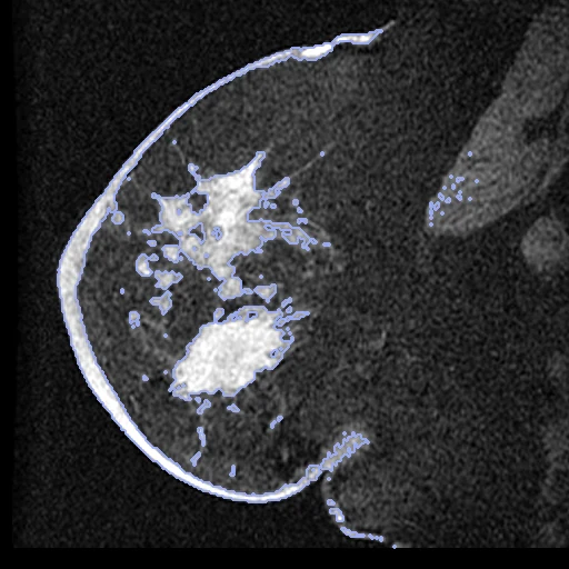
Fat-suppressed pre-contrast T1 MR of a breast with a tumour, before neoadjuvant chemotherapy (fixed) and after its first cycle (moving) (Breast-MRI-NACT-Pilot, UCSF).
Read before comparing (8)
- Truth. The breast tissue mask is the provider's analysis of each visit: the pre-contrast image above 60% of the 95th percentile inside a box an operator drew around the enhancing tumour. It keeps the fibroglandular tissue, not the outline of the breast, and no one drew or checked its boundary: graded automatic. Exclusion regions the operator drew are cut out (UCSF-BR-08 at both visits, UCSF-BR-23 at MRI 2).
- Threshold. Each visit's threshold comes from its own box: when the tumour fades it drops and the mask grows (UCSF-BR-50: 137 ml at MRI 1, 345 ml at MRI 2). Over the test pairs the mask changes by -35% to +152% between visits, and it takes in noise in the pectoral muscle and along the skin (UCSF-BR-49 holds 45 ml). No registration reaches a Dice of 1.
- Tumour. The provider's tumour mask, the tissue in the box that enhances by 70% or more on the early phase, is shown (dice_tumour) and never ranked. After one cycle its volume is a median 0.54 of the first visit's, and treatment lowers enhancement as well as size, so its overlap mostly measures the response.
- Visits. MRI 2, after the first of four cycles of adriamycin-cytoxan and 12 to 70 days after MRI 1 (median 29), is the moving visit: the later visit where tumour and breast have changed least. MRI 3 and MRI 4 are not used.
- Images. The pre-contrast phase of each visit's DCE series, the one the mask thresholds, whose contrast does not depend on enhancement. One breast a series, sagittal, 0.70 to 0.78 mm in the slice (0.35 mm on UCSF-BR-23's MRI 1) and 2.0 to 2.3 mm between slices, 1.5 T GE Signa, one site.
- Alignment. Each visit is in its own scanner frame, the patient prone in the coil; as delivered, the tumour centres of the first 13 pairs built were 8 to 38 mm apart. The moving header carries one elastix translation from the intensities, no annotation used; no voxel is resampled.
- Baseline. With no registration the ten test pairs start at a mean tissue Dice of 0.51 (0.21 to 0.79), lowest on UCSF-BR-20, where the breast lies differently in the coil, then UCSF-BR-49 (0.24) and UCSF-BR-50 (0.31). The tumour Dice starts at 0.00 to 0.66.
- Masks. Some of the provider's SEG frames sit half a pixel off their image; prepare.py puts each frame on its image's pixels, where the mask is the image above one value.
| Accuracy | Smoothness | Cost | ||||||||
|---|---|---|---|---|---|---|---|---|---|---|
| # | Method | Dice | Change | HD95 mm | SDlogJ | Folds % | Time s | RAM GB | GPU GB | Images |
| 11–2 | ConvexAdam (MIND)GPUapp | 0.656 ±0.139 | +0.147 | 14.24 ±6.48 | 0.089 | 0.40 | 9 | 2.3 | 1.6 | |
| 21–2 | DemonsCPUapp | 0.642 ±0.124 | +0.133 | 13.48 ±6.36 | 0.098 | 0.14 | 5 | 0.8 | ||
| No registration | 0.509 ±0.144 | 16.71 ±7.48 | 0.000 | 0.00 | ||||||
The order averages each method's ranks on Dice, HD95 mm, SDlogJ, so the first one is not always the best in every column. ± is the 95% interval of the mean over the 10 pairs. Two neighbours are tied when a paired Wilcoxon test on the shared pairs does not find the upper one better on Dice (5%): the places a method could hold are then shown under its own. The best value of each column is in colour. Click a row to see its images, a column title to sort by it. Time and memory measured on Laptop.
Dice: how much the organ contours overlap, 1 is perfect. HD95: distance between the contours' surfaces. SDlogJ: how irregular the deformation is, 0 is perfectly smooth. Folds: share of the image where the deformation folds over itself.
Pair by pair
Click a score to look at that result: the fixed image, and over it the moving image after registration, with the annotations. Underlined scores have their image; for the others you see the pair without the result. A green cell gained against no registration on that pair, a red one lost.
About the data
- Test pairs
- 10 test pairs drawn with seed 2026 among the 46 patients outside the first three development pairs, and 10 development pairs in all; the test pairs lie among the 39 outside development. The pool is the 49 of the 64 patients whose MRI 2 the provider analysed.
- Truth
- contours by a program tumour by a program the provider's breast tissue mask on each visit: the pre-contrast image above a threshold set from the operator's box around the tumour, which keeps the fibroglandular tissue
- Source
- www.cancerimagingarchive.net/collection/breast-mri-nact-pilot
doi.org/10.7937/K9/TCIA.2016.QHsyhJKy
www.cancerimagingarchive.net/wp-content/uploads/ISPY-1-DCE-MRI-Data-Sharing-Dictionary_v2.pdf - Licence
- CC BY 3.0
10 pairs for tuning, never tested
UCSF-BR-01, UCSF-BR-03, UCSF-BR-04, UCSF-BR-53, UCSF-BR-22, UCSF-BR-62, UCSF-BR-21, UCSF-BR-47, UCSF-BR-40, UCSF-BR-57