 fidopt
Head Neck CBCT CT (HUST)
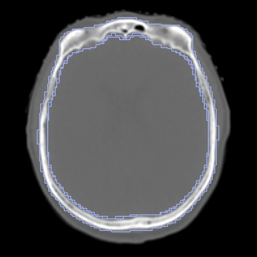
Planning CT (fixed) and first-day cone-beam CT (moving) of one head and neck radiotherapy patient at Huazhong University of Science and Technology.
Read before comparing (8)
- Truth. The archive holds the images only: the paper's expert structures and landmarks are not in it. The labels are TotalSegmentator 2.18.0's (task total) on each image, automatic truth: a method that registers on that network's features is scored against its own view of the anatomy, and the dataset is ranked apart from those people annotated.
- Values. Not Hounsfield units: the provider scaled each image to 0 to 255, air at 0, by a scale that changes between images (soft tissue reads 60 to 91 on the CTs, 17 to 90 on the CBCTs), kept as delivered. Fixed HU windows or thresholds and networks trained on HU see another contrast unless the method rescales; mutual information, NCC and MIND are not affected.
- Labels. TotalSegmentator read copies rescaled so that the background reads -1000 and the most frequent body value 40. All 17 test pairs share C1-C6, the spinal cord, the brain and the skull. The carotid arteries come out fragmented, the right in seven test pairs (median Dice 0.203), the left in twelve (0.129): weak truth.
- Field of view. The CT and the CBCT cover different superior and inferior levels. The CT's labels are cut to the CBCT's field through the starting transform, so only the common field is scored.
- Alignment. Each image keeps its scanner frame, on 240 x 240 x 64 grids of 1 x 1 x 3 mm whose centres lie 2 to 68 cm apart (median 23 cm). The moving image's and labels' headers carry one translation a patient from elastix mutual information on intensities and body masks, no annotation used; all voxels stay native.
- Baseline. With no registration from that start the 17 test pairs score a median Dice of 0.696 (0.434 to 0.820) and a median HD95 of 4.20 mm; per structure the median Dice is 0.96 for the brain, 0.878 for the skull, 0.838 for the spinal cord and 0.634 to 0.834 for C1-C6. The neck's posture is left to register.
- Images. The CT is from a Philips Brilliance CT Big Bore, resampled by the provider to 1 x 1 x 3 mm; the CBCT from a Varian Edge's On-Board Imager on the first day of treatment. Both were cropped to the head and neck, the shoulders and the couch removed. Patients 52, 60, 76, 99, 120 and 149 are missing from the archive.
- Outside numbers. None: the paper ranks rigid registrations only, on its own labels and landmarks, and does not say which patients form its test split.
| Accuracy | Smoothness | Cost | ||||||||
|---|---|---|---|---|---|---|---|---|---|---|
| # | Method | Dice | Change | HD95 mm | SDlogJ | Folds % | Time s | RAM GB | GPU GB | Images |
| 1 | ConvexAdam (MIND)GPUapp | 0.699 ±0.035 | +0.028 | 4.99 ±0.87 | 0.040 | 0.06 | 7 | 2.2 | 1.3 | |
| No registration | 0.671 ±0.045 | 4.61 ±0.82 | 0.000 | 0.00 | ||||||
The order averages each method's ranks on Dice, HD95 mm, SDlogJ, so the first one is not always the best in every column. ± is the 95% interval of the mean over the 17 pairs. Two neighbours are tied when a paired Wilcoxon test on the shared pairs does not find the upper one better on Dice (5%): the places a method could hold are then shown under its own. The best value of each column is in colour. Click a row to see its images, a column title to sort by it. Time and memory measured on Laptop.
Dice: how much the organ contours overlap, 1 is perfect. HD95: distance between the contours' surfaces. SDlogJ: how irregular the deformation is, 0 is perfectly smooth. Folds: share of the image where the deformation folds over itself.
Pair by pair
Click a score to look at that result: the fixed image, and over it the moving image after registration, with the annotations. Underlined scores have their image; for the others you see the pair without the result. A green cell gained against no registration on that pair, a red one lost.
About the data
- Test pairs
- 17 test patients drawn with seed 2026 and 15 development patients, from the 170 independent patients left once the 20 patients HNCBCTCT_Syn then held are reserved (draw.held_out). HNCBCTCT_Syn's nine later patients are none of these 32, so the two tasks use no image twice.
- Truth
- contours by a program TotalSegmentator (task total) run by us on each image, read on a copy rescaled towards Hounsfield units; the CT's labels cut to the CBCT's field of view
- Licence
- CC BY 4.0
15 pairs for tuning, never tested
case000, case001, case002, case063, case022, case127, case125, case187, case072, case038, case150, case046, case160, case039, case003