 fidopt
Mayo LDCT-and-Projection-data, low-dose to full-dose CT
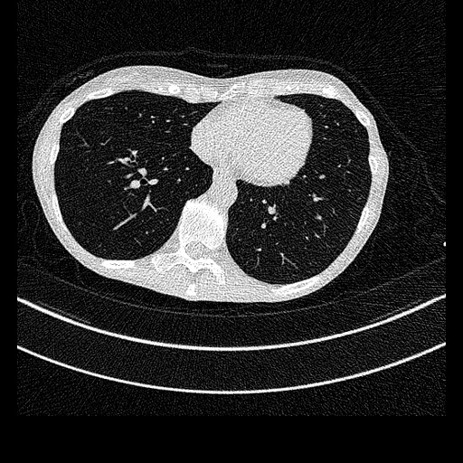
Low-dose (source) and full-dose (target) reconstructions of one chest or abdomen CT, the low dose made by Mayo Clinic inserting noise into the same projections (TCIA LDCT-and-Projection-data, Siemens exams).
Read before comparing (10)
- Dose. The low dose is the scan's own projections with noise inserted by a validated photon-counting model, to 10% of the dose for a chest and 25% for an abdomen; its measured noise matched the prediction within -2.6 ± 4.8% on the abdomens and stood 16.1 ± 8.9% above it on the chests.
- Reconstruction. Both doses by filtered back projection on one grid: kernel B50f and 1.5 mm slices every 1 mm for the chests, B30f and 5 mm slices every 3 mm for the abdomens.
- Alignment. Both reconstructions share their image positions, which prepare.py checks header by header; nothing is resampled or registered. The best translation between the images is at most 0.014 mm on the 20 cases, where the same check finds a 0.3 mm shift put in by hand.
- Target noise. The target is a routine-dose scan with noise of its own, a third of the inserted noise on a chest and 0.58 of it on an abdomen: even the noiseless image would score an MAE of roughly 30 to 100 HU on the test chests and 9 HU on the test abdomens, an estimate. Some abdomen targets hold inserted noise too; which ones is not said.
- Coverage. Scans cross regions: the chests run from C6 or C7 to L2, L3 or L4, six abdomens from the lung bases to the upper femurs, and L178, L277 and L150 from C6 to the upper femurs.
- Mask. The body on the full dose, the table left out. The field of view cuts the arms or sides of the body on nine of the ten test cases, all but L221, in 6% to 100% of their slices: what lies past the edge is in neither image.
- Baseline. Inside the mask on the ten test cases, with KonfAI's criteria at CT's default range, a constant at the full dose's median scores an MAE of 208 HU (125 to 361), a PSNR of 22.3 dB (18.0 to 25.4) and an SSIM of 0.21 (0.07 to 0.31); the low dose, the line to beat, 108 HU (15 to 299), 35.1 dB (20.4 to 46.6) and 0.70 (0.23 to 0.98).
- By region. The low dose's MAE is 201 HU (111 to 299) on the chests and 15.9 HU (14.8 to 16.8) on the abdomens, the constant's 266 and 149. On C267, the largest chest, the low dose loses to the constant on all three criteria (299 against 171 HU).
- Outside numbers. The collection was made for low-dose reconstruction and denoising, so a network trained on it may have seen these test cases. Thirteen abdomen patients were test cases of the 2016 Low Dose CT Grand Challenge: L145 here (the challenge's L136), and L123, L187 and L277 in development (L548, L631 and L106).
- Overlap. No patient of this collection is in another dataset of the corpus. A later dataset of its projections or of the 2016 challenge's cases must keep each of these patients on its side or leave it out.
No method has a result on every case of this dataset yet.
| Accuracy | Cost | ||||||||||
|---|---|---|---|---|---|---|---|---|---|---|---|
| # | Method | MAE | Change | PSNR dB | SSIM | Anatomy Dice | Anatomy HD95 mm | Time s | RAM GB | GPU GB | Images |
| Source as given | 108.4 ±77.5 | 35.11 ±8.38 | 0.700 ±0.223 | 0.860 ±0.090 | 7.00 ±5.41 | ||||||
MAE: mean absolute error of a synthetic image against its target, inside the body, in the image's units. PSNR: the same by signal-to-noise ratio, higher is better. SSIM: the same by structural similarity, 1 is perfect. Anatomy Dice and HD95: TotalSegmentator run on the synthetic and on the target image, the structures compared; shown, not ranked.
Case by case
Click a score to look at that result: the target image, and over it the method's synthetic image. Underlined scores have their image; for the others you see the case without the result.
About the data
- Test cases
- 10 test patients drawn with seed 2026 by exam type, 5 chests and 5 abdomens, among the 100 Siemens exams with both reconstructions (50 chest, 50 abdomen), then 10 development patients, 5 and 5. The GE exams, imaged at full dose only, and the head exams, under NIH controlled access, are left out.
- Truth
- target image as released the full-dose reconstruction of the scan, in Hounsfield units as delivered: the scanner's filtered back projection at the routine dose, as acquired or, for some abdomen scans acquired at a higher dose, brought down to it by the provider's noise insertion; the source is the provider's reconstruction of the same projections with more noise inserted, with the same kernel, slices and grid, so nothing is registered
- Source
- www.cancerimagingarchive.net/collection/ldct-and-projection-data
doi.org/10.7937/9npb-2637
doi.org/10.1002/mp.14594
pmc.ncbi.nlm.nih.gov/articles/PMC7985836
services.cancerimagingarchive.net/nbia-api/services/v1/getSeries?Collection=LDCT-and-Projection-data
aapm.app.box.com/s/eaw4jddb53keg1bptavvvd1sf4x3pe9h - Licence
- CC BY 4.0
10 cases for tuning, never tested
C099, L277, C158, L019, C004, L150, C067, L123, C268, L187