 fidopt
Spinal-Multiple-Myeloma-SEG, conventional CT to 40 keV monoenergetic image
Conventional CT (source) and 40 keV virtual monoenergetic image (target) of one low-dose dual-layer scan of a patient with multiple myeloma, cropped to the spine: two reconstructions of one acquisition on one grid (Spinal-Multiple-Myeloma-SEG, TCIA).
10 test casesspinesource CT, target CT
Read before comparing (9)
- Cohort. Patients with multiple myeloma scanned at the University Hospital Brno on a Philips IQon Spectral CT: whole-body low-dose scans without contrast agent, cropped by the provider to the spine. Patients without a lytic lesion of the spine, without spectral data or with heavy artefacts from spinal metal were excluded, so every case holds lesions.
- Alignment. Both images come from one helical acquisition: the conventional CT from both detector layers, the 40 keV image computed from the spectral data. prepare.py checks one frame of reference, slice positions within 0.01 mm, and the same pixel size, matrix, field of view and slice thickness; nothing is registered or resampled.
- Images. 512 x 512 pixels of 0.88 to 0.98 mm over the scanner's full field of view, arms and table included, slices every 0.6 mm from above C1 to below L5. They are noisy, as low-dose images are. At 40 keV calcium and iodine show brighter and soft tissue with more contrast.
- Clipping. Both images stop at 3071 HU, the top of their 12 bits: 0.02 to 0.20% of the target's body voxels sit there, dense bone and metal at 40 keV, against 0.06% at most in the source. Their true value is not in the files.
- Anatomy. The provider's table notes a metal implant in the spine of development patient 001, and an L6 in development patient 021 and test patient 060.
- Mask. The body of the conventional CT, above -500 HU, opened, its largest part, holes filled slice by slice: the trunk, without the table or arms that do not touch it.
- Baseline. Inside the mask on the ten test cases the source scores an MAE of 70.0 HU (57 to 84) against the target, and a constant at the target's median 219.7 HU (175 to 263).
- Geometry. TotalSegmentator reads the 40 keV image as it reads the conventional one, a mean vertebra Dice of 0.910 and 0.914 against the provider's on Myel_004, so the geometry shown beside the line means something here.
- Overlap. MyelomaSpineCT holds the same source images with the vertebrae and lesions; its 20 patients are left out. No patient of this collection is in another dataset of the corpus: VerSe is from Munich and other centres, Spine-Mets-CT-SEG from Boston, this one from Brno.
No method has a result on every case of this dataset yet.
Case by case
Click a score to look at that result: the target image, and over it the method's synthetic image. Underlined scores have their image; for the others you see the case without the result.
About the data
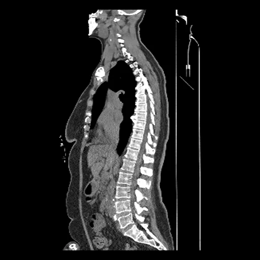
The first test caseClick to look at every case.
- Test cases
- 10 test patients drawn with seed 2026, then 10 development patients, the last of the same seeded order. The pool is the collection's 67 patients, one scan each, the earlier of two for 012, 018, 023, 043 and 052, less MyelomaSpineCT's 10 test and 10 development patients, whose conventional CT that dataset shows: 47 are left.
- Truth
- target image as released the virtual monoenergetic image at 40 keV the provider made from the scan's spectral data (Philips IntelliSpace Portal), in Hounsfield units as delivered, on the slices and pixels of the conventional CT of the same acquisition: nothing is registered or resampled
- Source
- www.cancerimagingarchive.net/collection/spinal-multiple-myeloma-seg
doi.org/10.7937/k4qv-hh78
services.cancerimagingarchive.net/nbia-api/services/v1/getSeries?Collection=Spinal-Multiple-Myeloma-SEG
www.cancerimagingarchive.net/wp-content/uploads/Spinal-Multiple-Myeloma-SEG_Table_of_image_data_description.tsv
www.cancerimagingarchive.net/wp-content/uploads/Spinal-Multiple-Myeloma-SEG_Table_of_acquisition_parameters.tsv
doi.org/10.59275/j.melba.2026-d741
github.com/MISAG-BUT/Spinal-Multiple-Myeloma-SEG - Licence
- CC BY 4.0
10 cases for tuning, never tested
Myel_080, Myel_026, Myel_027, Myel_079, Myel_001, Myel_009, Myel_068, Myel_028, Myel_021, Myel_062