 fidopt
HC18, the fetal head as a sonographer measured it
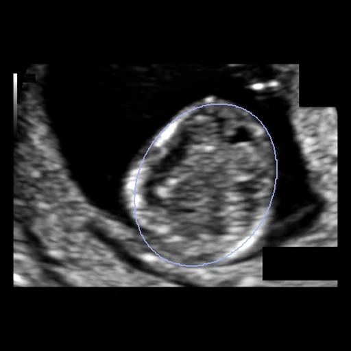
A 2D ultrasound image of the fetal head in its standard plane with the ellipse an experienced sonographer drew around the skull to measure the head circumference, filled: one examination of HC18 (Radboud University Medical Center, 2014 to 2015).
Read before comparing (10)
- Truth. A filled ellipse, not an outline: the clinical head circumference measurement, which the sonographer fitted to the skull and which the paper says never fits the skull's outer edge exactly. A method that traces the skull is scored against the ellipse. In the first trimester the skull is not yet bright and the ellipse follows a faint edge.
- Ellipse. The annotation is the ellipse's line drawn about 2.5 pixels wide; filled with its line, the label reaches about 1.3 pixels past the ellipse. The middle of the line gives the CSV's circumference to 0.01 mm on the four images checked (000_HC, 010_HC, 010_2HC, 500_HC).
- Agreement. On the provider's 335 test images a trained medical researcher's ellipses meet the sonographer's at a Dice of 0.968, 0.975 and 0.974 by trimester and a Hausdorff distance of 0.9, 1.8 and 3.3 mm (the paper's Table 4): a method near these is as close as a second person.
- Tolerance. 2 mm, about the second observer's Hausdorff distance in the second trimester, which holds most images.
- Start. On the 30 test images Otsu's dark pixels score a Dice of 0.43 and a surface Dice of 0.20 at 2 mm; one constant mask, the development ellipses' pixel majority, scores 0.81 and 0.31. The head fills the middle of most images, so the Dice says little below about 0.9 and the surface Dice separates methods.
- Images. 999 training images of the provider's 1334 from 551 pregnant women, GE Voluson E8 or 730, grey, mostly 800 x 540 pixels (test case 456_HC is 794 x 543, development case 500_HC 788 x 545), 0.052 to 0.326 mm a pixel as the CSV gives it, which the PNGs' own pixel size matches. Most screen text is blacked out in boxes.
- Subjects. The files name the examination (n in <n>_HC, <n>_2HC), numbered in the order of the head circumference, not the woman. Routine scans fall at 12 and 20 weeks and the 1334 images come from 551 women, so a test and a development case may be one woman's two examinations; nothing released tells them apart. The provider split by examination too.
- Repeats. 392_HC and 392_2HC are one file, and the annotations of examinations 243, 392, 507 and 788 repeat within their examination; one image an examination keeps one of each. SonoCorpus's copy (other file names) holds one pair of identical pixels with one annotation; in this deposit every repeated file lies within one examination.
- Outside numbers. The 999 training images and their ellipses are public since 2018; HC18's leaderboard scores the hidden test set. Networks trained on the training set (SonoBase on 698 of its images) have seen the test cases. SonoCorpus's nnU-Net, trained on HC18, scores a Dice of 0.977 on the 201 training images it held out, the second observer's level: little room is left.
- Overlap. No other dataset of the corpus holds fetal ultrasound, and SonoCorpus's MD5s match no HC18 image in another of its datasets.
No method has a result on every case of this dataset yet.
Under a licence that is not open
Methods whose code or weights are released for non-commercial use, or under share-alike or custom terms: shown for reference, ranked nowhere.
| Accuracy | Cost | ||||||||
|---|---|---|---|---|---|---|---|---|---|
| # | Method | Dice | Surface Dice | HD95 mm | Time s | RAM GB | GPU GB | Machine | Images |
| Medical SAM3 · fetal headGPUappSAM License (code); weights: MIT on the model card, a fine-tune of SAM 3's weights (SAM License)1 failedolder run | 0.896 ±0.064 | 0.666 ±0.092 | 4.13 ±1.11 | 6.6 | 6.1 | Laptop | |||
Dice: how much the organ contours overlap, 1 is perfect. Surface Dice: the share of the contours' surfaces within a tolerance of each other, 1 is perfect. HD95: distance between the contours' surfaces.
Case by case
| Dice | 1 | 2 | 3 | 4 | 5 | 6 | 7 | 8 | 9 | 10 | 11 | 12 | 13 | 14 | 15 | 16 | 17 | 18 | 19 | 20 | 21 | 22 | 23 | 24 | 25 | 26 | 27 | 28 | 29 | 30 |
|---|
Click a score to look at that result: the image, the truth in green and the method's map in red. Underlined scores have their image; for the others you see the case without the result.
About the data
- Test cases
- 30 test images drawn with seed 2026 among the 806 examinations of the provider's training set, and 30 development images from the rest; the provider's 335 test images have no published truth. One image an examination, its first (<n>_HC); one woman's two examinations cannot be told apart (notes).
- Truth
- contours by experts the ellipse that best fits the head circumference, drawn on the image by an experienced sonographer during the routine examination (van den Heuvel et al. 2018), released as a closed line about 2.5 pixels wide (<n>_HC_Annotation.png) and filled here, the line included; its circumference is the head circumference that training_set_pixel_size_and_HC.csv gives
- Source
- zenodo.org/records/1327317
doi.org/10.5281/zenodo.1327317
hc18.grand-challenge.org
doi.org/10.1371/journal.pone.0200412
pmc.ncbi.nlm.nih.gov/articles/PMC6107118 - Licence
- CC BY 4.0 (the Zenodo deposit's licence)
30 cases for tuning, never tested
146_HC, 791_HC, 689_HC, 042_HC, 037_HC, 197_HC, 434_HC, 703_HC, 458_HC, 621_HC, 685_HC, 730_HC, 786_HC, 119_HC, 632_HC, 668_HC, 001_HC, 640_HC, 608_HC, 029_HC, 487_HC, 707_HC, 802_HC, 341_HC, 741_HC, 500_HC, 561_HC, 618_HC, 696_HC, 550_HC