 fidopt
HQColon
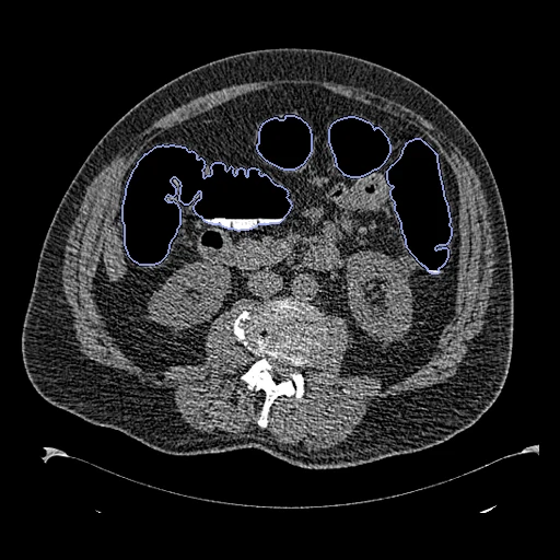
CT colonography of one patient, supine (fixed) and prone (moving) in one exam, the colon distended with gas and segmented whole on both scans (HQColon masks on TCIA's CT COLONOGRAPHY, ACRIN 6664).
Read before comparing (9)
- Motion. The patient turns over between the scans: the colon moves and changes shape, the gas moves to the side now up and the fluid to the side now down, and the organs shift. A large deformation, and content that differs between the two images.
- Truth. The gas by a threshold at -800 HU and region growing, the fluid by RootPainter, joined; a biomedical engineer reviewed every mask and corrected it by hand. A radiologist rated the reconstructions of 50 subjects as the patient's colon on all 50, disagreeing on four that the folds match the clinical software. No agreement between two people is given: graded semi-automatic.
- Label. One label, the whole colon from the rectum to the caecum with its gas and fluid: it says whether the colon lands on the colon, not each segment or fold. On the test pairs the colon holds 1.4 to 3.1 litres and its volume changes by -14 to +10% between the scans, so a field that matches the masks must change it.
- Selection. The authors dropped 51 scans that needed too much correction, the scans whose automatic seed failed and the masks with collapsed segments or running into another organ: the dataset favours well-distended colons.
- Organs. TotalSegmentator (task total), run by us on both scans with its colon taken out, gives the label set organs, 60 to 72 structures a test pair: shown, never ranked. A method that registers on that network's features is scored against its own view. The ribs, one label each, weigh in the mean Dice as much as the liver.
- Alignment. As delivered the sacrum lies 249 to 306 mm apart on most test pairs. The corpus gives each pair one rigid transform from elastix mutual information inside each body, no annotation used, written in the moving image's header; a translation alone left the bodies rolled by up to 22 degrees.
- Baseline. With no registration the 11 test pairs score a colon Dice of 0.52 (0.31 to 0.64), against 0.51 after a fit on labels and bone, and an HD95 of 30 mm (19 to 57); the organs a Dice of 0.47 (0.35 to 0.62) and an HD95 of 17 mm.
- Images. Native voxels in Hounsfield units as read; the corners outside the reconstructed circle become air (-1024). On the 40 scans built first each mask shares its CT's grid and its gas reads below -800 HU on every voxel.
- Outside numbers. None: the dataset was made for segmentation and the pairing is ours.
| Accuracy | Smoothness | Cost | ||||||||
|---|---|---|---|---|---|---|---|---|---|---|
| # | Method | Dice | Change | HD95 mm | SDlogJ | Folds % | Time s | RAM GB | GPU GB | Images |
| 1 | DemonsCPUapp | 0.695 ±0.093 | +0.178 | 26.12 ±9.68 | 0.074 | 0.17 | 149 | 18.1 | ||
| No registration | 0.517 ±0.087 | 30.17 ±7.80 | 0.000 | 0.00 | ||||||
The order averages each method's ranks on Dice, HD95 mm, SDlogJ, so the first one is not always the best in every column. ± is the 95% interval of the mean over the 11 pairs. Two neighbours are tied when a paired Wilcoxon test on the shared pairs does not find the upper one better on Dice (5%): the places a method could hold are then shown under its own. The best value of each column is in colour. Click a row to see its images, a column title to sort by it. Time and memory measured on Laptop.
1 result not ranked: an older run, pairs still missing, or a failure on every pair
| Accuracy | Smoothness | Cost | |||||||||
|---|---|---|---|---|---|---|---|---|---|---|---|
| # | Method | Dice | Change | HD95 mm | SDlogJ | Folds % | Time s | RAM GB | GPU GB | Machine | Images |
| ConvexAdam (MIND)GPUapp6/11 pairs | 0.586 ±0.134 | +0.069 | 35.94 ±15.29 | 0.133 | 1.39 | 78 | 9.8 | 22.5 | Laptop | ||
Dice: how much the organ contours overlap, 1 is perfect. HD95: distance between the contours' surfaces. SDlogJ: how irregular the deformation is, 0 is perfectly smooth. Folds: share of the image where the deformation folds over itself.
Pair by pair
Click a score to look at that result: the fixed image, and over it the moving image after registration, with the annotations. Underlined scores have their image; for the others you see the pair without the result. A green cell gained against no registration on that pair, a red one lost.
About the data
- Test pairs
- 11 test pairs drawn with seed 2026 among the 100 outside development, then 10 development pairs, the last of the same seeded order. The pool is the 113 patients HQColon segments in both positions, less three that CTColonography uses (1.3.6.1.4.1.9328.50.4.0080, 1.3.6.1.4.1.9328.50.4.0490 and CTC-3174825007), so that no scan is in both datasets.
- Truth
- contours by a program, verified by people organs by a program the whole colon (1), its gas- and fluid-filled parts, the dataset's mask on each scan
- Source
- doi.org/10.1038/s41597-025-06518-z
doi.org/10.17605/OSF.IO/8TKPM
github.com/horizon-europe-2023-ire/colon-segmentation-dataset
www.cancerimagingarchive.net/collection/ct-colonography
doi.org/10.7937/K9/TCIA.2015.NWTESAY1 - Licence
- masks CC BY 4.0, images CC BY 3.0
10 pairs for tuning, never tested
1.3.6.1.4.1.9328.50.4.0511, 1.3.6.1.4.1.9328.50.4.0776, 1.3.6.1.4.1.9328.50.4.0195, 1.3.6.1.4.1.9328.50.4.0423, 1.3.6.1.4.1.9328.50.4.0328, 1.3.6.1.4.1.9328.50.4.0659, CTC-3304961391, 1.3.6.1.4.1.9328.50.4.0007, 1.3.6.1.4.1.9328.50.4.0005, 1.3.6.1.4.1.9328.50.4.0060