 fidopt
Liver-DIR-QA
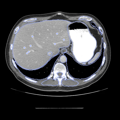
Contrast-enhanced CT of the abdomen and pelvis of one patient at two dates, 12 to 1499 days apart, with vessel bifurcations paired inside the liver (Liver-DIR-QA).
Read before comparing (5)
- Truth. Vessel bifurcations inside the liver, 17 to 140 a pair, detected on one image and carried to the other by two registration methods, pTVreg and GroupRegNet; a pair is kept when the two agree and observers accept it. The authors measure 1.3 mm against manual placement. A method close to those two may be favoured, and the score says nothing outside the liver.
- Alignment. The scans come on different grids with their origin at zero and keep their native voxels. The moving header carries one translation from elastix mutual information inside each body, the best of several starts, no annotation used. A rigid fit turns the scans 0.7 to 8.5 degrees on the test pairs, posture and breathing, so only the translation is kept.
- Baseline. The translation matches the whole body, so the liver can stay far off: on case25 the landmarks start 43 mm apart. With no registration the 10 test pairs score a landmark error of 17.5 mm (6.6 to 43.5), a label Dice of 0.44 (0.28 to 0.59) and an HD95 of 19 mm.
- Labels. TotalSegmentator (task total), run by us on each image, shown beside the landmarks, which stay the ranked truth. A method that registers on that network's features is scored against its own view of the anatomy.
- Values. Hounsfield units plus 1024, air sitting at 24 to 28, whatever the dataset's notes say of 1000.
| Accuracy | Smoothness | Cost | ||||||||||
|---|---|---|---|---|---|---|---|---|---|---|---|---|
| # | Method | Dice | Change | HD95 mm | TRE mm | rTRE | SDlogJ | Folds % | Time s | RAM GB | GPU GB | Images |
| 1 | ConvexAdam (MIND)GPUapp | 0.743 ±0.039 | +0.303 | 11.97 ±5.04 | 5.57 ±5.65 | 0.0077 ±0.0069 | 0.117 | 1.69 | 30 | 4.4 | 10.3 | |
| 2 | DemonsCPUapp | 0.658 ±0.052 | +0.219 | 14.93 ±6.57 | 11.68 ±5.59 | 0.0166 ±0.0065 | 0.100 | 1.36 | 35 | 5.7 | ||
| No registration | 0.439 ±0.084 | 19.48 ±6.69 | 17.46 ±7.78 | 0.0249 ±0.0092 | 0.000 | 0.00 | ||||||
The order averages each method's ranks on Dice, HD95 mm, TRE mm, SDlogJ, so the first one is not always the best in every column. ± is the 95% interval of the mean over the 10 pairs. Two neighbours are tied when a paired Wilcoxon test on the shared pairs does not find the upper one better on Dice (5%): the places a method could hold are then shown under its own. The best value of each column is in colour. Click a row to see its images, a column title to sort by it. Time and memory measured on Laptop.
Dice: how much the organ contours overlap, 1 is perfect. HD95: distance between the contours' surfaces. TRE: distance between matching landmarks. rTRE: the TRE over the length of the image's diagonal, to compare datasets of other sizes and units. SDlogJ: how irregular the deformation is, 0 is perfectly smooth. Folds: share of the image where the deformation folds over itself.
Pair by pair
Click a score to look at that result: the fixed image, and over it the moving image after registration, with the annotations. Underlined scores have their image; for the others you see the pair without the result. A green cell gained against no registration on that pair, a red one lost.
About the data
- Test pairs
- The 10 pairs outside development that AbdomenDIRQA does not hold, and 5 development pairs. The dataset's other 25 pairs are also pairs of Abdominal-DIR-QA, by the same authors, and are left to AbdomenDIRQA.
- Truth
- contours by a program landmarks by a program, verified by people TotalSegmentator (task total) run by us on each image, in Hounsfield units the dataset's vessel bifurcation pairs inside the liver, 17 to 140 a pair
- Licence
- CC BY 4.0
5 pairs for tuning, never tested
case01, case02, case03, case10, case22