 fidopt
PFUS1, the pelvic floor on transperineal ultrasound
The first frame of a transperineal ultrasound video of a woman's pelvic floor in the midsagittal plane, at rest, with eight structures outlined as polygons by sonographers under an expert: one woman of PFUS1 (Valme University Hospital, Seville, 2023).
10 test casespelvisimage US2D, distances in px
Read before comparing (9)
- Frames. All 14,852 frames carry eight polygons, but the paper says the sonographers drew the frames where the image changed and the rest were interpolated linearly; the deposit does not mark which. The first frame, where the tracks start, was drawn, so it is the case: the pelvic floor at rest, before the Valsalva manoeuvre.
- Repeats. The 110 patient folders hold 90 women. Six repeat another folder's video byte for byte (P011, P022, P024, P061, P090, P104). Fourteen hold the same woman recorded again: P006 and P017 (of P003), P012 (P007), P013 and P014 (P010), P100 (P041), P101 (P009), P102 (P096), P103 (P026), P105 (P092), P107 (P089), P108 (P049), P109 (P027), and probably P098 (P015). A reviewer found most by eye; grey thumbnails of three frames a folder then correlate at 0.89 to 0.95 for these pairs, 0.84 for P015 and P098, and at most 0.83 between any other two folders, the closest checked by eye. The later folder of each is left out (draw.held_out).
- Agreement. Five of the byte copies were outlined twice, apart (P003 and P011, P010 and P104, P013 and P022, P057 and P061, P087 and P090). On their first frame the two outlines meet at a Dice of 0.77 to 0.82 a video (0.44 to 0.94 a structure, the bladder the loosest) and a surface Dice of 0.59 at 10 pixels: a method near a Dice of 0.8 is as close as a second outline.
- Tolerance. 10 pixels, about 1% of the width: the polygons are coarse, 9 to 29 vertices a structure on P000.
- Start. On the 10 test cases Otsu's dark pixels score a mean Dice of 0.05 over the eight structures and a surface Dice of 0.05 at 10 pixels; one constant label map, the development cases' pixel majority on the commonest image size (empty on the others), scores 0.11 and 0.06.
- Images. Women seen in gynaecology consultations from April to July 2023, without pelvic floor dysfunction (110 by the paper, 90 distinct in the deposit), scanned by one expert on a Canon Aplio i700 with a PVT-675 MV probe. PNG frames of 499 x 345 to 1680 x 816 pixels on the drawn cases, some with the screen's scale beside the sector (P062), a tinted grey stored in colour. No pixel size is given, so distances are in pixels.
- Labels. 1 is the deposit's 'Pubis' (the pubic symphysis), 6 its 'Anus' (the anal canal); the rest of the sector is background. Polygons are filled with Pillow; neighbours share their edges, so up to 1,367 pixels a case (P047) lie under two and take the higher value.
- Outside numbers. SonoBase trained on 72 of the patients (SonoCorpus's split); the paper's 86 and 24 patients are not named. SonoCorpus's nnU-Net scores a mean Dice of 0.78 over the structures and frames of its 22 test patients, about the two outlines' agreement.
- Overlap. No other dataset of the corpus holds pelvic floor ultrasound. JNU-IFM (intrapartum videos, Jinan University, Youkey D8) shares no source with it, and SonoCorpus's MD5s match no PFUS frame in another of its datasets.
No method has a result on every case of this dataset yet.
Case by case
Click a score to look at that result: the image, the truth in green and the method's map in red. Underlined scores have their image; for the others you see the case without the result.
About the data
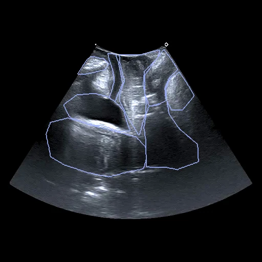
The first test caseClick to look at every case.
- Test cases
- 10 test women drawn with seed 2026 among the 90 the deposit holds, and 10 development women from the rest. The 20 patient folders that repeat another woman's recording, six byte for byte and 14 recorded again, are left out (draw.held_out). One frame a woman, the first.
- Truth
- contours by experts the deposit's polygons on the first frame of each video, drawn in CVAT by one of two sonographers under an expert pelvic floor sonographer (García-Mejido et al. 2024); the later frames, interpolated between drawn ones, are not used
- Source
- zenodo.org/records/10800787
doi.org/10.5281/zenodo.10800787
www.cs.us.es/~dsolis/en/pfus1.html
doi.org/10.1007/s00192-024-05841-0
pmc.ncbi.nlm.nih.gov/articles/PMC11732871 - Licence
- CC BY 4.0 (the Zenodo deposit's licence)
10 cases for tuning, never tested
P057, P058, P083, P026, P003, P092, P097, P005, P043, P009