 fidopt
BraTS 2021 adult glioma, T1, T2 and FLAIR to T1Gd
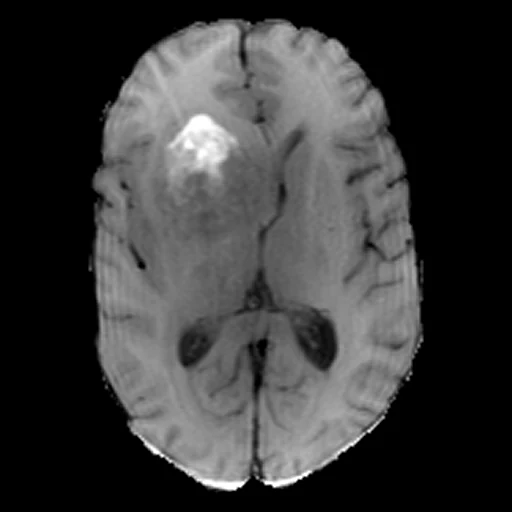
One pre-operative brain MR of an adult with a glioma, on the BraTS pipeline's skull-stripped 1 mm SRI24 grid: T1, T2 and FLAIR as three channels (source) and the post-contrast T1 (target) (BraTS 2021).
Read before comparing (11)
- Cohort. Adults with a glioma, one pre-operative examination each, scanned with different scanners and protocols at 24 sites (BraTS2021's notes). The label maps are not used.
- Task. As in BraSyn, a missing sequence is made from the others; here it is always the post-contrast T1, which needs a gadolinium injection. The source is one image with three channels, T1, T2 and FLAIR in that order.
- Alignment. The provider registered the T1, T2 and FLAIR rigidly to the T1Gd and the T1Gd to the SRI24 atlas; the corpus registers and resamples nothing. The four sequences share one header. What a rigid fit leaves, motion within a sequence or distortion, counts as error for every method.
- Intensities. Each scanner's units, as delivered. The target's median in the brain is 201 to 3,492 on the test cases, and the T1's median 0.31 to 2.25 times the T1Gd's: a method must infer the target's scale from the source. The MAE weighs the brightest scans most, above all 00638, 00044 and 00026.
- Range. truth.dynamic_range is 1635.5, the median of the 30 test targets' 99.9th percentiles inside the mask (464 to 12,650). The pooled 99.9th percentile, 8,127, would follow the three brightest scans alone.
- Mask. The brain: the voxels nonzero in any of the four skull-stripped sequences, 1.17 to 1.75 litres on the test cases.
- Baseline. Inside the mask on the 30 test cases, a constant at the target's median scores an MAE of 92.5 (21.5 to 659.1), a PSNR of 20.7 dB (3.3 to 33.9) and an SSIM of 0.49 (0.15 to 0.91). The T1 channel taken as the T1Gd scores 86.3 (33.8 to 1,965.6), 19.9 dB (-2.0 to 28.7) and 0.67 (0.10 to 0.90), losing on the MAE where its scale differs.
- Enhancement. What a method must find is the enhancing tumour, bright on the T1Gd and near the healthy brain on the T1. The target is a real acquisition with its noise and artefacts: no method reaches an MAE of 0.
- Overlap. The 60 subjects come from UPENN-GBM (19), UCSF-PDGM (15), TCGA-GBM (5), TCGA-LGG, IvyGAP and CPTAC-GBM (two each) and 15 UPenn and UCSF subjects new to TCIA; a later dataset of those collections should leave them out. The eight unnamed sites are left out because they could be the hospitals of GliomaUTSWMR, UCSDGliomaPostMR or MUGliomaPostMR, other examinations of the same patients.
- Image check. Each drawn subject's T1Gd correlates at most 0.39 with BraTSReg's 52 pairs and GliomaUTSWMR's 40 cases, and at most 0.37 with MUGliomaPostMR's 38, where one subject moved by a voxel keeps 0.79 to 0.94.
- Outside numbers. BraSyn scores its own hidden cases, each missing one sequence at random, with other measures: no published number is scored as here. The training images are public and BraSyn's data are based on BraTS 2021, so a model trained on BraTS data may have seen these subjects and their T1Gd.
No method has a result on every case of this dataset yet.
Case by case
Click a score to look at that result: the target image, and over it the method's synthetic image. Underlined scores have their image; for the others you see the case without the result.
About the data
- Test cases
- 30 test subjects drawn by contributing site with seed 2026, then 30 development subjects, the last of the same seeded order, the sites in proportion: 15 UPenn, 10 UCSF and one each from five others. The pool is the 1,065 BraTS 2021 training subjects outside BraTS2021's 30 test and 30 development subjects as first drawn (draw.held_out), whose T1Gd is public there, BraTSReg's five, and the 124 subjects of eight sites TCIA does not name; BraTS2021's later development subjects were drawn outside this dataset's cases.
- Truth
- target image as released the examination's post-contrast T1 (T1Gd) as delivered, on its source's grid through the provider's own rigid co-registration of the four sequences to the SRI24 atlas
- Source
- www.cancerimagingarchive.net/analysis-result/rsna-asnr-miccai-brats-2021
doi.org/10.7937/jc8x-9874
doi.org/10.48550/arXiv.2107.02314
cbica.github.io/CaPTk/preprocessing_brats.html
doi.org/10.48550/arXiv.2305.09011
www.cancerimagingarchive.net/wp-content/uploads/BraTS2021_MappingToTCIA.xlsx
doi.org/10.1109/tmi.2014.2377694
doi.org/10.1038/sdata.2017.117
doi.org/10.7937/K9/TCIA.2017.KLXWJJ1Q
doi.org/10.7937/K9/TCIA.2017.GJQ7R0EF
doi.org/10.7937/K9/TCIA.2016.RNYFUYE9
doi.org/10.7937/K9/TCIA.2016.L4LTD3TK
doi.org/10.7937/tcia.bdgf-8v37
doi.org/10.7937/TCIA.709X-DN49 - Licence
- CC BY 4.0
30 cases for tuning, never tested
BraTS2021_00220, BraTS2021_00718, BraTS2021_00273, BraTS2021_00533, BraTS2021_00412, BraTS2021_01364, BraTS2021_00523, BraTS2021_00837, BraTS2021_01406, BraTS2021_01542, BraTS2021_00162, BraTS2021_01514, BraTS2021_00630, BraTS2021_01300, BraTS2021_00746, BraTS2021_00459, BraTS2021_01336, BraTS2021_00583, BraTS2021_00736, BraTS2021_00231, BraTS2021_01381, BraTS2021_01547, BraTS2021_01360, BraTS2021_00759, BraTS2021_00139, BraTS2021_00275, BraTS2021_00626, BraTS2021_00131, BraTS2021_01077, BraTS2021_01278