 fidopt
Sunnybrook cine MR, left ventricle
Short-axis cine MR stack of the heart of one patient, at end diastole or end systole, with the left ventricular cavity drawn by a cardiologist (MICCAI 2009).
Read before comparing (1)
- Cases. Both images of each of Sunnybrook's pairs, as delivered, each on its pair's side of the split: a patient's two frames are two cases of the same split.
No method has a result on every case of this dataset yet.
Case by case
Click a score to look at that result: the image, the truth in green and the method's map in red. Underlined scores have their image; for the others you see the case without the result.
About the data
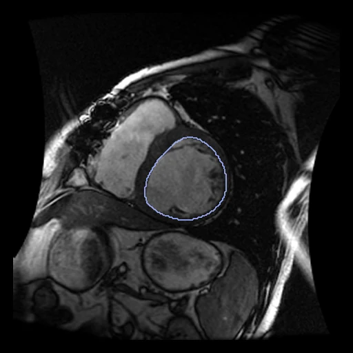
- Truth
- contours by experts the left ventricular cavity inside the endocardial contours a cardiologist drew on each slice of the two frames
- Licence
- CC0 1.0 (the authors ask that Radau et al., MIDAS Journal 2009, be cited)
20 cases for tuning, never tested
SC-HF-I-01/fixed, SC-HF-I-01/moving, SC-HF-I-02/fixed, SC-HF-I-02/moving, SC-HF-I-04/fixed, SC-HF-I-04/moving, SC-HF-NI-31/fixed, SC-HF-NI-31/moving, SC-HYP-09/fixed, SC-HYP-09/moving, SC-HF-NI-12/fixed, SC-HF-NI-12/moving, SC-HYP-06/fixed, SC-HYP-06/moving, SC-HF-I-05/fixed, SC-HF-I-05/moving, SC-HF-NI-13/fixed, SC-HF-NI-13/moving, SC-HYP-08/fixed, SC-HYP-08/moving