 fidopt
Cine MR of pigs at 7 T, left ventricle
Short-axis cine MR stack of one pig's heart at 7 T, at end diastole or end systole, 12 days after a myocardial infarction or sham procedure, the left ventricular cavity and myocardium drawn by hand.
20 test casesheartimage MR
Read before comparing (1)
- Cases. Both images of each of PigCardiacMR's pairs, as delivered: a test pair's images are test cases, a development pair's development cases, so the two frames of one pig are always on the same side.
No method has a result on every case of this dataset yet.
Case by case
Click a score to look at that result: the image, the truth in green and the method's map in red. Underlined scores have their image; for the others you see the case without the result.
About the data
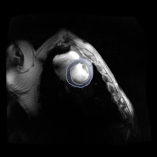
The first test caseClick to look at every case.
- Truth
- contours by experts the left ventricular cavity (1, the papillary muscles inside) and the myocardium (2), within the endocardial and epicardial contours observer one drew by hand on the short-axis slices of the two frames (the provider's masks of that reading)
- Source
- zenodo.org/records/7684034
github.com/chfc-cmi/cmr-cine-sscrofa
doi.org/10.1038/s41598-024-61417-4 - Licence
- CC BY 4.0 (the repository asks that the corresponding publication be cited: Kollmann et al., Scientific Reports 2024)
2 cases for tuning, never tested
A26/fixed, A26/moving