 fidopt
Cardiac CT of pigs, heart and left ventricle
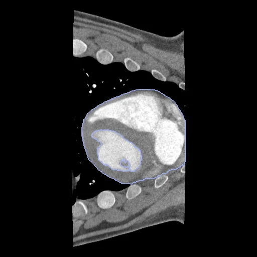
Contrast-enhanced ECG-gated cardiac CT of one growing pig, the first phase of the 4D scan, with the heart and its left ventricular cavity drawn by hand.
Read before comparing (4)
- Cases. The fixed images of PigCardiacCT's pairs, each on its pair's side of the split. The moving images are left out.
- Truth. The provider says the masks were drawn by hand on the contrast-enhanced images, not by whom, with which tool or how far two readers differ. The left ventricle (2) is cut out of the heart (1), and its intensities (median 243 to 327 HU on three scans checked) make it the contrast-filled cavity: label 1 holds the myocardium and the other chambers.
- Images. ECG-gated 4D CT of 10 or 11 phases on two Canon scanners. The first phase (IM00001) is the one the provider says typically corresponds to diastole; on 60 of the 62 annotated scans the left ventricle is larger there than at the fifth. Hounsfield units as delivered, -2048 outside the reconstruction circle.
- Coverage. Two large labels: the board says nothing of the chambers, valves or vessels one by one.
No method has a result on every case of this dataset yet.
1 result not ranked: an older run, cases still missing, or a failure on every case
| Accuracy | Cost | |||||||
|---|---|---|---|---|---|---|---|---|
| # | Method | Dice | Surface Dice | Time s | RAM GB | GPU GB | Machine | Images |
| MONAI wholeBody CTGPUapp1 of 2 structuresfailed on every case | 0.000 ±0.000 | 0.000 ±0.000 | Workstation | |||||
Dice: how much the organ contours overlap, 1 is perfect. Surface Dice: the share of the contours' surfaces within a tolerance of each other, 1 is perfect.
Case by case
| Dice | 1 | 2 | 3 | 4 | 5 | 6 | 7 | 8 | 9 | 10 |
|---|
Click a score to look at that result: the image, the truth in green and the method's map in red. Underlined scores have their image; for the others you see the case without the result.
About the data
- Truth
- contours by experts the provider's masks of the first phase of each scan, drawn by hand on the contrast-enhanced images: 1 the heart outside the left ventricle, 2 the left ventricle (its contrast-filled cavity)
- Source
- doi.org/10.25452/figshare.plus.30398977.v2
plus.figshare.com/articles/dataset/A_spatially_and_temporally_aligned_contrast-non-contrast_cardiac_CT_dataset_of_pigs/30398977 - Licence
- CC BY 4.0
7 cases for tuning, never tested
Control_1_7569_rounds_1_2/fixed, Control_1_7569_rounds_2_3/fixed, Control_1_7569_rounds_4_5/fixed, Stent_1_7547_rounds_1_2/fixed, Stent_1_7547_rounds_2_3/fixed, Stent_1_7547_rounds_4_5/fixed, Control_1_7569_rounds_5_6/fixed