 fidopt
OpenEar, inter-specimen temporal-bone CBCT
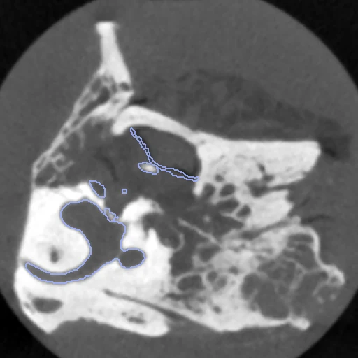
A pair of distinct cadaver temporal bones, CBCT to CBCT, scored on the nine small middle- and inner-ear structures both specimens hold whole, drawn by the provider (OpenEar).
Read before comparing (7)
- Images. The provider's registered CBCT of each full, unembedded specimen, at 0.125 mm, as delivered. ALPHA has no such scan: its only CBCT is of the specimen cut for embedding, a 39 x 39 x 62 mm field, which still holds every scored structure.
- Labels. Each segment of the provider's Slicer segmentation, found by name, is sampled by nearest neighbour onto its CBCT through the provider's registration in the headers. A voxel two kept structures claim, 0 to 38 a specimen, is left at 0.
- Scored set. A structure is kept when no voxel touches a face of the provider's segmentation box, checked on every build, and it looks whole in three planes on all six specimens: the ossicles, scalae, chorda tympani and tympanic membrane, the facial nerve filling its canal and the vestibulocochlear nerve the internal auditory canal. DELTA's vestibulocochlear nerve touches the box, so DELTA__ZETA has eight.
- Not scored. The temporal bone, whose mask stops at the box and at ALPHA's saw cut; sinus and dura, a block drawn over empty space; the external auditory canal and the internal carotid artery, closed by flat planes where the lumen goes on, so a pair's two masks cover different lengths.
- Alignment. Both specimens keep their native voxels. The moving image's and labels' headers carry one rigid transform from elastix mutual information inside each body, no label used, run from each of the 24 axis-aligned rotations of the moving specimen and kept where the densest tenth of each body, the bone, overlaps most. The specimens were scanned 167 degrees apart on ALPHA__THETA, 78 on DELTA__ZETA and 22 on EPSILON__ETA, further than mutual information turns from a single start. Size and shape are left to the methods.
- Baseline. With no registration the two test pairs start at a label Dice of 0.23 (0.22 to 0.23) and an HD95 of 2.3 mm (2.0 to 2.6), the development pair at 0.16 and 2.0 mm. The structures sit within a few millimetres of each other but overlap little; the stapes, facial nerve and chorda tympani score 0 to 0.10 on the test pairs.
- Ranking. Two test pairs from at most four donors: results are shown, not ranked. The specimens are OpenEar_Seg's, and post-mortem specimens on a 0.125 mm CBCT do not stand for clinical scans of the ear.
| Accuracy | Smoothness | Cost | ||||||||
|---|---|---|---|---|---|---|---|---|---|---|
| # | Method | Dice | Change | HD95 mm | SDlogJ | Folds % | Time s | RAM GB | GPU GB | Images |
| 11–4 | ConvexAdam (MIND)GPUapp | 0.596 ±0.699 | +0.114 | 0.94 ±5.12 | 0.128 | 0.74 | 54 | 8.2 | 15.7 | |
| 21–4 | NiftyRegCPUapp | 0.469 ±0.751 | −0.014 | 2.20 ±2.73 | 2.190 | 25.87 | 773 | 12.7 | ||
| 31–4 | multiGradICONGPUapp | 0.150 ±1.901 | −0.332 | 7.23 ±56.95 | 0.289 | 0.35 | 116 | 9.0 | 14.0 | |
| 41–4 | uniGradICONGPUapp | 0.161 ±2.050 | −0.321 | 8.77 ±80.47 | 0.350 | 1.78 | 132 | 9.0 | 14.0 | |
| No registration | 0.482 ±0.530 | 0.97 ±1.06 | 0.000 | 0.00 | ||||||
The order averages each method's ranks on Dice, HD95 mm, SDlogJ, so the first one is not always the best in every column. ± is the 95% interval of the mean over the 2 pairs. Two neighbours are tied when a paired Wilcoxon test on the shared pairs does not find the upper one better on Dice (5%): the places a method could hold are then shown under its own. The best value of each column is in colour. Click a row to see its images, a column title to sort by it. Time and memory measured on Laptop.
Dice: how much the organ contours overlap, 1 is perfect. HD95: distance between the contours' surfaces. SDlogJ: how irregular the deformation is, 0 is perfectly smooth. Folds: share of the image where the deformation folds over itself.
Pair by pair
Click a score to look at that result: the fixed image, and over it the moving image after registration, with the annotations. Underlined scores have their image; for the others you see the pair without the result. A green cell gained against no registration on that pair, a red one lost.
About the data
- Test pairs
- Two test pairs and one development pair, kept within the OpenEar_Seg specimen partitions; each specimen is in one pair only, and GAMMA is unused. The eight specimens come from four donors and the deposit gives no donor crosswalk, so a pair's two specimens may be one donor's two ears.
- Truth
- contours by experts The provider's manual, threshold-assisted Slicer segments of the structures both images of a pair hold whole, as one label map: 1 scala tympani, 2 scala vestibuli (with the vestibule and the semicircular canals, as the provider drew it), 3 malleus, 4 incus, 5 stapes, 6 vestibulocochlear nerve (the content of the internal auditory canal), 7 facial nerve, 8 chorda tympani, 9 tympanic membrane. A structure cut by the provider's segmentation box in one specimen is left out of both maps of its pair: the vestibulocochlear nerve of DELTA, so DELTA__ZETA has eight. The temporal bone, sinus and dura, the external auditory canal and the internal carotid artery are not scored (notes).
- Licence
- CC BY 4.0
1 pair for tuning, never tested
DELTA__ZETA