 fidopt
Uterine Myoma MRI
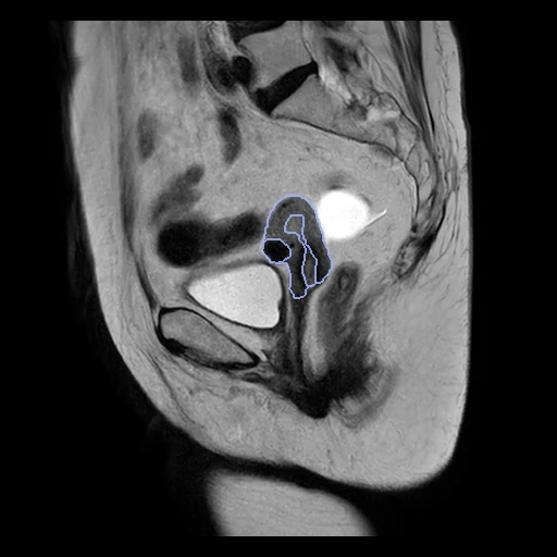
Sagittal T2-weighted MRI of the pelvis of a woman with uterine myomas, with the uterine wall, cavity, myomas and nabothian cysts outlined by hand (UMD, Beijing Shijitan Hospital, Pan et al. 2024).
Read before comparing (7)
- Cohort. 300 women with myomas confirmed at surgery and by pathology, scanned from 2015 to 2023 on a Philips Ingenia 3.0 T; malignancy, adenomyosis, pregnancy, menstruation and severe motion were excluded. Values are the files' own, 16-bit integers (32-bit floats on 18 of the 267), no unit; the headers' axes are right.
- Truth. Two of eight experienced clinicians outlined each case in ITK-SNAP until they agreed, then a radiologist reviewed it; a doubtful myoma was checked on the other sequences. No agreement between annotators is published, so the truth's own error is unknown. It meets the bar of a main leaderboard: drawn by people on every scan, with room left.
- Labels. The cavity takes in the junctional zone, so it is not the endometrium alone; the wall runs down the cervix, where the cysts lie. Labels 1 to 3 are in every case, label 4 in 11 of the 26 test cases; a case without a cyst does not score label 4 unless a method writes it.
- Sizes. A case holds a median of 97 ml of wall, 17 ml of myoma and 4 ml of cavity, and 0.2 ml of cyst where there is one; the test myomas range from 0.24 to 918 ml (UMD_221129_016's fills the pelvis).
- Slices. 20 to 32 sagittal slices 4.4 to 7.5 mm apart, pixels of 0.25 to 0.78 mm, several protocols over the years. Across the slices a surface is known to a slice, so the HD95 and the surface Dice (at 1 mm, the provider giving no tolerance) move by whole slices there.
- Strata. The archive gives no FIGO type a case, so the draw has no strata.
- Room. An nnU-Net trained on 246 of the 300 cases reached a Dice of 0.86 for the wall, 0.79 for the cavity, 0.70 for the myomas and 0.68 for the cysts (Saleem and Yaqub 2025), on another split whose training cases may hold these test cases: a network trained on UMD may have seen them. TotalSegmentator makes none of the four structures.
No method has a result on every case of this dataset yet.
Case by case
Click a score to look at that result: the image, the truth in green and the method's map in red. Underlined scores have their image; for the others you see the case without the result.
About the data
- Test cases
- 26 test cases drawn with seed 2026 among the 243 outside development, and 24 development cases, one scan a patient, from a pool of 267. The 33 cases whose label map came without geometry, 30 of them also with stray marks, are left out.
- Truth
- contours by experts the dataset's outlines of the uterine wall, the uterine cavity, the myomas and the nabothian cysts, drawn in ITK-SNAP by two clinicians to consensus and reviewed by a radiologist
- Source
- doi.org/10.6084/m9.figshare.23541312.v3
doi.org/10.1038/s41597-024-03170-x
arxiv.org/abs/2508.11010 - Licence
- CC BY 4.0
24 cases for tuning, never tested
UMD_221129_168, UMD_221129_101, UMD_221129_279, UMD_221129_265, UMD_221129_012, UMD_221129_245, UMD_221129_149, UMD_221129_277, UMD_221129_051, UMD_221129_246, UMD_221129_283, UMD_221129_210, UMD_221129_102, UMD_221129_125, UMD_221129_231, UMD_221129_056, UMD_221129_143, UMD_221129_026, UMD_221129_212, UMD_221129_275, UMD_221129_289, UMD_221129_261, UMD_221129_047, UMD_221129_178