 fidopt
NCI-ISBI 2013 prostate
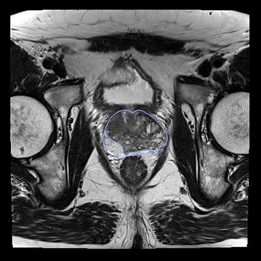
Transverse T2 MR of one patient's prostate, at 3 T without an endorectal coil (Radboud) or at 1.5 T with one (Boston Medical Center), its central gland and peripheral zone outlined by experts (NCI-ISBI 2013).
10 test casesprostateimage MR
Read before comparing (8)
- Truth. Experts of Boston University, Case Western and Radboud outlined both zones slice by slice; the pages do not say who drew which case. They are the challenge's reference, held by its organisers for scoring, then released. No second reading is published, so how far two experts would differ is not known.
- Labels. Two labels that do not overlap make the gland: 1 the peripheral zone, 2 the central gland, the rest of the gland. The pages do not say which value is which, and the challenge once had them swapped on ProstateDx-01-0006. On the 20 held-out maps label 1 is the posterior crescent, its centre 8 to 20 mm behind label 2's.
- Size. In the test cases the gland holds 16 to 73 mL (median 37), the peripheral zone 15 to 49% of it, over 8 to 15 slices.
- TotalSegmentator. Its total_mr task writes the prostate whole, which counts as neither zone: a method that writes only the gland scores 0 on both. On Prostate3T-03-0001 and ProstateDx-03-0001 its prostate overlaps the union of the two labels at a Dice of 0.83 and 0.84.
- Images. The ten held-out Prostate-3T series come from a Siemens Skyra, 3 T, by their DICOM headers: voxels of 0.5 x 0.5 x 3 mm, tilted 4 to 17 degrees. PROSTATE-DIAGNOSIS comes from a Philips Achieva, 1.5 T, voxels of 0.4 x 0.4 x 3 mm, the endorectal coil's balloon a dark disc behind the gland. Each image keeps its own grid.
- Values. As the DICOM reader gives them: Siemens' 16-bit integers, and Philips' stored values times each series' rescale slope, kept as float32. The two collections share no scale, nor do two Philips series.
- Grid. The Prostate-3T NRRDs place their grid half a voxel off the DICOM's; prepare.py writes every label map on its T2's grid.
- Overlap. PROMISE12's Radboud images include 18 of the 30 Prostate-3T training images, matched by size, spacing and origin to 0.01 mm; this dataset holds none of them. Its 20 held-out images match none of PROMISE12's 100 images nor QINProstateRepeat's: no image is in the corpus twice.
No method has a result on every case of this dataset yet.
Case by case
Click a score to look at that result: the image, the truth in green and the method's map in red. Underlined scores have their image; for the others you see the case without the result.
About the data
The first test caseClick to look at every case.
- Test cases
- The challenge's 10 test cases, 5 from each collection, are the test cases, and its 10 leaderboard cases, 5 from each, the development cases; the draw (seed 2026, by collection) takes all ten. The 60 training cases are left out: they are what models of this task are trained on, and 18 of their 30 Prostate-3T images are in PROMISE12.
- Truth
- contours by experts the peripheral zone and the central gland (the gland outside its peripheral zone), outlined slice by slice by experts of Boston University, Case Western and Radboud, the challenge's reference
- Source
- www.cancerimagingarchive.net/analysis-result/isbi-mr-prostate-2013
doi.org/10.7937/K9/TCIA.2015.zF0vlOPv
wiki.cancerimagingarchive.net/display/Public/NCI-ISBI+2013+Challenge+-+Automated+Segmentation+of+Prostate+Structures
www.cancerimagingarchive.net/collection/prostate-3t
www.cancerimagingarchive.net/collection/prostate-diagnosis - Licence
- CC BY 3.0
10 cases for tuning, never tested
Prostate3T-02-0001, Prostate3T-02-0002, Prostate3T-02-0003, Prostate3T-02-0004, Prostate3T-02-0005, ProstateDx-02-0001, ProstateDx-02-0002, ProstateDx-02-0003, ProstateDx-02-0004, ProstateDx-02-0005